
 ?Mathematical formulae have been encoded as MathML and are displayed in this HTML version using MathJax in order to improve their display. Uncheck the box to turn MathJax off. This feature requires Javascript. Click on a formula to zoom.
Abstract
Magnetic Fe3O4 nanoparticles were prepared via a simple hydrothermal method and utilized to load paclitaxel. The average particle size of Fe3O4 nanoparticles was found to be 20.2 ± 3.0 nm, and the calculated saturation magnetization reached 129.38 emu/g, verifying superparamagnetism of nanomaterials. The specific surface area and pore volume were 84.756 m2/g and 0.265 cm3/g, respectively. Subsequently, Fe3O4@mSiO2 nanoparticles were successfully fabricated using the Fe3O4 nanoparticles as precursors with an average size of 27.81 nm. The relevant saturation magnetization, zeta potential, and specific surface area of Fe3O4@mSiO2-NH2-FA were respectively 76.3 emu/g, −14.1 mV, and 324.410 m2/g. The pore volume and average adsorption pore size were 0.369 cm3/g and 4.548 nm, respectively. Compared to free paclitaxel, the solubility and stability of nanoparticles loaded with paclitaxel were improved. The drug loading efficiency and drug load of the nanoformulation were 44.26 and 11.38%, respectively. The Fe3O4@mSiO2-NH2-FA nanocomposites were easy to construct with excellent active targeting performance, pH sensitivity, and sustained-release effect. The nanoformulation also showed good biocompatibility, where the cell viability remained at 73.8% when the concentration reached 1200 μg/mL. The nanoformulation induced cell death through apoptosis, as confirmed by AO/EB staining and flow cytometry. Western blotting results suggested that the nanoformulation could induce iron death by inhibiting Glutathione Peroxidase 4 (GPX4) activity or decreasing Ferritin Heavy Chain 1 (FTH1) expression. Subsequently, the expression of HIF-1α was upregulated owing to the accumulation of reactive oxygen species (ROS), thus affecting the expression of apoptosis-related proteins regulated by p53, inducing cell apoptosis.
1. Introduction
Breast cancer is a systemic disease with a strong malignancy and a complicated cause (Mubarik et al., Citation2022; Yin et al., Citation2022; Xie et al., Citation2022a). As of 2020, breast cancer has surpassed lung cancer as the most common cancer worldwide. According to GLOBOCAN statistics in 2020, new cases of breast cancer in women worldwide accounted for 24.5% of all tumors. Breast cancer is caused by numerous factors including the environment (Cheng et al., Citation2022), heredity (Lee et al., Citation2022), and other factors (Ro et al., Citation2022). According to a summary of relevant papers published by Chinese clinicians on the treatment of breast cancer (Killelea et al., Citation2020; He et al., Citation2021), the current clinical treatment methods (Dinparvar et al., Citation2020; Liu et al., Citation2020) are mainly a combination of surgical treatment (Efetov et al., Citation2020; Jarvis et al., Citation2020; Webster et al., Citation2020) and chemotherapy (Guay et al., Citation2022; Karampinis et al., Citation2022), or radiotherapy (Yuan et al., Citation2020; Hashida et al., Citation2021; Hata et al., Citation2021; Wei et al., 2021) and targeted therapy (Chen et al., Citation2020). Currently, the chemotherapeutic drugs anthracycline and taxane are widely used in breast cancer and are the benchmark for future targeted therapies (Wala et al., Citation2022).
Paclitaxel (PTX), also known as taxol, is one of the most outstanding natural anticancer drugs that has been found (Yu et al., Citation2022a). It has been widely used in the clinical treatment of breast, ovarian, and lung cancer. As a diterpenoid alkaloid compound with anticancer activity, PTX has been greatly favored by botanists, chemists, pharmacologists, and molecular biologists owing to its novel and complex chemical structure, extensive and significant biological activity, and unique mechanisms of action. Hence, it has become an anti-cancer drug and a research focus since the second half of the 20th century (Vergote et al., Citation2020). However, the chemical structure of PTX is composed of several hydrophobic groups, making it extremely insoluble in water and difficult to inject intravenously. Hence, improving the bioavailability of PTX has become the focus of current research (Gade et al., Citation2022).
At present, the most commonly used clinical drug preparation is a mixture of polyoxyethylene castor oil and ethanol, which tends to precipitate as a result of solvent conversion. Furthermore, polyoxyethylene castor oil may cause severe allergic reactions, nephrotoxicity, and neurotoxicity when degraded in vivo, posing a great risk to clinical use. Therefore, research on new dosage forms of taxol with high efficiency and low toxicity has been the focus of new drug research in recent years (Suzuki et al., Citation2022; Vemuri et al., Citation2022; Zhang et al., Citation2022a).
Currently, new dosage forms of PTX mainly include emulsions (Li et al., Citation2022a), micelles (Abdallah et al., Citation2020), precursor drugs (Al-Hilfi & Walker, Citation2022), inclusion compounds, liposomes (Duan et al., Citation2022), nanoparticles (Gulsu et al., Citation2022; Chen et al., Citation2022b; Li et al., Citation2022c), and drug-releasing scaffolds (Obayemi et al., Citation2020). PTX nanoemulsion has many advantages, such as improved therapeutic effects, reduced side effects, maintained drug activity, and enhanced cell uptake and biological activity; however, their effectiveness and safety remain to be confirmed. Liposomes and micelles are promising drug carriers; however, their targeting, stability, and encapsulation efficiency remain to be improved (Alavi & Nokhodchi, Citation2022). The Cyclodextrin inclusion complex provided a feasible scheme for the oral preparation of PTX. The synthesis of precursor drugs is a complicated and expensive process, and their pharmacodynamics and pharmacokinetic properties may change unexpectedly. Regarding PTX preparation, nanoparticles have the advantages of high drug loading, large specific surface area, significantly increasing water solubility and reduced toxic and side effects (Fu et al., Citation2022; Gulsu et al., Citation2022; Sakhi et al., Citation2022). Magnetic nanodrug delivery systems have attracted increasing attention because of their good biocompatibility and multifunctional carrying capacity. Magnetic iron oxide nanoparticles have been applied to magnetic drug delivery systems by many researchers owing to their perfect preparation technology and good biocompatibility (Işıklan et al. Citation2022).
Magnetic nanomaterials have many unique advantages (Liu et al., Citation2022; Zhang et al., Citation2022b) and have been used in many aspects of production and life (Ni et al., Citation2022; Yu et al., Citation2022b). Magnetic nanoparticles are primarily composed of Fe, Ni, Co, Cu, and their oxides. Fe and its oxides are widely used in the field of biomedicine owing to their good biocompatibility and application prospects in targeted delivery, biological separation, medical imaging, and other fields (Singh & Amiji, Citation2022; Zhao et al., Citation2022; Wang et al., Citation2022a). Fe3O4 stands out from other magnetic nanoparticles because of its simple and convenient preparation and unique physiological and catalytic properties. The size, surface characteristics, and magnetism of magnetic Fe3O4 nanoparticles are considered when they are applied in the biomedical field. For instance, size mainly determines how the particle enters cells and how long it circulates in the body, while the surface characteristics determine their ability to bind to drugs, and the active targeting effect of a drug is influenced by its magnetic properties. Simple magnetic Fe3O4 nanomaterials often cannot meet the delivery requirements for various drugs owing to their single surface groups. Therefore, various surface modification methods have been proposed to improve surface characteristics to achieve higher drug loading or other requirements (Dutta et al., Citation2022; Kovrigina et al., Citation2022; Vieira et al., Citation2022). For example, it has been reported that multicomponent composite nanomaterials have been prepared by microwave-assisted synthesis (Kumar et al., Citation2018; Citation2020; Kalia et al., Citation2022) or chemical coprecipitation method (Tan et al., Citation2020), which significantly improves their electrochemical performance and promotes the development of high-performance advanced energy storage materials. Compared with the traditional physical method, the chemical method is preferred because of its advantages of mass production and low cost. Hence, chemical synthesis is the most commonly used preparation method at present, which includes coprecipitation (Arumugam et al., Citation2022; Caldera et al., Citation2022; Sulaiman et al., Citation2022), high temperature pyrolysis (Li et al., Citation2022d), as well as hydrothermal (Yoko et al., Citation2022), microemulsion and green biosynthesis methods (Zheng et al., Citation2019; Chen et al., Citation2022a), etc. Compared with other methods, the hydrothermal method utilized in this study can improve the magnetic properties of Fe3O4 nanomaterials at high temperature. Meanwhile, the volatilization of each component can be reduced in the high-pressure environment, thus improving the purity and magnetism of the product. Hydrothermal method has the advantages of simple operation, low raw materials and low pollution. To reduce the self-agglutination phenomenon of magnetic Fe3O4 nanoparticles and overcome their shortcomings of being easily oxidized and corroded, various surface modification methods have been applied to modify iron oxide nanoparticles. Common modification materials can be classified as organic or inorganic, each is applied to meet different modification requirements owing to their different characteristics. The relevant materials for surface modification utilized in the biomedical field mainly include metals and metal oxides, silicon dioxide (Michałowska & Kudelski, Citation2022), carbon materials (Fukushima et al., Citation2022), high polymers (Wang et al., Citation2022b), biological materials (Li et al., Citation2022b), and so on. Polysaccharides (Gong et al., Citation2022), proteins, and peptides are the most commonly used biomaterials for surface modification (Jiang et al., Citation2022). Polysaccharide, a biopolymer, is used to modify the surface of the nanoparticles by embedding covalent coupling. Hence, a hydrophilic coating can improve the biocompatibility and stability of nanoparticles (Deng et al., Citation2022). Polysaccharides are classified as neutral, positively charged, or negatively charged owing to their surface charge properties. Chitosan coating is commonly used for surface modification because the positive charge on its surface can absorb negatively charged tumor cells (Resen et al., Citation2022; Shahdeo et al., Citation2022). Hyaluronic acid (Dong et al., Citation2022), a ligand of CD44, has also been used to modify the surface of targeted delivery systems and improve their targeting performance. The protein most widely used for surface modification is bovine serum albumin (Araya-Sibaja et al., Citation2022), which can prolong circulation time in the body. Other materials have been shown to improve the targeting of nanomaterials, including folic acid (Shams et al., Citation2022), glucose transporter, short peptide of arginine-glycine-aspartate sequence, and sialic acid (Wu & Roy, Citation2022).
In this study, Fe3O4 nanoparticles were prepared via a hydrothermal process using ferric chloride, sodium acetate, and propylene glycol as raw materials. No complex operation was required and economical materials with little toxicity were employed in this method. The surface modification of Fe3O4 nanoparticles was completed to improve their agglomeration and targeting performance of the nanodrug delivery platform. The loading of the hydrophobic drug PTX was realized, and a nanodrug delivery platform was utilized for the loading of hydrophobic drugs. The antitumor effect of the targeted delivery platform under a magnetic field and the anti-tumor mechanism of magnetic Fe3O4 nanoparticles loaded with PTX at the molecular level were explored. PTX could achieve a high-throughput load and slow release effect through a targeted delivery platform. The chemotherapy effect of PTX was enhanced, and the side effects of chemotherapy were reduced, thus providing new ideas for the application of other antitumor drugs and the treatment of breast cancer.
2. Experimental details
2.1. Preparation of magnetic Fe3O4 nanoparticles
Ferric chloride (1.5 g) and sodium acetate (4.1 g) were placed in 40 mL of propanediol. The mixture was then completely dissolved in propanediol by ultrasound for 10 min and transferred to a glass conical flask. The mixture was stirred vigorously in a water bath at 95 °C for 30 min. The stirred mixture was then transferred to a Teflon-lined stainless steel reaction kettle and heated at 170 °C for 24 h. The resulting black product was separated by an external magnetic field, cleaned three times with ethanol and water, and dried overnight in a dynamic vacuum at 90 °C to obtain Fe3O4 nanoparticles.
The morphology and microstructure of magnetic Fe3O4 nanoparticles were characterized by scanning electron microscopy (SEM) and transmission electron microscopy (TEM). At the same time, the energy dispersive spectroscopy (EDS) was employed to evaluate the elemental composition of the nanoparticles. The crystal properties of the samples were measured using X-ray diffraction (XRD), and the content of each phase was analyzed by comparison with the XRD data of the standard phase. The crystal sizes of the samples were calculated using the Debye-Scherrer formula. The chemical compositions of the samples were qualitatively analyzed using Fourier transform infrared spectroscopy (FTIR). The specific surface area and pore properties of the samples were characterized using a Brunauer-Emmett-Teller (BET) analyzer, and the hydrodynamic particle size distribution of the samples was measured using a laser particle size analyzer.
2.2. Synthesis of magnetic Fe3O4@mSiO2 nanoparticles
Cetyltrimethylammonium bromide (CTAB) (1.0 g) was added into 180 mL of deionized water containing 2 mL ammonium hydroxide and dissolved completely using ultrasound. Next, 35 mL diethyl ether and 50 mL ethanol were added into the mixture and stirred continuously at room temperature (25 °C). Magnetic Fe3O4 nanoparticles (0.2 g) were dispersed in anhydrous ethanol (10 mL) by ultrasound. Tetraethyl orthosilicate (TEOS) (2 mL) was added after stirring for 10 min, followed by vigorous stirring at room temperature for 2 h. The obtained products were separated under an external magnetic field, washed with deionized water and ethanol, and vacuum dried at 70 °C for 12 h. The products were then dispersed in 50 mL of acidic ethanol and refluxed for 8 h (Fang et al. Citation2019). This process was repeated three times. Magnetic Fe3O4@mSiO2 nanocomposites were obtained via centrifugation, washing, and drying. Acidic ethanol contained 1% glacial acetic acid. The magnetic Fe3O4@mSiO2 nanocomposites were characterized using FTIR, dynamic light scattering (DLS), SEM, TEM, BET, and vibrating sample magnetometer (VSM).
2.3. Construct of magnetic Fe3O4@mSiO2 -NH2-FA nanocomposites
Fe3O4@mSiO2 (0.2 g) was added into ethanol (40 mL ethanol containing 0.5 mL aminopropyltriethoxysilane) and stirred at room temperature for 24 h. Fe3O4@mSiO2-NH2 nanocomposites were successfully fabricated after being alcohol-washed for 3 h and dynamically dried at 80 °C. 1-ethyl-3-(3-dimethylaminopropyl) carbodiimide hydrochloride (EDC) (86 mg) and N-hydroxysuccinimide (NHS) (77 mg) were dissolved in 27 mL DMF/DI (3:1) solution and stirred for 24 h. Next, 300 mg FA was added into activate folic acid (FA). Fe3O4@mSiO2-NH2 (250 mg) nanocomposite was added into a solution of activated folic acid and stirred overnight at room temperature under anhydrous conditions. Fe3O4@mSiO2-NH2-FA nanocomposites were obtained after washing with water and ethanol several times, and were characterized by FTIR, DLS, SEM, TEM, BET, and VSM.
2.4. Loading and release of PTX carrier
In the process of drug loading, 20 mg of Fe3O4@mSiO2-NH2-FA nanocomposites was dispersed in 10 mL PBS by ultrasound for 10 min. Next, 2 mg PTX dispersed in 10 mL anhydrous ethanol was added, stirred at room temperature for 12 h, centrifuged under an external magnetic field, and washed with PBS three times. The supernatant and washing solution were combined and the ratio of PBS to anhydrous ethanol was adjusted to 1:1. The absorbance of residual PTX in the mixture was measured by UV-Vis spectrophotometer at 270 nm. Drug loading and encapsulation rates were calculated as follows:
(1)
(1)
(2)
(2)
where L was the drug loading amount of PTX (μg/mg) on the prepared nanocomposites, E was the encapsulation efficiency of PTX, M0 was the initial amount of PTX (μg), c was the concentration of PTX in the mixture (μg/mL), V was the volume of the mixed liquid (mL), and M was the mass of the magnetic Fe3O4@mSiO2-NH2-FA nanocomposite (mg).
Magnetic Fe3O4@mSiO2-NH2-FA nanocomposites (10 mg) were transferred to a dialysis bag with a molecular weight of 2000 Da. The dialysis bag was placed in a triangular flask and 20 mL of PBS solution (pH 7.4) was added. The triangular flask was oscillated in a water bath at 37 °C, and 2 mL of the dialysate was removed for UV-vis analysis according to the time interval. The PTX release curve was plotted according to the measured changes in PTX concentration. The pH value of the PBS solution was adjusted to 5.5, 6.5, and the above process was repeated.
2.5. Cell culture
Frozen MCF-7 cells were quickly rewarmed in a water bath at 37 °C. The cells were then transferred to a plate after centrifugation. The medium was renewed one day later to remove the influence of dimethyl sulfoxide (DMSO) on cells as much as possible. Cell culture was performed when the cells reached 80–85% of the dish. The cells were digested with trypsin until they became round and bright under a microscope. Digestion was terminated with a serum-containing medium. The collected cells were centrifuged at 1000 rpm for 5 min, resuspended again, and kept in a CO2 incubator for cultivation.
2.6. In vitro toxicity assay of nanoparticles
To verify the low toxicity of nanomaterials, MCF-7 cells were seeded in a 96-well cell culture plate and cultured with the nanomaterials and nanoformulation at various concentrations in a CO2 incubator for 24 h. Different concentrations of drugs or materials were dispersed in the medium to examine their effects on cell viability. After the culture medium was removed using an oil pump, 100 μL of hyclone medium was added into each well and cultured at 37 °C for various times (24, 48, 72, and 96 h). Then, 20 μL 3-(4,5-Dimethylthiazol-2-yl)-2,5-diphenyltetrazolium bromide (MTT) solution (5 mg/mL) was added into each well and incubated at 37 °C for 4 h. DMSO (100 μL) was added into each well to dissolve formazan after discarding the culture medium and shaking for 15 min under dark conditions until the violet crystals were fully dissolved. Optical density (O.D.) was measured at 570 nm using a microplate reader. A blank control was set in each experiment with a cell activity of 100%. The cell viability was calculated based on the O.D. value.
2.7. Cellular uptake test
MCF-7 cells were co-incubated with magnetic Fe3O4@mSiO2-NH2-FA nanocomposites (40 μg/mL) in a petri dish for 24 h. Subsequently, Prussian blue staining was performed. After the culture medium was carefully discarded, the cells were washed 3–5 times with PBS to eliminate the influence of the material itself on the staining results. The cells were treated with 4% paraformaldehyde for 15 min, washed to 3 − 5 times with deionized water, stained with Prussian blue staining reagent for 20 min, and then washed with deionized water for 3 min. Finally, the cells were washed with deionized water after staining with a nuclear fast red solution for 15 min. The uptake of the intracellular nanocarriers was observed using an optical microscope.
Three dishes of MCF-7 cells in the same generation were cultured under different conditions: normal culture and 40 μg/mL Fe3O4@mSiO2-NH2-FA medium with or without a magnetic field. After incubation for 24 h, the material was digested with trypsin and centrifuged. The supernatant was discarded and resuspended in 10 μL of deionized water. The electrochemical behavior of the materials was evaluated by cyclic voltammetry. Cyclic voltammetry has a scanning range of −0.3 to 0.6 V, a scanning rate of 100 mV/s, and a sensitivity of 1 0 −5 A/V.
2.8. Detection of intracellular ROS level
The 2′,7′-Dichlorofluorescin diacetate (DCFH-DA) probe was used to analyze the intracellular ROS levels. MCF-7 cells were seeded in a 6-well plate at a density of 1 × 10⁴ cells per well and cultured in a 5% CO2 incubator at 37 °C. Cells were cultured for 24 h under various conditions: control, Fe3O4, Fe3O4@mSiO2-NH2-FA, Fe3O4@mSiO2-NH2-FA-TAX (without magnetic field), free TAX, and Fe3O4@mSiO2-NH2-FA-TAX (magnetic field). The cells were then washed with PBS for 3 times after removing the medium, stained with DCFH-DA, and incubated for 30 min at 37 °C in the dark. Nuclei were stained with Hoechst 33342 dye at 37 °C for 20 min. The cells were observed by fluorescence microscopy after the background was removed using PBS.
2.9. Apoptotic analysis
MCF-7 cells in logarithmic growth were seeded in 6-well plates to investigate the influence of the control group and 40 μM drug dose group on MCF-7 cells, which were incubated for 24 h. The cells were washed twice with PBS buffer to remove any residual medium and unattached cells. Next, 1 mL of 4% paraformaldehyde fixative was added and allowed to react for 10 min before removal. Working solution (60 μL) (AO and EB solutions mixed equally) was added after rinsing the cells twice with PBS buffer. Cells were stained for 5 min at room temperature and observed under a fluorescence microscope.
MCF-7 cells in the logarithmic growth phase were selected and seeded in different media: blank medium, Fe3O4, Fe3O4@mSiO2-NH2-FA, Fe3O4@mSiO2-NH2-FA-TAX (without magnetic field), free TAX, and Fe3O4@mSiO2-NH2-FA-TAX (magnetic field). After culturing for 24 h, the cells were digested with trypsin and washed with PBS 3 times after collection. Next, 100 μL 1× binding buffer was added into the resuspended cells. The cell suspension was mixed with 5 μL FITC-Annexin V at 37 °C for 30 min in the dark. Then 10 μL PI was added for 5 min. The level of apoptosis was monitored by flow cytometry within 2 h.
2.10. Western blotting analysis
MCF-7 cells were seeded on 6-well plates and incubated with medium containing the Fe3O4@mSiO2-NH2-FA-TAX nanocomposite at 37 °C for 24 h. After the culture medium was carefully discarded, 100 μL of lysate was added and the cells were shaken for 30 min at 4 °C. The collected lysate was centrifuged at 12000 rpm for 15 min and the total protein concentration was measured using a BCA protein assay kit. Protein samples were separated by 10% SDS-PAGE and electrophoretically transferred onto a polyvinylidene difluoride (PVDF) membrane. The PVDF membrane was blocked with block solution (2.5 g skim milk powder + 50 mL TBST) for 2 h and then incubated with 3 mL primary antibodies overnight. The membrane was washed with TBST three times to remove the antibodies and then incubated with a secondary antibody (1:1000) for 1 h. After incubation, the strips were washed several times with TBST and subjected to the exposure solution. The target proteins were detected by western blotting.
2.11. Analysis of intracellular SOD and MDA levels
The cells were treated with the 100 μg/mL carrier and 40 μM preparation group for 24 h. After removing the cell culture fluid, the cells were washed with pre-cooled PBS and digested with trypsin. The cells were then lysed using a cell crusher and centrifuged at 10000 rpm at 4 °C for 5 min. The supernatant was then collected for further testing. The activity of antioxidant enzymes and content of lipid peroxidation products were determined strictly in accordance with the manufacturer’s instructions.
2.12. Statistical analysis
All results are expressed as mean ± standard deviation ( ± S). All experiments were repeated at least thrice under the same conditions. A standard t-test was used to compare the mean of the control group. The symbols ‘*’ and ‘**’ stood for the comparison with the control group while the symbols ‘#’ and ‘##’ stood for comparison between experimental groups. The difference was relatively significant when P < .05 and extremely significant when P < .01.
3. Results and discussion
3.1. Characteristics of magnetic Fe3O4 nanoparticles
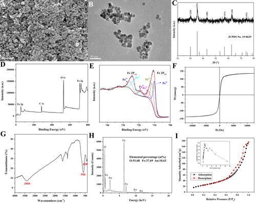
As shown in , the prepared nanoparticles were approximately spherical with an average particle size of 20.2 ± 3.0 nm, and the size distribution was uniform with agglomeration (Figure S1). The XRD pattern of the prepared nanoparticles in showed that the diffraction peaks at the (220), (311), (400), (511), and (533) crystal planes correspond to the diffraction peaks of the standard card NO.19-0629, which proved that the prepared nanoparticles were composed of Fe3O4. The XPS spectra in suggested that the elements in the substance contained Fe and O. The valence state of iron in the magnetic products was further analyzed, and the results of peak fitting were shown in . The Fe 2p peaks corresponding to Fe 2p3/2 and Fe 2p1/2 were located at 710.6 and 724.1 eV, respectively. The mean relative areas of each constituent peak assigned to Fe2+ and Fe3+ were calculated. The ratio of Fe2+:Fe3+ was theoretically 1:2 because the stoichiometric magnetite phase could be expressed as FeO·Fe2O3. The results of the deconvoluted peaks showed that the ratio of Fe2+:Fe3+ was calculated to be 0.37:0.63. This value was clearly that of the stoichiometric oxide within the uncertainty of the calculations. The above experimental results verified the existence of Fe3O4. The saturation magnetization of Fe3O4 nanoparticles in was 129.38 emu/g, suggesting that it was a kind of superparamagnetic material. According to the FTIR spectrum analysis of nanoparticles in , it could be seen that the vibration peak at 3404 cm−1 belonged to the hydroxyl group while the vibration peak at 590 and 438 cm−1 were attributed to the Fe-O bond. The absorption band at 1624 cm−1 was assigned to bending vibration of OH bonds (Santos et al., Citation2021). The elements of the nanoparticles shown in included Fe and O, among which Fe accounted for 37.69% and O accounted for 51.68% (the characteristic peak of Au was due to the gold-spraying treatment during the scanning electron microscopy to increase the conductivity). The nitrogen adsorption-desorption curve and pore size distribution of the magnetic Fe3O4 nanoparticles were depicted in . The adsorption-desorption nitrogen isotherm of the nanoparticles at low pressure exhibited a type II adsorption isotherm. The calculated specific surface area and pore volume were found to be 84.756 and 0.265 cm3/g, respectively, and the average adsorption pore size was 11.973 nm, which was close to the particle size of the nanoparticles. This might be due to the agglomeration of the nanoparticles. The curve showed a rapid upward trend under high pressure, and the nitrogen adsorption type was H3. The above characterization verified that the prepared magnetic nanoparticles were Fe3O4 nanoparticles, and their saturation magnetization reached 129.38 emu/g with the characteristic of agglomeration. To successfully load insoluble drugs, it was necessary to modify the surface of nanoparticles to improve their agglomeration and enhance their ability to load drugs.
Figure 1. SEM morphology (A), TEM image (B), XRD pattern (C), XPS survey scan spectrum (D), XPS spectra of Fe2p (E), Hysteresis loop (F), FTIR spectra (G), EDS spectrum (H), and BET measurement (I) of magnetic Fe3O4 nanoparticles heated at 170 °C for 24 h with a heating rate of 3 °C/min.
3.2. Modification of magnetic Fe3O4 nanoparticles
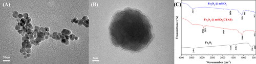
From the TEM images of Fe3O4 nanoparticles and Fe3O4@mSiO2 nanocomposite in , it could be seen that the surface of the nanoparticles was successfully coated with a silica shell. The mesoporous silica shell did not change the original morphology of the magnetic nanoparticles, and the nanocomposites coated with silica were still spherical with an average particle size of 27.81 nm, which was larger than that of magnetic Fe3O4 nanoparticles. The increased particle size was attributed to the mesoporous silica shell, and the dispersion of the magnetic Fe3O4@mSiO2 nanocomposites coated with mesoporous silica was improved. showed the infrared spectra of the different nanoparticles during the preparation process. A template agent (CTAB) was added to the mesoporous silica coating process. Subsequently, the template agent should be removed to make the silica coating appear mesoporous. In the infrared spectrum of Fe3O4, the characteristic peaks at 590 and 438 cm−1 belonged to Fe-O bonds, while the characteristic peak at 3404 cm−1 was attributed to the hydroxyl group. The infrared spectra of the nanocomposites with CTAB showed that the characteristic peaks at 2922 cm−1 and 2851 cm−1 were attributed to CTAB, and the peaks at 1066 cm−1 were attributed to Si-O bonds, indicating the successful coating of silica. After 24 h of acidic ethanol reflux, the characteristic peak of CTAB disappeared, indicating its successful removal. It was evident that the characteristic peak at 1084 cm−1 was due to the Si-O bond and the characteristic peak at 462 cm−1 was attributed to the Fe − O bond, verifying the successful preparation of Fe3O4@mSiO2 nanocomposites with the removal of CTAB.
Figure 2. TEM image of Fe3O4 nanoparticles heated at 170 °C for 24 h (A) and Fe3O4@mSiO2 nanoparticles (B), and the FTIR spectra of the process of magnetic Fe3O4 nanoparticles coated with silica (C).
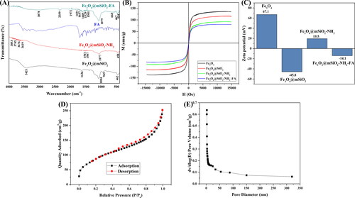
The surface amination and folic acid activation of Fe3O4@mSiO2 nanocomposites were successively carried out to improve the possibility of subsequent drug loading and targeting of the nanodrug delivery system (Liu et al., Citation2021). As shown in the infrared spectrum in , the characteristic peaks at 1541 and 1507 cm−1 belonged to the amino group. After the activation of folic acid, the characteristic peak at 3078 cm−1 was attributed to the amide group. The characteristic peaks at 668 and 1529 cm−1 were respectively attributed to the amide I and amide III bands, respectively. This reason was that an amide bond was formed during the crosslinking of folic acid. The magnetic properties in also confirmed the success of the surface modification of the nanoparticles. The saturation magnetization decreased from 129.38 to 76.3 emu/g after the activation of folic acid. During the surface modification process, the zeta potential was measured at each step of the product. The results in also verified the success of the surface modification. The zeta potential of Fe3O4 was positive under acidic conditions (Zhang et al., Citation2022). In , the zeta potential of Fe3O4 nanoparticles was 67.1 mv, which might be because acidic raw materials were utilized to prepare Fe3O4 nanoparticles. Magnetic Fe3O4 nanoparticles changed from positive charge to negative charge after coating with silica, which might be caused by the hydroxyl groups on the surface of silica. After modification by amination with APTES, the nanocomposites exhibited positive electric properties owing to the presence of amino groups. After the activation of folic acid, the nanocomposite became electronegative because folic acid was electronegative. The above characterization results verified the success of the surface modification of the nanoparticles. Although the stability decreased slightly as the absolute value of the zeta potential decreased, the agglomeration process was clearly improved. showed the nitrogen adsorption-desorption curve and pore size distribution of the Fe3O4@mSiO2-NH2-FA nanocomposites. The calculated specific surface area was 324.410 m2/g. The pore volume was 0.369 cm3/g and the average pore size was 4.548 nm. The nitrogen adsorption-desorption curve of Fe3O4@mSiO2-NH2-FA nanocomposites could be classified as type IV adsorption isotherms, which belonged to mesoporous materials (Calzaferri et al., Citation2021). The specific surface area and pore volume of Fe3O4@mSiO2-NH2-FA nanocomposites experienced significantly higher increases than those of Fe3O4 nanoparticles. The average pore size of the Fe3O4 nanoparticles was 11.973 nm, which was close to the particle size. This might be caused by the agglomeration of the nanoparticles. After surface modification, the nanoparticles formed a mesoporous structure. Hence, the improved agglomeration phenomenon and increased specific surface area provided a material basis for subsequent drug loading.
Figure 3. FTIR spectra (A), Hysteresis loop (B), and the change of surface zeta potential (C) of Fe3O4, Fe3O4@mSiO2, Fe3O4@mSiO2-NH2, Fe3O4@mSiO2- NH2-FA; The Nitrogen adsorption-desorption curve (D), and pore size distribution (E) of Fe3O4@mSiO2-NH2-FA.
3.3. The loading and release of PTX
The standard curve of the corresponding absorbances of PTX at various concentrations measured at an ultraviolet wavelength of 270 nm was shown in Figure S2. The drug loading efficiency and drug loading capacity of the nanocomposites were 44.26 and 11.375%, respectively. As depicted in , the cumulative PTX release rates of the Fe3O4@mSiO2-NH2-FA-TAX nanodrug increased by 53.2, 76.4, and 80.1% at pH 7.4, 6.5, and 5.5, respectively, indicating that the nanocomposite exhibited a certain pH sensitivity. The cumulative release rate in the pH of tumor microenvironment was more than 80%. The drug release of the nanocomposite showed a sustained release effect according to the curve analysis, which might be caused by the interaction between the drug and the nanocarrier. The higher drug release efficiency of nanomaterials in acidic environments might be attributed to the destruction of the carrier structure of nanomaterials, resulting in a higher release rate of PTX. As an anti-tumor drug, PTX could be released more in the tumor microenvironment, which was conducive to its enhanced anti-tumor effect.
3.4. The assay of cytotoxicity and magnetic targeting of carriers
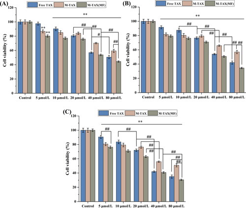
The viability of MCF-7 cells after treatment was determined by the MTT assay to evaluate the effect of nanomaterials. As shown in , when the concentration of Fe3O4 reached 800 μg/mL and the incubation time exceeded 72 h, the cell viability was 56.6%, verifying the effects of nanomaterials on cell viability. After incubation for 48 h, cell viability was still above 95%. As depicted in , cell viability was still greater than 95% when the concentration reached 1200 μg/mL and the incubation time was 48 h. Cell viability remained at 73.8% after incubation for 72 h. Because the silica coating and folic acid improved the biocompatibility of the nanocomposites, the cell viability was enhanced with an increase in the concentration. To study the effect of the magnetic field on the toxicity of the nanomaterials, breast cancer cells were co-incubated with Fe3O4@mSiO2-NH2-FA with or without a magnetic field. After incubation for 24 h, the results in showed that the toxicity of nanomaterials was enhanced by the magnetic field. Most importantly, although the nanomaterials exhibited biological toxicity when incubated at a certain concentration for a certain time, these concentrations were far beyond the material concentration required for subsequent experiments. Therefore, it was feasible to utilize nanomaterials with good biocompatibility for drug loading.
Figure 4. The toxicity of Fe3O4 (A) and Fe3O4@mSiO2-NH2-FA (B) to cells at different time points and the toxicity of Fe3O4@mSiO2-NH2-FA to cells with or without an external magnetic field (C) (n = 3, * P < .05, ** P < .01, compared with the control group, # P < .05, ## P < .01, comparison between groups).
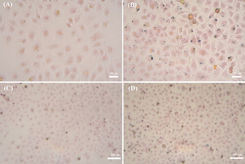
Prussian blue staining could reflect the uptake capacity of magnetic Fe3O4@mSiO2-NH2-FA nanocomposites. Compared with the blank control group (), blue particles were observed in the cytoplasm of the group containing Fe3O4@mSiO2-NH2-FA and MCF-7 cells (), because trivalent iron could react with ferrous potassium to produce insoluble blue products (Guari et al., Citation2022). The nanoparticles could aggregate within the cells after co-incubation, theoretically releasing the drug into the cytoplasm.
Figure 5. Prussian blue staining of MCF-7 cells: blank group (A, C), 40 μg/mL magnetic Fe3O4@mSiO2-NH2-FA nanocomposites (B, D).
The electrochemical method could also be used to detect the cellular uptake of nanoparticles (Figure S3). Whether an applied magnetic field promoted cellular uptake could also be assessed electrochemically. Cyclic voltammetry was the most common electrochemical method for assessing the electron conductivity. The peak current of the bare electrode was larger than that of the Fe3O4@mSiO2-NH2-FA nanocomposite because the Fe3O4@mSiO2-NH2-FA nanocomposite prevented Fe(CN)63- from reaching the electrode surface. The peak current decreased after the co-incubation of Fe3O4@mSiO2-NH2-FA and MCF-7 cells, which was attributed to the deposition of cells on the electrode surface and obstruction of electron conduction. The current of the applied magnetic field decreased further compared with that without a magnetic field, indicating that more nanomaterials were absorbed by cells with a magnetic field.
3.5. Cell cytotoxicity and apoptotic assay
respectively depicted the viability of MCF-7 cells at different times with different doses of magnetic Fe3O4@mSiO2-NH2-FA-TAX nanoformulation. As shown in , when the drug concentration was higher than 10 μM, the cell viability increased from high to low in Fe3O4@mSiO2-NH2-FA-TAX nanoformulation without a magnetic field, free TAX, and Fe3O4@mSiO2-NH2-FA-TAX nanoformulation with a magnetic field. The cell viability of the nanoformulation group without a magnetic field was higher than that of the free TAX group because of the slow release effect of the nanoformulation. The killing rate of the nanoformulation on cells was more than 50% at 24 h with a magnetic field, indicating that the nanoformulation had good anti-tumor effects. The applied magnetic field effectively increased the targeting of nanoformulation and the uptake and intracellular release of nanomaterials. The cell viability of the free TAX group was the lowest when the concentration was lower than 10 μM, which might be due to the precipitation of PTX in the medium. The killing effect of MCF-7 cells in all groups gradually increased over time (). When the concentration of the drug was 80 μM and the incubation time was 72 h, the killing rate of cells co-incubated with the nanoformulation reached nearly 70% under an external magnetic field, indicating the good antitumor effect of the nanoformulation.
Figure 6. Cytotoxicity of free TAX and magnetic Fe3O4@mSiO2-NH2-FA-TAX nanocomposites (with and without a magnetic field) to MCF-7 cells at 24 h (A), 48 h (B), 72 h (C) (n = 3, ** P < .01, compared with the control group, # P < .05, ## P < .01, comparison between groups).
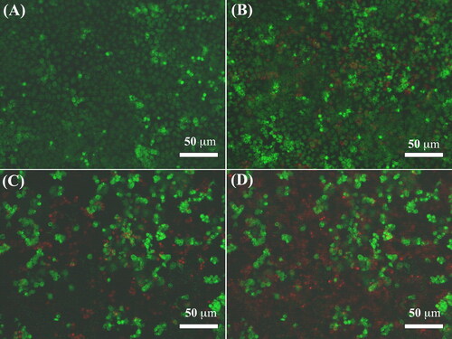
The main modes of cell death included apoptosis, iron death, autophagy (Ketelut-Carneiro & Fitzgerald, Citation2022; Wang et al., Citation2021b), and autophagy. As shown in , AO/EB staining was used to evaluate cell apoptosis during treatment with the nanoformulation. After staining, the cells were divided into four groups under the microscope: living cells, early apoptotic cells, late apoptotic cells, and dead cells (Kuang et al., Citation2021). No dead cells were observed in the control group (), while the cells treated with Fe3O4@mSiO2-NH2-FA-TAX showed apoptosis and were orange-red (). The cells exhibited extensive apoptosis under a microscope after treatment with free PTX () and Fe3O4@mSiO2-NH2-FA-TAX () under a magnetic field. Free PTX showed a stronger killing effect on cells than the nanoformulation, which might be attributed to the slow-release effect of the nanoformulation. These results were consistent with the above results for cell viability. In addition, the killing effect of nanomaterials under a magnetic field was better than that of free drugs, which was consistent with the above cytotoxicity results. This phenomenon might be caused by an increase in the targeting of nanomaterials under a magnetic field.
Figure 7. The AO/EB double staining fluorescence images of MCF-7 cells treated with blank control (A), Fe3O4@mSiO2-NH2-FA-TAX nanocomposites without MF (B), free TAX (C), and Fe3O4@mSiO2-NH2-FA-TAX nanocomposites with MF (D) for 24 h.
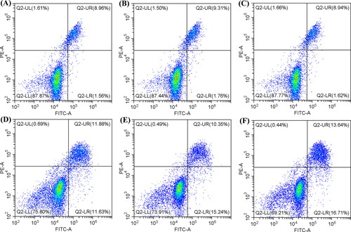
AO/EB staining, as a qualitative test for the detection of apoptosis, had certain limitations. Hence, flow cytometry was used to quantitatively evaluate the level of apoptosis (Kumari et al., Citation2022). When the cells were in an apoptotic state, PS flipped to the surface of the cell membrane, which could bind to FITC-annexin V and emit strong fluorescence. However, since this probe could also be labeled on the surface of necrotic cells, another probe should be introduced to distinguish cells at different apoptotic stages from necrotic cells. PI could not pass through the living cell membrane, and only when the membrane permeability increased, it could enter the cell and bind to DNA. Therefore, Annexin V-FITC/PI double staining divided cells into 4 categories: live cells (-/-), early apoptotic cells (±), late apoptotic cells (+/+) and necrotic cells (-/+). As shown in , the overall apoptotic rate in the control group reached 10.52%, owing to the utilization of trypsin. Trypsin induced apoptosis. However, this ratio was within the acceptable range. As depicted in , after co-incubating Fe3O4 and Fe3O4@mSiO2-NH2-FA with cells, the overall apoptotic rates were 11.07 and 10.56%, respectively. The apoptotic rate was not significantly different from that in the control group, and the slightly lower overall apoptosis rate in the Fe3O4@mSiO2-NH2-FA group might be due to the improved biocompatibility of the material by surface modification. After co-incubation with Fe3O4@mSiO2-NH2-FA-TAX in , the overall apoptotic rate reached 23.51%, which was higher than that of the control group, indicating that the nanoformulation could induce cell death through the apoptotic pathway. The apoptosis rate of the free TAX group in was higher than that of the nanoformulation group, reaching 25.59%, which might be attributed to the sustained-release effect of the nanoformulation. However, the apoptosis rate of the nanoformulation group with the applied magnetic field was higher than that of the free TAX group (30.35%), reflecting the effect of magnetic field-enhanced targeting ().
Figure 8. Flow cytometry scatter plots of Annexin V-FITC/PI apoptosis staining: (A) Control, (B) Fe3O4, (C) Fe3O4@mSiO2-NH2-FA, (D) Fe3O4@mSiO2-NH2-FA-TAX, (E) Free TAX, and (F) Fe3O4@mSiO2-NH2-FA-TAX + MF.
3.6. The detection of intracellular ROS, SOD, and MDA levels
Nanoformulation, an exogenous substance, might stimulate the production of ROS in cells, leading to physiological and pathological reactions in cells (Patel et al., Citation2020). As a result, the level of intracellular oxidative stress might change, leading to cytotoxicity. Hoechst 33342, a nucleic acid dye, induced a blue fluorescence in the nucleus. The DCFH-DA fluorescent probe could enter cells and be hydrolyzed into DCFH by intracellular enzymes. However, DCFH could not cross the cell membrane and was oxidized to DCF in the presence of reactive oxygen species (ROS), emitting green fluorescence. The fluorescence level of DCF positively correlated with the level of intracellular ROS. Therefore, the DCFH-DA fluorescence probe could be used to detect ROS accumulation in MCF-7 cells. As shown in , the nuclei showed blue fluorescence with uniform staining, and no green fluorescence was observed in the control, Fe3O4, and Fe3O4@mSiO2-NH2-FA treated cells. However, the cells treated with Fe3O4@mSiO2-NH2-FA-TAX () showed obvious green fluorescence, however its intensity was weaker than that of the free PTX group (), which might have been caused by the slow-release effect of the nanoformulation. Under an external magnetic field, the fluorescence intensity of the cells treated with Fe3O4@mSiO2-NH2-FA-TAX was higher than that of the free drug group, indicating that the magnetic field enhanced the targeting effect of the nanoformulation ().
Figure 9. Detection of ROS in MCF-7 cells treated by blank control group (A1), Fe3O4 (A2), Fe3O4@mSiO2-NH2-FA (A3), Fe3O4@mSiO2-NH2-FA-TAX (A4), free TAX (A5), and Fe3O4@mSiO2-NH2-FA-TAX with magnetic field (A6), respectively. Detection of SOD level (B1) and MDA level (B2) in MCF-7 cells treated by blank control group, Fe3O4 (M), Fe3O4@mSiO2-NH2-FA (M-FA), free TAX, Fe3O4@mSiO2-NH2-FA-TAX (M-FA-TAX), and Fe3O4@mSiO2-NH2-FA-TAX with MF, respectively (n = 3, ** P < .01, compared with the control group, ## P < .01, comparison between groups).
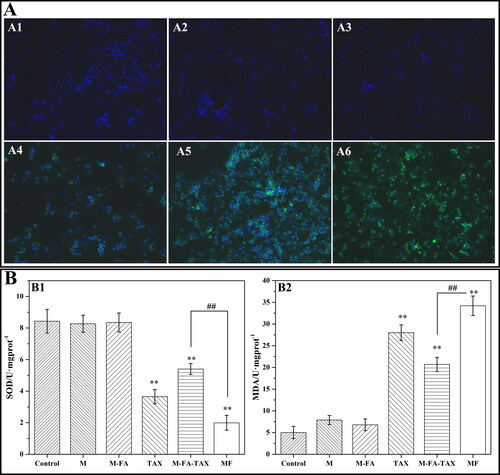
SOD, an active substance with antioxidant properties, could catalyze the disproportionation reaction of free radicals and generate oxygen and hydrogen peroxide, which played a critical role in the oxidative balance of the body (Kumar & Clair, Citation2021). The decrease in SOD levels reflected the weakened antioxidant and free radical scavenging capacity of the cells, indicating that the cells were damaged. As shown in , the SOD activities of the control, Fe3O4, and Fe3O4@mSiO2-NH2-FA groups were similar without significant differences, indicating that Fe3O4 and Fe3O4@mSiO2-NH2-FA did not cause cell damage. However, SOD activity decreased significantly after treatment with free PTX and the nanoformulations with/without a magnetic field. This led to accumulation of free radicals and cell damage. The SOD activity of the nanoformulation group without a magnetic field was higher than that of the free PTX group because of the slow release effect of the nanoformulation. In contrast, the SOD activity of the nanoformulation group under a magnetic field was lower than that of the free PTX group, because the magnetic field increased the targeting effect of the nanoformulation. MDA, an indicator of the degree of cell membrane damage, could reflect the degree of lipid oxidation (Wang et al., Citation2018). As shown in , compared to the control group, the level of MDA in the Fe3O4 group and Fe3O4@mSiO2-NH2-FA group showed no significant difference, indicating that Fe3O4 and Fe3O4@mSiO2-NH2-FA did not cause cell damage. However, MDA levels were significantly increased after treatment with free PTX and the nanoformulation with/without magnetic field, indicating that the degree of cell membrane damage was aggravated after drug administration. The above experimental results demonstrated that the levels of ROS and MDA increased, while the level of SOD decreased after drug administration, indicating that the nanoformulation could affect the viability of MCF-7 cells by changing the level of oxidative stress.
3.7. Influence of nanoformulation on protein expression
showed the effects of the nanoformulation on the expression of apoptotic proteins in MCF-7 cells. Apoptosis, which was characterized by cell shrinkage, nucleosome formation, and DNA fragmentation, was a type of programmed cell death. Caspase, a member of the cysteine protease family, was the core regulatory factor in apoptosis (Gan et al., Citation2022). Caspase 3 was the executor of apoptosis and could directly degrade proteins that caused apoptosis. Caspase 8 and caspase 9 were the promoters of apoptosis, which could be activated by self-shearing after receiving the signal, and then caused a cascade reaction to amplify the apoptotic signal. Caspase 8 and caspase 9 could bind closely to pro-apoptotic signals when apoptotic signal transduction occurred, with cleaved caspase 9 being the activated form. After self-shear activation, caspase 8 and caspase 9 could activate the downstream executor caspase 3, which forms cleaved caspase 3. Subsequently, apoptosis was induced by protein degradation. As shown in , caspase 8 was downregulated after continuous cleavage and activation, while cleaved caspase 9 was upregulated after introducing the apoptosis signal. As a result, cell apoptosis was induced and that cleaved caspase 3 was continuously up-regulated by cleaved caspase 9.
Figure 10. Effects of apoptosis related protein expression levels in MCF-7 cells treated with different groups (A). Where M represents Fe3O4@mSiO2-NH2-FA, M-TAX represents Fe3O4@mSiO2-NH2-FA, and MF represents magnetic field action (n = 3, ** P < .01, compared with the control group, # P < .05, ## P < .01, comparison between groups). Effects of ferroptosis related protein expression levels in MCF-7 cells treated with different groups (B). Where M represents Fe3O4@mSiO2-NH2-FA, M-TAX represents Fe3O4@mSiO2-NH2-FA, MF means magnetic field action (n = 3, ** P < .01, compared with the control group, # P < .05, ## P < .01, comparison between groups).
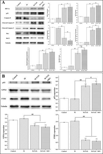
P53 protein was a transcription factor that was widely present in tumor cells and played a critical role in the initiation of apoptosis. When cell damage was irreversible, p53 played different roles, depending on the degree of DNA variation. When the degree of DNA variation was controlled, the p53 gene promoted cell repair, acting as a repairman. In contrast, the p53 gene induced apoptosis if the DNA changed too much to be salvaged. These two functions were regulated by multiple upstream and downstream genes in dynamic equilibrium (Xie et al., Citation2022b). The expression levels of HIF-1α and p53 increased after MCF-7 cells were treated with the nanoformulation (). P53 could regulate various apoptosis-related cytokines, such as the apoptosis precursor protein Bax and anti-apoptotic cytokine Bcl-2. As shown in , the expression of Bax increased, while the expression of Bcl-2 decreased owing to the upregulation of P53, and the ratio of Bax/Bcl-2 increased after treatment with the nanoformulation, indicating that the nanoformulation could induce cell apoptosis through the apoptotic pathway. The ratio of Bax/Bcl-2 increased when a magnetic field was applied, suggesting that the applied magnetic field could enhance the apoptotic effect of the nanoformulation on cells by targeting the nanoformulation.
Iron death, distinguished from other cell death modes, was an iron-dependent, non-apoptotic form of cell death characterized by iron overload and lipid peroxidation, which could be inhibited by various antioxidants (Wang et al., Citation2021a). The phospholipid hydrogen peroxide glutathione peroxidase (GPX4), a key regulator of iron death, could reduce the toxicity of lipid peroxides to maintain cell membrane homeostasis (Zhu et al., Citation2021). When GPX4 was inhibited or inactivated, the accumulation of lipid peroxides in the cells induced iron death. P53 could inhibit the activity of GPX4, which was associated with iron death. Ferritin (FTH1), the stored form of iron in the body, participated in iron metabolism. However, iron death occured when the amount of ferritin in the body was reduced, and the intracellular iron balance was disturbed under physiological or pathological conditions (Kong et al., Citation2021).
The results of Prussian blue staining showed that the nanoformulation could release Fe3+, which was associated with cell iron death. To further explore the mechanism of cell death induced by the nanoformulation, the expression levels of iron-death-related proteins were evaluated. As shown in , the expression level of p53 was upregulated, whereas those of GPX4 and FTH1 were downregulated. Downregulation of GPX4 expression resulted in the inhibition of intracellular lipid peroxide clearance and accumulation of intracellular ROS, leading to iron death. P53 could inhibit GPX4 expression to induce iron death. FTH1, ferritin in the human body, induced iron death if its expression level was downregulated. The changes in the levels of these three proteins suggested that the nanoformulation could induce cell death through the iron death pathway. The results also indicated that this effect could be enhanced by an applied magnetic field, which was associated with the targeting of magnetic fields.
3.8. The antitumor mechanism of magnetic nanomaterials on MCF-7 cells
According to the experimental results of intracellular ROS, MDA, and SOD levels, the nanoformulation could change the intracellular oxidative stress level. The accumulation of ROS in cells was necessary for iron death. ROS levels in cells increased abnormally under hypoxia and mitochondria were the main source of ROS production. Mitochondrial ROS not only participated in the activation of the HIF-1α upstream signal but also activated the PI3K/AKT (phosphatidylinositol 3 kinase-protein kinase B) signaling pathway, thus upregulating HIF-1α. ROS also played an important role in apoptosis and iron death. To confirm that the change in the oxidative stress level in vivo was the molecular mechanism of cell death induced by the nanoformulation, further experiments were needed.
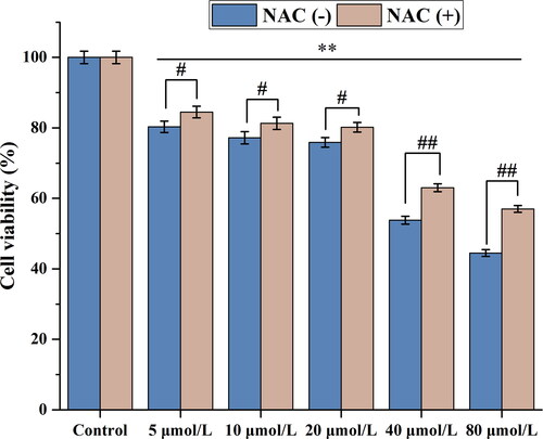
To prove the relationship between ROS and HIF-1α, p53, Bax, Bcl-2, GPX4, and FTH1, related protein levels were detected after ROS levels were downregulated by the ROS scavenger N-acetylcysteine (NAC). MCF-7 cells were treated with the Fe3O4@mSiO2-NH2-FA-TAX nanocomposite under the same conditions after the addition of the ROS scavenger NAC, and the cell viability increased significantly (). The cell viability of the control group did not change before and after NAC incorporation, whereas that of the nanoformulation group increased significantly, indicating that NAC could enhance cell viability by scavenging ROS.
Figure 11. Effect of MCF-7 cell activity treated by Fe3O4@mSiO2-NH2-FA-TAX under the action of magnetic fields with and without NAC treatment (n = 3, ** P < .01, compared with the control group, # P < .05, ## P < .01, comparison between groups).
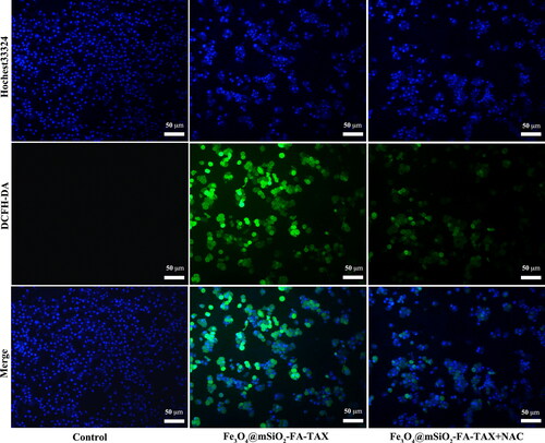
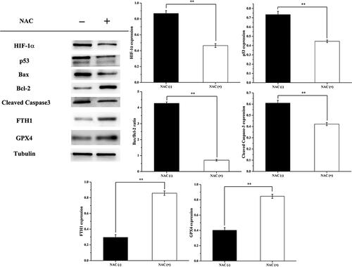
The experimental results shown in demonstrated that the incorporation of NAC significantly reduced intracellular ROS levels. With the addition of the ROS scavenger, NAC, the intracellular ROS expression level of MCF-7 cells treated with Fe3O4@mSiO2-NH2-FA-TAX was significantly reduced.
Figure 12. Level of ROS detected by DCFH-DA probe.
The effects of the Fe3O4@mSiO2-NH2-FA-TAX nanocomposite on the expression levels of related proteins in MCF-7 cells with a magnetic field before and after NAC treatment were shown in . The expression level of HIF-1α in the NAC-treated group was downregulated compared with that in the non-NAC-treated group, suggesting that NAC could downregulate the expression level of HIF-1α by scavenging ROS. Since mitochondrial ROS could upregulate the expression of HIF-1α by activating the upstream signals of HIF-1α, the level of HIF-1α was downregulated after ROS clearance. As shown in , NAC down-regulated the expression of p53, possibly because p53 was regulated by HIF-1α under hypoxic conditions. After NAC treatment, the levels of Bax and cleaved caspase 3 were downregulated, whereas the level of Bcl-2 was upregulated due to the downregulation of p53 after ROS clearance. These results indicated that the antioxidant NAC could reduce the promoting effect of the nanoformulation on MCF-7 cell apoptosis, which verified that the nanoformulation could indeed induce cell death through the apoptotic pathway. The levels of GPX4 and FTH1 were upregulated after NAC treatment, which was related to the stabilization of intracellular iron content and decrease in intracellular lipid peroxides after ROS clearance. The above results indicated that the antioxidant NAC could reduce the promoting effect of the nanoformulation on iron death in MCF-7 cells, which confirmed that the nanoformulation could indeed induce cell death through the iron death pathway. The results of intracellular oxidative stress-related indicators and changes in cell viability and protein expression levels after incorporation of NAC suggested that the molecular mechanism of cell death induced by nanoformulation might be the change in the oxidative stress level.
Figure 13. Effects of apoptosis and ferroptosis related protein expression levels in MCF-7 cells treated by Fe3O4@mSiO2-NH2-FA-TAX under the action of magnetic fields with and without NAC treatment. (−) means not treated by NAC, (+) means treated by NAC (n = 3, **P < .01, comparison between groups).
4. Conclusions
In this study, magnetic Fe3O4 nanoparticles were successfully fabricated, and their biocompatibility was improved by surface modification. A magnetic nano-delivery system was successfully constructed by loading PTX onto the surface of the nanocomposite, thus improving the stability and solubility of PTX. The mechanism of induction of breast cancer cell death by the magnetic Fe3O4@mSiO2-NH2-FA-TAX nanocomposites was elucidated. The main conclusions were as follows.
Fe3O4 nanoparticles were successfully prepared by a hydrothermal method using ferric chloride, sodium acetate, and propylene glycol as raw materials. The average size of the nanoparticles was found to be 20.2 ± 3.0 nm. The saturation magnetization reached 129.38 emu/g, showing superparamagnetism. The specific surface area and pore volume were 84.756 m2/g and 0.265 cm3/g, respectively.
Fe3O4@mSiO2-NH2-FA nanocomposites were constructed with a saturation magnetization of 76.3 emu/g and zeta potential of −14.1 mV. The drug loading efficiency and drug loading capacity of the nanocarriers were 44.26 and 11.375%, respectively. The cumulative release rates of PTX in PBS were 53.2, 76.4, and 80.1% at pH 7.4, 6.5, and 5.5, respectively. The nanocarriers exhibited pH sensitivity and sustained release.
Fe3O4@mSiO2-NH2-FA nanocomposites had the advantages of good biocompatibility and excellent active targeting performance in magnetic fields. The nanoformulation also showed a good killing effect on MCF-7 cells, which could have a slow-release effect, and its targeting ability could be enhanced by an external magnetic field.
Fe3O4@mSiO2-NH2-FA-TAX nanoparticles inhibited GPX4 activity or reduced FTH1 expression, resulting in ROS accumulation and iron death. At the same time, ROS accumulation could upregulate HIF-1α, affected the expression of apoptosis-related proteins regulated by p53, and induced cell apoptosis, thus showing an anti-tumor effect.
Author contributions
Yun Ni: Conceptualization, Methodology, Writing-Original Draft. Peng Deng: Investigation, Data Curation, Formal analysis. Ruitong Yin: Visualization, Project administration. Ziye Zhu: Validation, Software. Chen Ling: Resources, Visualization. Mingyi Ma: Investigation, Project administration. Jie Wang: Investigation, Resources. Shasha Li: Validation, Writing - Review & Editing. Ruijiang Liu: Conceptualization, Writing - Review & Editing, Supervision, Funding acquisition.
Supplemental Material
Download MS Word (746.4 KB)Disclosure statement
The authors declare no competing financial interest.
Data availability statement
The data and materials generated and analyzed during the current study are available from the corresponding author on reasonable request.
Additional information
Funding
References
- Abdallah QM, Kazi M, Khaleel MA, et al. (2020). Utilization of novel self-nanoemulsifying formulations (SNEFs) loaded paclitaxel for the treatment prosperity of bladder cancer. J Drug Deliv Sci Tec 56:101514.
- Alavi M, Nokhodchi A. (2022). Micro- and nanoformulations of paclitaxel based on micelles, liposomes, cubosomes, and lipid nanoparticles: recent advances and challenges. Drug Discov Today 27:576–84.
- Al-Hilfi A, Walker KD. (2022). Biocatalysis of precursors to new-generation SB-T-taxanes effective against paclitaxel-resistant cancer cells. Arch Biochem Biophys 719:109165.
- Araya-Sibaja AM, Wilhelm-Romero K, Quiros-Fallas MI, et al. (2022). Bovine serum albumin-based nanoparticles: preparation, characterization, and antioxidant activity enhancement of three main curcuminoids from Curcuma longa. Molecules 27:2758.
- Arumugam B, Nagarajan V, Perumal KN, et al. (2022). Fabrication of wurtzite ZnO embedded functionalized carbon black as sustainable electrocatalyst for detecting endocrine disruptor trichlorophenol. Microchem J 175:107202.
- Caldera F, Nistico R, Magnacca G, et al. (2022). Magnetic composites of dextrin-based carbonate nanosponges and iron oxide nanoparticles with potential application in targeted drug delivery. Nanomaterials 12:754.
- Calzaferri G, Gallagher SH, Brühwiler D. (2021). Argon and nitrogen adsorption isotherms of nonporous, microporous, and mesoporous materials. Microporous Mesoporous Mater 312:110744.
- Chen S, Liu Q, He XY, et al. (2022a). One-step synthesis of nanoscale anhydrous calcium sulfate whiskers: direct conversion of calcium carbonate by mixed acid with microemulsion method. J Nanopart Res 24:4.
- Chen X, Wang M, Hu Y, et al. (2020). Low-dose paclitaxel via hyaluronan functionalized bovine serum albumin nanoparticulate assembly for metastatic melanoma treatment. J Mater Chem B 8:2139–47.
- Chen XL, Ji JB, Zhou K, et al. (2022b). A novel multifunctional nanoparticles formed by molecular recognition between AS1411 aptamer and redox-responsive paclitaxel-nucleoside analogue prodrug for combination treatment of β-lapachone and paclitaxel. Colloids Surf B Biointerfaces 212:112345.
- Cheng L, Yang J, Tseng CC, et al. (2022). Outdoor ambient air pollution and breast cancer survival among California participants of the Multiethnic Cohort Study. Environ Int 161:102088.
- Deng YQ, Li J, Sun CY, et al. (2022). Rational development of a polysaccharide-protein-conjugated nanoparticle vaccine against SARS-CoV-2 variants and Streptococcus pneumoniae. Adv Mater 34:2200443.
- Dinparvar S, Bagirova M, Allahverdiyev AM, et al. (2020). A nanotechnology-based new approach in the treatment of breast cancer: biosynthesized silver nanoparticles using Cuminum cyminum L. seed extract. J Photochem Photobiol B 208:111902.
- Dong SY, Bi Y, Sun XS, et al. (2022). Dual-loaded liposomes tagged with hyaluronic acid have synergistic effects in triple-negative breast cancer. Small 18:2107690.
- Duan HX, Liu C, Hou Y, et al. (2022). Sequential delivery of quercetin and paclitaxel for the fibrotic tumor microenvironment remodeling and chemotherapy potentiation via a dual-targeting hybrid micelle-in-liposome system. ACS Appl Mater Interfaces 14:10102–16.
- Dutta B, Shelar SB, Rajan V, et al. (2022). Gelatin grafted Fe3O4 based curcumin nanoformulation for cancer therapy. J Drug Deliv Sci Tec 67:102974.
- Efetov SK, Zubayraeva AA, Nekoval VM, et al. (2020). Extended colectomy followed by cecorectal anastomosis as a surgical treatment modality in synchronous colorectal cancer. Case Rep Oncol 13:813–21.
- Fang ZZ, Li XY, Xu ZY, et al. (2019). Hyaluronic acid-modified mesoporous silica-coated superparamagnetic Fe3O4 nanoparticles for targeted drug delivery. Int J Nanomedicine 14:5785–97.
- Fu Y, Yang SY, Liu YN, et al. (2022). Peptide modified albumin-paclitaxel nanoparticles for improving chemotherapy and preventing metastasis. Macromol Biosci 22:2100404.
- Fukushima K, Lee SY, Tanaka K, et al. (2022). Effect of surface modification for carbon cathode materials on charge-discharge performance of li-air batteries. Materials 15:3270.
- Gade JV, Sharma PP, Jain B, et al. (2022). Synthesis and characterization of paclitaxel nanoparticles for drug delivery. Mater Today: Proc 51:445–50.
- Gan YH, Zhang J, Lei SL, et al. (2022). Atomically precise multi-domain GdxFe3-xO4 nanoclusters with modulated contrast properties for T2-weighted magnetic resonance imaging of early orthotopic cancer. Chem Eng J 429:132255.
- Gong HX, Li WA, Sun JL, et al. (2022). A review on plant polysaccharide based on drug delivery system for construction and application, with emphasis on traditional Chinese medicine polysaccharide. Int J Biol Macromol 211:711–28.
- Guari Y, Cahu M, Félix G, et al. (2022). Nanoheterostructures based on nanosized Prussian blue and its analogues: design, properties and applications. Coordin Chem Rev 461:214479.
- Guay E, Cordeiro E, Roberts A. (2022). ASO visual abstract: young women with breast cancer: chemotherapy or surgery first? An evaluation of time to treatment for invasive breast cancer. Ann Surg Oncol 29:2262.
- Gulsu A, Killi B, Alper M. (2022). Paclitaxel delivery by cationic gelatin nanoparticles. Chemistryselect 7:e202103495.
- Hashida N, Shamoto H, Maeda K, et al. (2021). Impact of geniohyoid and masseter muscle masses on dysphagia after salvage surgery and radiotherapy in head and neck cancer. Sci Rep 11:2278.
- Hata S, Shin T, Abe S, et al. (2021). Degarelix as a neoadjuvant hormonal therapy for acute urinary tract toxicity associated with external beam radiotherapy for intermediate- and high-risk prostate cancer: a propensity score matched analysis. Jpn J Clin Oncol 51:478–483.
- He CY, Majd MH, Shiri F, et al. (2021). Palladium and platinum complexes of folic acid as new drug delivery systems for treatment of breast cancer cells. J Mol Struct 1229:129806.
- Işıklan N, Hussien NA, Türk M. (2022). Multifunctional aptamer-conjugated magnetite graphene oxide/chlorin e6 nanocomposite for combined chemo-phototherapy. Colloids Surface A 645:128841.
- Jarvis CA, Bonney PA, Ding L, et al. (2020). Readmission with venous thromboembolism after surgical treatment by primary cancer site. Surg Oncol 35:268–275.
- Jiang HF, Chen WJ, Wang J, et al. (2022). Selective N-terminal modification of peptides and proteins: recent progresses and applications. Chinese Chem Lett 33:80–88.
- Kalia S, Rana DS, Thakur N, et al. (2022). Two-dimensional layered molybdenum disulfide (MoS2)-reduced graphene oxide (rGO) heterostructures modified with Fe3O4 for electrochemical sensing of epinephrine. Mater Chem Phys 287:126274.
- Karampinis L, Dionysopoulou A, Galata C, et al. (2022). Hyperthermic intrathoracic chemotherapy for the treatment of malignant pleural effusion caused by breast and ovarian cancer: a systematic literature review and pooled analysis. Thorac Cancer 13:883–888.
- Ketelut-Carneiro N, Fitzgerald KA. (2022). Apoptosis, pyroptosis, and necroptosis—oh my! The many ways a cell can die. J Mol Biol 434:167378.
- Killelea BK, Jeph H, Soulos PR, et al. (2020). Income disparities in needle biopsy patients prior to breast cancer surgery across physician peer groups. Breast Cancer 27:381–388.
- Kong N, Chen X, Feng J, et al. (2021). Baicalin induces ferroptosis in bladder cancer cells by downregulating FTH1. Acta Pharm Sin B 11:4045–4054.
- Kovrigina E, Chubarov A, Dmitrienko E. (2022). High drug capacity doxorubicin-loaded iron oxide nanocomposites for cancer therapy. Magnetochemistry 8:54.
- Kuang WW, Hu WL, Ren H, et al. (2021). Plant derived coumestrol phytochemical targets human skin carcinoma cells by inducing mitochondrial-mediated apoptosis, cell cycle arrest, inhibition of cell migration and invasion and modulation of m-TOR/PI3K/AKT signalling pathway. Saudi J Biol Sci 28:2739–2746.
- Kumar R, Singh RK, Alaferdov AV, et al. (2018). Rapid and controllable synthesis of Fe3O4 octahedral nanocrystals embedded-reduced graphene oxide using microwave irradiation for high performance lithium-ion batteries. Electrochim Acta 281:78–87.
- Kumar R, Youssry SM, Ya KZ, et al. (2020). Microwave-assisted synthesis of Mn3O4-Fe2O3/Fe3O4@rGO ternary hybrids and electrochemical performance for supercapacitor electrode. Diam Relat Mater 101:107622.
- Kumar S, Clair DS. (2021). Radioresistance in prostate cancer: focus on the interplay between NF-κB and SOD. Antioxidants 10:1925.
- Kumari M, Kamat S, Jayabaskaran C. (2022). Usnic acid induced changes in biomolecules and their association with apoptosis in squamous carcinoma (A-431) cells: a flow cytometry, FTIR and DLS spectroscopic study. Spectrochim Acta A 274:121098.
- Lee J, Ham JY, Park HY, et al. (2022). Feasibility of targeted cascade genetic testing in the family members of BRCA1/2 gene pathogenic variant/likely pathogenic variant carriers. Sci Rep 12:1842.
- Li B, Tan TF, Chu WW, et al. (2022a). Co-delivery of paclitaxel (PTX) and docosahexaenoic acid (DHA) by targeting lipid nanoemulsions for cancer therapy. Drug Deliv 29:75–88.
- Li S, Huan YF, Zhu B, et al. (2022b). Research progress on the biological modifications of implant materials in 3D printed intervertebral fusion cages. J Mater Sci-Mater M 33:2.
- Li XQ, Ma FH, Yang MH, et al. (2022c). Nanomaterial based analytical methods for breast cancer biomarker detection. Mater Today Adv 14:100219.
- Li YH, Wang XF, Zou SJ, et al. (2022d). Nanocomposites of immobilized nano-zirconia on low-cost activated carbon derived from hazelnut shell for enhanced removal of 3-nitro-4-hydroxy-phenylarsonic acid from water. Environ Res 209:112851.
- Liu RJ, Rong GX, Liu YH, et al. (2021). Delivery of apigenin-loaded magnetic Fe2O3/Fe3O4@mSiO2 nanocomposites to A549 cells and their antitumor mechanism. Mater Sci Eng C Mater Biol Appl 120:111719.
- Liu RJ, Zhang YL, Deng P, et al. (2022). Construction of targeted delivery system for curcumin loaded on magnetic α-Fe2O3/Fe3O4 heterogeneous nanotubes and its apoptosis mechanism on MCF-7 cell. Biomater Adv 136:212783.
- Liu TI, Lu TY, Yang YC, et al. (2020). New combination treatment from ROS-induced sensitized radiotherapy with nanophototherapeutics to fully eradicate orthotopic breast cancer and inhibit metastasis. Biomaterials 257:120229.
- Michałowska A, Kudelski A. (2022). The first silver-based plasmonic nanomaterial for shell-isolated nanoparticle-enhanced raman spectroscopy with magnetic properties. Molecules 27:3081.
- Mubarik S, Sharma R, Hussain SR. (2022). Breast cancer mortality trends and predictions to 2030 and its attributable risk factors in East and South Asian Countries. Front Nutr 9:847920.
- Ni Y, Lv ZX, Wang Z, et al. (2022). Immobilization and evaluation of penicillin G acylase on hydroxy and aldehyde functionalized magnetic α-Fe2O3/Fe3O4 heterostructure nanosheets. Front Bioeng Biotech 9:812403.
- Obayemi JD, Jusu SM, Salifu AA, et al. (2020). Degradable porous drug-loaded polymer scaffolds for localized cancer drug delivery and breast cell/tissue growth. Mater Sci Eng C Mater Biol Appl 112:110794.
- Patel A, Kosmacek EA, Fisher KW, et al. (2020). MnTnBuOE-2-PyP treatment protects from radioactive iodine (I-131) treatment-related side effects in thyroid cancer. Radiat Environ Biophys 59:99–109.
- Resen AK, Atiroğlu A, Atiroğlu V, et al. (2022). Effectiveness of 5-Fluorouracil and gemcitabine hydrochloride loaded iron-based chitosan-coated MIL-100 composite as an advanced, biocompatible, pH-sensitive and smart drug delivery system on breast cancer therapy. Int J Biol Macromol 198:175–186.
- Ro E, Vu V, Wei YD. (2022). Ambient air emissions of endocrine-disrupting metals and the incidence of hormone receptor- and HER2-dependent female breast cancer in USA. Med Oncol 39:69.
- Sakhi M, Khan A, Iqbal Z, et al. (2022). Design and characterization of paclitaxel-loaded polymeric nanoparticles decorated with trastuzumab for the effective treatment of breast cancer. Front Pharmacol 13:855294.
- Santos JG, Lopes H, Moreno H, et al. (2021). Towards anti-angiogenic activity of NiFe2O4 nanoparticles. Ceram Int 47:16152–16161.
- Shahdeo D, Roberts A, Kesarwani V, et al. (2022). Polymeric biocompatible iron oxide nanoparticles labeled with peptides for imaging in ovarian cancer. Bioscience Rep 42:BSR20212622.
- Shams A, Shabani R, Asgari H, et al. (2022). In vitro elimination of EL4 cancer cells from spermatogonia stem cells by miRNA-143-and 206-loaded folic acid conjugated PLGA nanoparticles. Nanomedicine (Lond) 17:531–545.
- Singh A, Amiji MM. (2022). Application of nanotechnology in medical diagnosis and imaging. Curr Opin Biotechnol 74:241–246.
- Sulaiman S, Ahmad S, Naz SS, et al. (2022). Synthesis of zinc oxide based etoricoxib and montelukast nanoformulations and their evaluation through analgesic, anti-inflammatory, anti-pyretic and acute toxicity activities. J King Saud Univ Sci 34:101938.
- Suzuki M, Takebe G, Takagi T, et al. (2022). Characterization of novel paclitaxel nanoparticles prepared by laser irradiation. Chem Pharm Bull (Tokyo) 70:269–276.
- Tan WK, Asami K, Maegawa K, et al. (2020). Fe3O4-embedded rGO composites as anode for rechargeable FeOx-air batteries. Mater Today Commun 25:101540.
- Vemuri SK, Halder S, Banala RR, et al. (2022). Modulatory effects of biosynthesized gold nanoparticles conjugated with curcumin and paclitaxel on tumorigenesis and metastatic pathways-in vitro and in vivo studies. IJMS 23:2150.
- Vergote I, Bergfeldt K, Franquet A, et al. (2020). A randomized phase III trial in patients with recurrent platinum sensitive ovarian cancer comparing efficacy and safety of paclitaxel micellar and Cremophor EL-paclitaxel. Gynecol Oncol 156:293–300.
- Vieira J, Maurmann N, Venturini J, et al. (2022). PCL-coated magnetic Fe3O4 nanoparticles: production, characterization and viability on stem cells. Mater. Today Commun 31:103416.
- Wala K, Szlasa W, Sauer N, et al. (2022). Anticancer efficacy of 6-gingerol with paclitaxel against wild type of human breast adenocarcinoma. Molecules 27:2693.
- Wang HY, Cheng Y, Mao C, et al. (2021a). Emerging mechanisms and targeted therapy of ferroptosis in cancer. Mol Ther 29:2185–2208.
- Wang K, Shen RY, Meng TT, et al. (2022a). Nano-drug delivery systems based on different targeting mechanisms in the targeted therapy of colorectal cancer. Molecules 27:2981.
- Wang XP, Wang GM, Li JB, et al. (2022b). A simple and straightforward polymer post-modification method for wearable difluoroboron beta-diketonate luminescent sensors. Polymer 239:124449.
- Wang YK, Chiang WC, Kuo FC, et al. (2018). Levels of malondialdehyde in the gastric juice: its association with Helicobacter pylori infection and stomach diseases. Helicobacter 23:e12460.
- Wang YQ, Kanneganti T. (2021b). From pyroptosis, apoptosis and necroptosis to PANoptosis: a mechanistic compendium of programmed cell death pathways. Comput Struct Biotechnol J 19:4641–4657.
- Webster EM, Dugan KB, Mcnamara B, et al. (2020). Surgical approach for interval debulking after neoadjuvant chemotherapy for treatment of advanced ovarian cancer: a single-institution retrospective cohort study. Gynecol Oncol 159:131.
- Wei M, Wang XC, Zimmerman DN, et al. (2021b). Endocrine therapy and radiotherapy use among older women with hormone receptor-positive, clinically node-negative breast cancer. Breast Cancer Res Treat 187:287–294.
- Wu WN, Roy P. (2022). Sialic acid binding sites in VP2 of bluetongue virus and their use during virus entry. J Virol 96:e01677.
- Xie YX, Gou QH, Zhang YJ, et al. (2022a). Association between age at initial diagnosis and post-metastasis mortality among women with recurrent metastatic breast cancer in China. BMC Cancer 22:385.
- Xie Z, Lu G, Zhou R, Ma Y. (2022b). Thiacloprid-induced hepatotoxicity in zebrafish: activation of the extrinsic and intrinsic apoptosis pathways regulated by p53 signaling pathway. Aquat Toxicol 246:106147.
- Yin XL, Zhang TC, Zhang Y. (2022). The global, regional, and national disease burden of breast cancer attributable to low physical activity from 1990 to 2019: an analysis of the Global Burden of Disease Study 2019. Int J Behav Nutr Phy 19:42.
- Yoko A, Kamonvarapitak T, Seong G, et al. (2022). Supercritical hydrothermal synthesis of organic-modified Ce1-xZrxO2-δ (0 ≤ x ≤ 1) nanoparticles as a low-temperature oxygen carrier. Chemnanomat 8:e202100495.
- Yu DL, Lou ZP, Ma FY, Najafi M. (2022a). The interactions of paclitaxel with tumour microenvironment. Int Immunopharmacol 105:108555.
- Yu LL, Liu M, Zhang YL, et al. (2022b). Magnetically induced self-assembly DNAzyme electrochemical biosensor based on gold-modified α-Fe2O3/Fe3O4 heterogeneous nanoparticles for sensitive detection of Ni2. +. Nanotechnology 33:095601.
- Yuan C, Wang D, Zhang N, et al. (2020). DNA damage/cGAS-triggered up-regulation of MALAT1 promotes undesirable inflammatory responses in radiotherapy of cancer. Biochem Biophys Res Commun 528:746–752.
- Zhang HP, Chen SY, Shan YH, et al. (2022). Highly effective lead ion adsorption by manganese-dioxide-supported core-shell structured magnetite. Front Env Sci 10:925205.
- Zhang W, Chen Y, Wang B, et al. (2022a). Facile preparation of paclitaxel nano-suspensions to treat lung cancer. j Biomater Tissue Eng 12:690–694.
- Zhang YL, Liu M, Pan S, et al. (2022b). A magnetically induced self-assembled and label-free electrochemical aptasensor based on magnetic Fe3O4/Fe2O3@Au nanoparticles for VEGF165 protein detection. Appl Surf Sci 580:152362.
- Zhao YL, Ma GZ, Wang SP. (2022). Magnetic nanoparticle tracking for one-step protein separation and binding kinetics analysis. J Electrochem Soc 169:057509.
- Zheng YH, Huang Y, Shi HB, Fu L. (2019). Green biosynthesis of ZnO nanoparticles by plectranthus amboinicus leaf extract and their application for electrochemical determination of norfloxacin. Inorg Nano-Met Chem 49:277–282.
- Zhu LJ, Chen DZ, Zhu Y, et al. (2021). GPX4-regulated ferroptosis mediates S100-induced experimental autoimmune hepatitis associated with the Nrf2/HO-1 signaling pathway. Oxid Med Cell Longev 2021:6551069.