
Abstract
The goal of the present study was to investigate the antibacterial properties, enzyme production, and metabolic profiling of a new Ceratorhiza hydrophila strain isolated from the submerged aquatic plant Myriophyllum spicatum. Furthermore, the fungus' morphological characterization and DNA sequencing have been described. The fungus has been identified and submitted to the GenBank as Ceratorhiza hydrophila isolate EG19 and the fungus ID is MK387081. The enzyme analyses showed its ability to produce protease and cellulase enzymes. According to the CSLI standard, the ethyl acetate extract of C. hydrophila showed intermediate antibacterial activity against Streptococcus pneumonia, Micrococcus luteus, and Staphylococcus aureus. Metabolic profiling has been carried out using 700 MHz NMR spectroscopy. Based on the 1H and 1H-13C heteronuclear single quantum coherence (HSQC) NMR data and NMR databases, 23 compounds have been identified. The identified metabolites include 31% amino acids, 9% sugars, 9% amines, 4% sugar alcohols, and 4% alkaloids. This is the first report for the metabolic characterization of C. hydrophila, which gave preliminary information about the fungus. It is expected that our findings not only will pave the way to other perspectives in enormous applications using C. hydrophila as a new promising source of antimicrobial agents and essential metabolites, but also it will be valuable in the classification and chemotaxonomy of the species.
1. Introduction
Aquatic habitat is a rich environment for the growth of huge number of microorganisms. Myriophyllum spicatum L. (F. Haloragaceae) is a submersed aquatic plant that grows in both fresh and brackish water. In weedy situations, it is fast growing, forming dense mats of foliage that interfere with the normal usage of water courses [Citation1]. It was believed that M. spicatum was introduced to Egypt by migratory birds [Citation2]. The first record of M. spicatum in Egypt was found in Lake Qarun [Citation3]. The introduction of M. spicatum to the Delta was detected and started to spread during the past few decades [Citation4]. M. spicatum has a key role in producing oxygen, stability of sediments, cycling of nutrition, and providing shelter and habitat for aquatic life [Citation5]. In addition, there are extensive studies which show the use of M. spicatum as suitable sorbents of heavy metals from sediment and water. M. spicatum plant species may be a useful vehicle for absorbing heavy elements from nutrient-rich sediments [Citation5].
Ceratorhiza hydrophila (Sclerotium hydrophilum) is an anamorphic basidiomycetous species that belongs to the family Ceratobasidiaceae. It is a phytopathogen that infects aquatic or semi-aquatic plants of wet meadows and marshes [Citation6]. The fungus has been reported in different areas of the world, including Northeast China [Citation7], Myanmar [Citation8], and in southeastern Australia [Citation9]. It does not produce asexual or sexual spores but produces small sclerotia on the vegetative mycelium that begin white in color, then upon maturation, become darkened [Citation10]. Morphologically, it has a well-defined dolipore septum [Citation11]. The fungus has been reported to infect a wide variety of plants including, Zizania aquatica L. [Citation11], Oryza sativa L. [Citation12], and Nymphaea odorata Aiton [Citation13]. In addition, it was isolated from Myriophyllum spicatum L. (or Eurasian water milfoil) in the Afritzer See Lake of Carinthia in Austria [Citation14]. In the past, Ceratorhiza hydrophila was called S. hydrophilum based on the phenotypes. Recently, based on rDNA analysis and morphology, S. hydrophilum was transferred to the genus Ceratorhiza, which is the anamorph of the Ceratobasidium species [Citation15]. Although, there are no available data about the biotechnological potential of Ceratorhiza hydrophila, other related basidiomycetous species showed the ability to produce various metabolic products. Rhizocotonia solani was found to produce various metabolites including amino acids, fatty acids, sugar alcohols, monosaccharides and carboxylic acids [Citation16]. In addition, Diplodia zeae produced many pectolytic and cellulolytic enzymes [Citation17].
Nowadays, it is important to discover new and alternative sources for active metabolites needed in various application fields, with the continuous need for alternatives to the commercial use products. So, in this research, we describe the isolation of C. hydrophila from M. spicatum from the shore of the river Nile in Helwan, Cairo, Egypt. To our knowledge this is the first report of C. hydrophila isolated from this plant in Egypt. Morphological and molecular characterizations of the fungus have been studied. Furthermore, we study its antibacterial activity. We also describe the metabolic profiling of the polar extract of the C. hydrophila.
2. Materials and methods
2.1. Plant material
The submerged aquatic plant Myriophyllum spicatum was collected from the banks of the river Nile in Helwan, Cairo, Egypt and authenticated by Prof. Tarek Galal, Professor of Plant taxonomy, Botany and Microbiology Dept. Helwan University.
2.2. Fungus isolation
For fungus isolation, the plant was washed gently under tap water. Surface sterilization was carried out using 70% ethanol for 1 min, then washed with sterile distilled water for 15 min. The stems and leaves of the Myriophyllum spicatum plant were cut into cubes (1 cm) and then were incubated on wet sterilized filter paper in 15 cm diameter Petri dishes at 25 °C for five to seven days [Citation18]. The appearance of white like crystals on the surface of the plant parts indicated the growth of the fungus which was further isolated and incubated for a week at 25 °C on potato dextrose agar plates. The fungus was purified and kept on potato dextrose agar slants at 4 °C.
2.3. Fungus identification
2.3.1. Morphological identification
The morphological characteristics of the fungus were studied by observing the color and texture of the colonies, as well as the color and shape of the sclerotia produced. Half cm diameter agar disk from 7-day-old fungal culture inoculated at the center of 9 cm diameter Petri dish using different culture media including Czapek-dox agar [Citation19], Potato dextrose agar (PDA) [Citation20], and Malt agar [Citation21], then incubated for 7–10 days at 25 °C, all the used chemicals were purchased from (Sigma-Aldrich, St. Louis, MO, USA). Colony texture, the shape, and color of sclerotia were examined with the visual inspection and recorded after four to six days of incubation. Moreover, the fungus' microscopic characterization, including branching patterns and septation of mycelium, was also examined under a light microscope.
2.3.2. Molecular identification
DNA isolation
The fungus was cultured for seven days onto potato dextrose broth medium. The mycelium was separated and ground and the total genomic DNA was isolated from the fungal mycelium according to [Citation22]. The isolated DNA was resolved on 1% agarose gel (Sigma-Aldrich) prepared in 1 X TAE (Tris-acetate-ethylenediaminetetra acetic acid) buffer containing 0.5 µg ml−1 ethidium bromide (Sigma-Aldrich). Ethidium bromide-stained gel was visualized using a UV-transilluminator (Vilber Lourmat, Collégien, France).
PCR reaction and sequencing
Polymerase chain reaction was performed using Mytaq Red DNA polymerase master mix (BIOLINE Cat. No. BIO-21108, London, UK) according to the manufacturer’s instructions. Briefly, the reaction contained 1 X PCR red master mix buffer, 2.0 µl of 10 pmol µl−1 of ITS1 and ITS4 primers which were designed according to White et al. [Citation23], 1.0 µl of DNA template (∼30 ng), and 0.25 µl of MyTaqTM DNA polymerase (5 Uµl−1). The total volume was adjusted to 50 µl using sterile distilled, demineralized water. The amplification reactions were performed using a Thermal Cycler (Biometra, Göttingen, Germany) as follows: 1st cycle of 3 min at 95 °C for initial denaturation, followed by 35 cycles of 20 s at 95 °C (denaturation), 20 s at 55 °C (annealing), 30 s at 72 °C (extension), and then a final extension was carried out for 10 min at 72 °C, after which the reaction was held at 4 °C. The PCR products were separated on 1% agarose gel. The PCR products were purified using PCR-M clean up system (VIOGENE Cat. No. PF1001, Taipei, Taiwan) according to the manufacturer's protocol. The purified samples were sequenced by GATC Company using ABI 3730xl DNA sequencer and ITS4 primer.
Sequencing data analysis
Blast was performed for the obtained nucleotide sequences using Geneious pro software 11.1.5, where the retrieved sequence from NCBI that has high similarity was aligned using MUSCLE tools and the phylogenetic tree was built using maximum likelihood methods [Citation24,Citation25].
2.4. Enzyme production
C. hydrophila was investigated for the production of various industrial enzymes, including protease, asparaginase, cellulase, and amylase. All media were inoculated with a 0.5 cm fungal disk from 7 days old fungal culture and incubated at 25 °C for 72 h. All measurements were made in triplicates.
2.4.1. Protease
Analysis of proteolytic activity was determined using gelatin agar medium [Citation26] containing g l−1: 20.0 gelatin; 20.0 agar in 0.2 M phosphate buffer pH = 7. Protease production was shown by a clear zone formation after the addition of acidic mercuric chloride solution [Citation27].
2.4.2. Asparaginase
Modified Czapek Dox medium [Citation28] was prepared, containing g l−1: 6.0 Na2HPO4; 2.0 KH2PO4; 0.5 NaCl; 0.2 MgSO4.7H2O; 0.005 CaCl2.2H2O; 20.0 L-asparagine in dist. H2O adjusted to pH 5.5. This medium was supplemented with 2% agar and 0.009% phenol red (Sigma-Aldrich). A change in the color of the medium from yellow to red indicated asparaginase production.
2.4.3. Cellulase
The ability to produce cellulase was tested by using cellulose medium [Citation29], containing g l−1: 1.0 carboxymethylcellulose; 0.5 NaNO3; 1.0 K2HPO4; 0.5 MgSO4·7H2O; 0.001 FeSO4·7H2O; 1.0 yeast extract; 15.0 agar (Sigma-Aldrich). After incubation, plates were flooded with 0.2 g l−1 potassium iodide for 5 min. Clear zone formation indicated cellulase production.
2.4.4. Amylase
Amylase activity was assayed using starch agar medium [Citation30], containing g l−1: 2.0 starch; 1.0 peptone; 20.0 agar (Sigma-Aldrich). After incubation, the plates were flooded with 0.1 g l−1 potassium iodide solution. Clear zone formation indicated amylase production.
2.5. Assessment of antibacterial activity
2.5.1. Preparation of crude fungal extracts
For preparation of crude fungal extracts, C. hydrophila was cultured into Potato dextrose broth media for seven days at 25 ± 2 °C. Extraction was carried out for both mycelium and filtrate using ethyl acetate by the ratio 1:1 V/V for 24 h and then filtered using Whatman filter paper followed by centrifugation at 10,000 rpm for 10 min at 4 °C. The ethyl acetate extract was evaporated under reduced pressure using rotary evaporator (IKA RV10 Staufen, Germany) to obtain the crude extract [Citation31].
2.5.2. Test bacteria
Five bacterial strains were used including Staphylococcus aureus (ATCC 25923), Escherichia coli (NBRC102203), which obtained from our lab., whereas the other strains in Streptococcus pneumonia, Micrococcus luteus and Proteus sp. are clinical pathogens which were kindly provided from Pharmaceutical Control Authority, Giza, Egypt. All strains were inoculated on nutrient agar medium and incubated at 37 °C for 24 h. After incubation, bacteria were suspended in sterile nutrient broth media and the cell suspensions were adjusted to a concentration of 0.5 McFarland (1.5 × 10⁸ CFU ml−1).
2.5.3. Antibacterial activity by well diffusion method
The antibacterial activity of the ethyl acetate extract from C. hydrophila against the five tested bacteria was determined by the well diffusion method [Citation32]. About100 µl of bacteria inoculum was streaked onto nutrient agar plates (NA) using sterile swabs. Wells were made on the agar surface with sterilized 6 mm cork borer, then 100 µl of the extract (55.0 mg ml−1 dissolved in ethyl acetate) was inoculated into the well. The bacteria seeded plates were placed in the refrigerator for 8 h at 4 °C before being incubated at 37 °C for 24 h. Clindamycin (2.0 µg/disk) and ciprofloxacin (5.0 µg/disk) were used as the positive control antibiotics according to the Clinical Laboratory Standards Institute (CLSI) [Citation33,Citation34], whereas ethyl acetate was used as the negative control. The plates were observed for the inhibition zone formation around the wells. The inhibition zone (mm) was determined by measuring the diameter of the inhibition zone around the well and expressed as means ± SD. All measurements were recorded in triplicates.
2.6. Statistical analysis
Data were analyzed using Minitab 17 software by one-way ANOVA. Fisher least significant difference (LSD) method with 95% confidence level.
2.7. Metabolic profiling using NMR spectroscopy
2.7.1. Fungal culturing and sample collection
C. hydrophila suspention culture was initiated by inoculating 0.5 cm disk of seven days old culture in 50 ml Potato dextrose broth medium and incubating at 25 °C for a week. The mycelia were collected (six replicates were collected from six different culturing flasks, i.e. n = 6). and immediately immersed in liquid nitrogen then stored at −80 °C for 24 h. For water removal from the fungal tissues, samples were lyophilized for 24 h. The dried mycelial samples were homogenized and ground to a fine powder, then 50 mg was used for metabolites extraction.
2.7.2. NMR metabolite extraction
The extraction was carried out based on the ratio between dry mass and water loss [Citation35,Citation36] using 2:2:1 methanol: chloroform: water according to Kim et al. [Citation37]. The hydrophilic fraction was removed from the extract and dried under vacuum using vacuum concentrator (Labconco, Kansas, USA) for 24 h.
2.7.3. NMR sample preparation, data collection, and metabolites identification
The dry polar fraction was dissolved in 620 µl of NMR buffer (1.0 mM TMSP (internal standard (3-trimethylsilyl)-2, 2′, 3, 3′-tetradeuteropropionic acid, 100 mM sodium phosphate buffer at pH 7.3 and 0.1% sodium azide, in 99.9 atom % D2O). NMR data collection and processing were carried out according to Abdelsalam et al. [Citation38]. Briefly, a 700 MHz Bruker AvanceTM III spectrometer (BrukerBioSpin, Billerica, MA) was used for the 1 D and 2 D NMR measurement. The data were collected with a spectral width of 16.0 ppm and 64 K points, providing a collection duration of 2.9 s. The first increment of the presat-noesy spectra for solvent suppression was collected using 120 scans, 4 dummy scans, 3 s relaxation delay, and on-resonance pre-saturation at the residual water frequency. The 90° pulse widths for each sample were measured using the automatic pulse calculation experiment (pulsecal). At 700 MHz, the Bruker hsqcedetgpsisp2.2 pulse sequence was utilized to collect two-dimensional 1H-13C HSQC (Heteronuclear Single Quantum Correlation) data.
The 1H was detected with a spectral width of 11 ppm in the F2 channel, whereas the 13C was detected in the F1 channel with a spectral width of 180 ppm.
The metabolites were identified using the Chenomx Suite library of compounds (Chenomx Inc., Edmonton, Alberta, Canada). Coupling constants (J value) were measured using Mnova software. Compounds identification was verified by comparing the measured chemical shifts (δ), multiplicities, and the coupling constants (J value) of the 1H and 1H-13C (HSQC) NMR data with those previously published and with the values of standard compounds reported in the Human Metabolome Database (HMDB) (https://hmdb.ca/).
3. Results
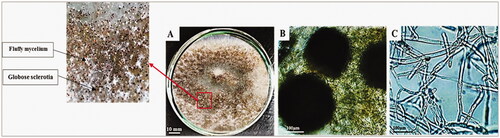
In the present study, C. hydrophila was isolated from the aquatic plant Myriophyllum spicatum growing along the banks of the river Nile in Helwan, Cairo, Egypt (Coordinates: 29.864° N and 31.291° E). Morphological examination of the fungus carried out on different growth media, including Czapeks-dox agar, Potato dextrose agar and Malt agar media. On the potato dextrose agar, the colonies initially appeared as white fluffy mycelium within the first four days of incubation then gradually large numbers of small and globose scattered sclerotia were observed on the surface of the colonies. The sclerotia were initially white in color, then turned brown to black over time. On Czapeks-dox agar medium, the colonies appeared as white mycelium without sclerotia until the sixth day of incubation. Starting from the eighth day, few white sclerotia were gradually developed, which acquired dark brown color over time. In addition, on malt agar medium, the fungus appeared as white mycelium until the end of the incubation. Sclerotia were absent on this growth medium. Microscopic examination for the fungus showed a septate and branched mycelium while the sclerotia were globose in shape ().
Figure 1. Morphological and microscopic examination of Ceratorhiza hydrophila. (a) growth on Potato dextrose agar media after seven days of incubation at 25ºC, (b) brown globose sclerotia (4X), (c) septate branched mycelium (40×).
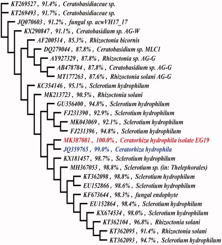
In addition, the resulting nucleotide sequence of fungal DNA was analyzed by GenBank database using advanced BLAST (Megablast) searches from the National Center for Biotechnology Information (NCBI). The fungal isolate was identified as Ceratorhiza hydrophila (synonym: Sclerotium hydrophilum) and submitted to the Genbank as Ceratorhiza hydrophila isolate EG19 with the fungus ID MK387081. Furthermore, the designed phylogenetic tree of the partial sequence with corresponding sequences from other fungi in GenBank showed that Ceratorhiza hydrophila isolate EG19 (MK387081) have high similarity toward Ceratorhiza hydrophila isolate SR6 (JQ359765) of 99% ().
Figure 2. Maximum likelihood tree showed that Ceratorhiza hydrophila isolate EG19 (MK387081) had a high similarity of 99% toward Ceratorhiza hydrophila isolate SR6 (JQ359765).
3.1. Enzyme production
The production of protease, asparaginase, cellulase, and amylase enzymes from C. hydrophila has been investigated. The results showed that the isolated fungus was capable to produce only protease and cellulase ( and ) but not asparaginase, and amylase.
Figure 3. Ceratorhiza hydrophila enzymatic activities after incubation for 72 h at 25ºC. (a) protease, (b) cellulase.
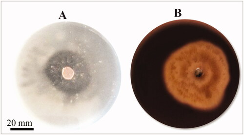
Table 1. Enzyme production by Ceratorhiza hydrophila.
3.2. Assessment of antibacterial activity
The ethyl acetate extract of C. hydrophila was assayed for its antibacterial activity against the tested bacteria () according to CLSI standards. The results reveal that the fungal extract had an intermediate inhibitory response against the Gram-positive bacteria Streptococcus pneumonia, Staphylococcus aureus, and Micrococcus luteus, recording inhibition zones 18 ± 1.4 mm, 20 ± 0.7 mm and 16 ± 0.7 mm, respectively. On the other hand, resistance response was shown by the tested Gram-negative bacteria; Escherichia coli and Proteus sp. toward the fungal extract with inhibition zones of 17 ± 2.1 mm and 0.0 mm, respectively.
Table 2. Antibacterial activity of the crude extracts of Ceratorhiza hydrophila against different tested bacterial strains.
3.3. Metabolic profiling using NMR spectroscopy
NMR spectroscopic analysis of the polar extract of C. hydrophila led to the identification of 23 metabolites. The chemical shifts, multiplicities, and coupling constants were retrieved from the 1H NMR spectra using Topspin and MestReNova software for spectral analysis, processing, and peak annotation (, ). By defining the 1H-13C correlation, the HSQC spectra verified the structure identification of the metabolites (, in Appendices). A variety of chemical classes have been recognized, including 31% amino acids, 9% sugars, 9% amines, 4% sugar alcohols, and 4% alkaloids. The presence of acetate, pyruvate, dimethylamine, trimethylamine, and betaine was revealed by singlet signals integrating for three protons, i.e. CH3, in the upfield section of the 1H NMR spectrum at δH 1.92 (δC 25.6), 2.38 (δC 29.3), 2.72 (δC 37.0), 2.90 (δC 47.4), and 3.28 (δC 56.2). The anomeric protons of α-, β-glucose, and trehalose have also been observed at δH 5.24 (d, J = 3.67), 4.64 (d, J = 7.88), and 5.20 (d, J = 3.73), respectively. Analysis of the cross peaks in the HSQC spectrum was used to correlate carbons to these protons (, in the appendices). The remaining protons of this sugars were defined in the region δH (δ 3.0–5.5 ppm). Furthermore, the amino acid glycine was identified by the methylene protons resonating at δH 3.55 (2H, s, δC 44.3), whereas alanine traced by its corresponding signals at δHα 3.77 (3H, q, J = 7.20, δC 53.5) and δHβ 1.48 (1H, d, J = 7.25, δC 18.6). The relatively deshielded methine protons detected downfield of the spectrum at δH 6.52 (1H, s, δC 137.3), δHα 7.96 (1H, s, δC 140.3), and δH 8.46 (1H, s) were attributed to the presence of fumarate, xanthine, and formate. Eventually, the assignment of all metabolites was further confirmed using Chenomx NMR suite and verified by comparing the generated spectral data with those reported in HMDB and other earlier literature studies.
Figure 4. 1H NMR spectra of the polar extract of Ceratorhiza hydrophila. (a) Aliphatic region, (b) sugars abundance signals region, (c) aromatic region.
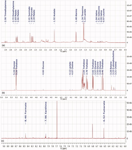
Table 3. The identified compounds in the polar extract of Ceratorhiza hydrophila with their chemical formulas, chemical shifts, multiplicity and coupling constants.
4. Discussion
This study was performed on the basidiomycetous fungus Ceratorhiza hydrophila, which was isolated from the aquatic plant Myriophyllum spicatum in Egypt. The aim of the study was to investigate the antibacterial properties, enzymes production, and metabolic profiling of the fungus as a promising and new source of metabolites, which can be used in various application fields.
To our knowledge, this is the first report of enzyme investigation from C. hydrophila, and it was found to have the ability to produce both protease and cellulase enzymes. Similarly, protease production was detected in Rhizoctonia solani [Citation39]. In addition, the proteolytic activity was also detected in Aspergillus tubingensis, Talaromyces purpureogenus, Acremonium flavum, Lichtheimia ramosa and Hormographiella aspergillata [Citation40]. In industrial applications, proteases used as anticlotting agent [Citation41], anticancer agent [Citation42] and in waste management [Citation43]. On the other hand, cellulase which has an important role in biofuel production, minimization of energy crisis, environmental pollution [Citation44], was also reported to be produced by Pleurotus ostreatus [Citation45], Rhizoctonia solani, Cephalosporium acremonium and Fusarium moniliforme [Citation46].
The ethyl acetate extract of C. hydrophila was found to have intermediate antibacterial activity against the tested Gram-positive bacteria, Staphylococcus aureus, Streptococcus pneumonia and Micrococcus luteus, whereas the Gram-negative bacteria, including Escherichia coli, and Proteus sp. showed resistance to the crude extract. Similar study reported the antibacterial activity of an extract from the mycelia and basidiocarp of some basidiomycetous fungi including Ganoderma applanatum, Trametes corrugata and Tricholoma crissum, which were found to have inhibitory activity against various pathogenic bacteria including, Micrococcus roseus, Mycobacterium phlei and Staphylococcus aureus [Citation47]. In addition, different Australian basidiomycetous macrofungi, including Amanita ochrophylla, Fomitopsis lilacinogilva as well as some Agaricus sp. showed excellent inhibition against S. aureus and E. coli [Citation48].
NMR spectroscopy is a crucial analytical tool in the field of metabolomic research due to its capacity to determine metabolite quantities, experimental reproducibility, nondestructive nature, and exceptional structure elucidation capabilities [Citation49]. Using both 1H and 1H-13C HSQC NMR data to study the metabolic profile of biological samples is a solid and dependable technique [Citation50]. In the present study, the metabolic profiling of the C. hydrophila polar extract was performed using the NMR spectroscopy. The data showed a presence of twenty-three metabolites. Six aliphatic amino acids (e.g. alanine, betaine, isoleucine) have been identified in the aliphatic region δ 0.5–3.0 ppm; the identification of these metabolites was confirmed by comparing our 1 D and 2 D data with HMDB and data published in the literature [Citation51–54].
The presence of amino acids alanine, glutamate, glutamine and arginine has been reported in the wild edible Boletus mushroom [Citation55]. Valine and glycine were identified in other members of the family Basidiomycota (Pleurotus tuoliensis) [Citation56]. Betaine is used as a source of carbon and energy source by fungi as reported by [Citation57]. Furthermore, the antibacterial activity of betaine has been reported [Citation58] as well as its activity against skin-associated fungus [Citation59]. In addition, betaine has an important medicinal value in several human diseases, such as obesity, diabetes, cancer, Alzheimer’s and gastrointestinal diseases [Citation60]. Moreover, alkyl betaines are used as antistatic and viscosity-increasing agents in cosmetic products such as shampoo [Citation61]. Betaine is used as a surfactant to remove various environmental pollutants such as toluene [Citation62]. In addition, betaine can improve the PCR reaction during DNA amplification by reducing the formation of secondary structure in GC-rich regions [Citation63].
In the present work, trehalose and glucose signals were clearly distinguished in the spectral region δ 3.0–5.5 ppm [Citation64]. Trehalose is a non-reducing disaccharide produced by many organisms, including bacteria, fungi, and plants [Citation65]. Trehalose was reported as an essential energy source in filamentous fungi and yeast [Citation66], and it has a key role during fermentation in Saccharomyces cerevisiae [Citation67]. The metabolic profiling of three strains of Grifola frondosa showed that trehalose, glycerol, and glucose were the most abundant metabolites [Citation68].
Moreover, the organic acids like acetic acid, formate, fumarate, lactate, malate, malonate, succinate and pyruvate have been identified. In other studies, acetic acid has been reported in the Antarctic yeasts metabolic profiling [Citation69]. Fumarate, lactate, malate, succinate and pyruvate have been identified in Grifola frondosa [Citation70]. The detection of malonate in the culture medium of the ectomycorhhizal fungus Suillus luteus was also reported [Citation71]. Moreover, through organic transformations, malonates can be used to produce fragrances, vitamins, agrochemicals, and pharmaceutical compounds [Citation71].
Choline was also identified in the C. hydrophila extract. It acts as a synthetic precursor for various important compounds including, the amino acid betaine, the neurotransmitter acetylcholine, and membrane phospholipids [Citation72].
Xanthine was identified in the fungal polar extract. It is an alkaloid with well-known pharmacological properties mainly in treatment of respiratory disease [Citation73], it also possesses anti-inflammatory, and anti-cancer activities. It is a precursor of several alkaloids and used as drug manufactory [Citation74]. Xanthine tablets dispensed in the market for treatment of pulmonary disorder (e.g. Xanthin Plus Tablets 20's® manufactured by Vega Pharmaceuticals (Pvt) Ltd).
In conclusion, this is the first report for isolation of C. hydrophila a basidiomycetous fungus from Myriophyllum spicatum plant in Egypt. All the results obtained from this study were reported for the first time. C. hydrophila showed the ability to produce cellulase and protease enzymes. The ethyl acetate extract of C. hydrophila showed intermediate antibacterial activity against all tested Gram-positive bacteria. Metabolic profiling using NMR spectroscopy showed that the fungal metabolite contents include carbohydrates, organic acids, amino acid, amines, and alkaloids. Some metabolites as choline and xanthine are used as nutritional supplement and in drug industry. It is expected that the results obtained in this study will pave the way to other perspective in enormous applications using C. hydrophila as a new promising source of antimicrobial agents and essential metabolites. Also, our findings about C. hydrophila can be valuable in classification and chemotaxonomy of the species.
Acknowledgements
The authors are very grateful to SC-INBRE (2 P20 GM103499) and NSF HBCU-UP (HRD-1332516) for providing NMR facilities. The authors are also grateful to Prof. Dr Tarek Galal, Professor of Plant Taxonomy, Botany and Microbiology Dept. Helwan University for helping in authenticating the plant material.
Disclosure statement
The authors declared that there is no conflict of interest.
References
- Harvey JL, Varley DR. Evaluation of european pathogens for the control of Myriophyllum spicatum in the United States of america. Proceedings of the IX International Symposium on Biological Control of Weeds. Moran VC, Hoffmann H. 1st edn. Stellenbosch, South Africa: University of Cape Town; 1996.
- Fayed AA. The distribution of Myriophyllum spicatum L. in the inland waters of Egypt. Folia Geobot Phytotax. 1985;20(2):197–199.
- Ascherson P, Schweinfurth G. Illustration de la flore d’Egypt. Mém. Inst Egypt. 1889;2:2.
- Abd El-Ghani M, El-Fiky AM, Soliman A, et al. Environmental relationships of aquatic vegetation in the fresh water ecosystem of the nile Delta, Egypt. Afr J Ecol. 2011;49(1):103–118.
- Galal TM, Shehata HS. Evaluation of the invasive macrophyte Myriophyllum spicatum L. as a bioaccumulator for heavy metals in some water courses of Egypt. Ecol Indic. 2014;41:209–214.
- Farr DF, Rossman AY, Palm ME, et al. Fungal databases. Systematic botany and mycology laboratory, ARS, USDA; 2008. Available from: http://nt.ars-grin.gov.
- Zhong QY, Song S, Yang MX, et al. First report of Sclerotium hydrophilum causing stem rot of rice in North East China. Plant Dis. 2018;102(3):681–681.
- Aye SS, Myint YY, Lwin T, et al. Stem rot of rice caused by Sclerotium hydrophilum isolated in Myanmar. Plant Pathol. 2009;58(4):799–799.
- Lanoiselet VM, Cother EJ, Ash GJ, et al. First report of Sclerotium hydrophilum on leaf sheath of rice (Oryza sativa) in South-Eastern Australia. Plant Pathol. 2002;51(6):813–813.
- Hausner G, Reid J. Factors influencing the production of sclerotia in the wild rice (Zizania aquatica) pathogen Sclerotium hydrophilum. Mycoscience. 1999;40(5):393–400.
- Punter D, Reid J, Hopkin AA. Notes on sclerotium-forming fungi from Zizania aquatica (wildrice) and other hosts. Mycologia. 1984;76(4):722–732.
- Qu SH. Rice diseases. Kew, England: Commonwealth Mycological Institute; 1972.
- Bowerman L, Goos RD. Physiological studies of two fungi isolated from Nymphaea odorata. Mycologia. 1991;83(5):624–632.
- Harvey JL, Evan HV. Assessment of fungal pathogens as biocontrol agents of Myriophyllum spicatum. Waterways experiment station, Vicksburg, MS. Miscellaneous Paper; 1997. A-97-1.
- Xu Z, Harrington TC, Gleason ML, et al. Phylogenetic placement of plant pathogenic Sclerotium species among teleomorph genera. Mycologia. 2010;102(2):337–346.
- Aliferis KA, Jabaji SH, Nmr G-MS. Metabolic fingerprinting of developmental stages of Rhizoctonia solani sclerotia. Metabolomics. 2010;6(1):96–108.
- Cappellini R, Peterson J. Production, in vitro, of certain pectolytic and cellulolytic enzymes by fungi associated with corn stalk rot. Bull Torrey Botanical Club. 1966;93(1):52–55.
- Toma FM, Abdulla NQF. Isolation and identification of fungi from spices and medicinal plants. RJEES. 2013;5(3):131–138.
- Waksman SA. The genus Streptomyces. In: Sebek OK, editor. The actinomycetes. A summary of current knowledge. New York: The Ronald Press Co.; 1967. Chapter 9, p. 101–112.
- da Silva N, Taniwaki M, Junqueira V, et al. Microbiological examination methods of food and water: a laboratory manual. London: Taylor and Francis Group; 2013.
- Zouari-Mechichi H, Mechichi T, Dhouib A, et al. Laccase purification and characterization from Trametes trogii isolated in Tunisia: decolorization of textile dyes by the purified enzyme. Enzyme Microb Technol. 2006;39(1):141–148.
- Prabha TR, Revathi K, Vinod MS, et al. A simple method for total genomic DNA extraction from water moulds. Curr Sci. 2013;104(3):10.
- White TJ, Bruns T, Lee S, et al. Amplification and direct sequencing of fungal ribosomal RNA genes for phylogenetics. San Diego, CA: Academic Press; 1990.
- Guindon S, Gascuel O. A simple, fast, and accurate algorithm to estimate large phylogenies by maximum likelihood. Syst Biol. 2003;52(5):696–704.
- Guindon S, Dufayard JF, Lefort V, et al. New algorithms and methods to estimate maximum-likelihood phylogenies: assessing the performance of PhyML 3.0. Syst Biol. 2010;59(3):307–321.
- Ammar MS, Louboudy SS, Abdul-Raouf UM. Distribution, total viable bacteria and identification of the most potent proteolytic bacterial strains isolated from Aswan city. Al-Azhar J Microbiol. 1991;11:224–238.
- Cowan ST. Cowan and steel's manual for the identification of medical bacteria. 2nd edn. England: Cambridge University Press; 1974. p. 193–227.
- Gulati R, Saxena RK, Gupta R. A rapid plate assay for screening L-asparaginase producing micro-organisms. Lett Appl Microbiol. 1997;24(1):23–26.
- Kasana RC, Salwan R, Dhar H, et al. A rapid and easy method for the detection of microbial cellulases on agar plates using gram's iodine. Curr Microbiol. 2008;57(5):503–507.
- Gessner RV. Degradative enzyme production by salt march fungi. Botanica Marina. 1980;23(2):133–139.
- Kjer J, Debbab A, Aly AH, et al. Methods for isolation of marine-derived endophytic fungi and their bioactive secondary products. Nat Protoc. 2010;5(3):479–490.
- Balakumar R, Sivaprakasam E, Kavitha D, et al. Antibacterial and antifungal activity of fruit bodies of Phellinus mushroom extract. Int J Biosci. 2011;1:72–77.
- CLSI. Methods for antimicrobial dilution and disk susceptibility testing of infrequently isolated or fastidious bacteria. 3rd edn. CLSI guideline M45. Wayne, PA: Clinical and Laboratory Standards Institute; 2015.
- CLSI. Performance standards for antimicrobial susceptibility testing. CLSI supplement M100. 29th edn. Wayne, PA: Clinical and Laboratory Standards Institute; 2019.
- Bligh EG, Dyer WJ. A rapid method of total lipid extraction and purification. Can J Biochem Physiol. 1959;37(8):911–917.
- Wu H, Southam AD, Hines A, et al. High-throughput tissue extraction protocol for NMR- and MS-based metabolomics. Anal Biochem. 2008;372(2):204–212.
- Kim HK, Choi YH, Verpoorte R. NMR-based metabolomic analysis of plants. Nat Protoc. 2010;5(3):536–549.
- Abdelsalam A, Mahran E, Chowdhury K, et al. NMR-based metabolomic analysis of wild, greenhouse, and in vitro regenerated shoots of Cymbopogon schoenanthus subsp. proximus with GC-MS assessment of proximadiol. Physiol Mol Biol Plants. 2017;23(2):369–383.
- Gvozdeva EL, Volotskaya AV, Sof’in AV, et al. Interaction of proteinases secreted by the fungal plant pathogen Rhizoctonia solani with natural proteinase inhibitors produced by plants. Appl Biochem Microbiol. 2006;42(5):502–507.
- Robledo-Mahón T, Calvo C, Aranda E. Enzymatic potential of bacteria and fungi isolates from the sewage sludge composting process. Appl Sci. 2020;10(21):7763.
- Osman ME, Khattab OH, Elsaba YM. Aspergillus terreus proteases: characterization and applications. J Chem Biol Phys Sci. 2014;4(3):2333–2346.
- Białas A, Kafarski P. Proteases as anti-Cancer targets-molecular and biological basis for development of inhibitor-like drugs against cancer. Anticancer Agents Med Chem. 2009;9(7):728–762.
- Lasekan A, Bakar FA, Hashim D. Potential of chicken by-products as sources of useful biological resources. Waste Manag. 2013;33(3):552–565.
- Sharada R, Venkateswarlu G, Venkateswar S, et al. Applications of cellulases: review. Int J Pharm Chem Biol Sci. 2014;4:424–437.
- da Silva IF, da Luz JMR, Oliveira SF, et al. High - yield cellulase and LiP production after SSF of agricultural wastes by Pleurotus ostreatus using different surfactants. Biocatal Agric Biotechnol. 2019;22:101428.
- Ahmad Y, Hameed A, Ghaffar A. Enzymatic activity of fungal pathogens in corn. Pak J Bot. 2006;38(4):1305–1316.
- Bhattacharyya C, De S, Basak A, et al. Antimicrobial activities of some basidiomycetous fungi. J Mycopathol Res. 2006;44:129–135.
- Neeraj B, Elizabeth ABA, Nigel F, et al. Evaluation of antibacterial activity of australian basidiomycetous macrofungi using a high-throughput 96-well plate assay. Pharm Biol. 2011;49(5):492–500.
- Emwas AH, Roy R, McKay RT, et al. NMR spectroscopy for metabolomics research. Metabolites. 2019;9(7):123.
- Zhang B, Powers R, O’Day EM. Evaluation of Non-Uniform sampling 2D 1H–13C HSQC spectra for semi-quantitative metabolomics. Metabolites. 2020;10(5):203.
- Qi S, Ouyang X, Wang L, et al. A pilot metabolic profiling study in serum of patients with chronic kidney disease based on (1) H-NMR-spectroscopy . Clin Transl Sci. 2012;5(5):379–385.
- Abdusalam KB, Yee LS, Mediani A, et al. 1H NMR-based metabolomics profiling of Syzygium grande and Oenanthe javanica and relationship between their metabolite compositions and antimicrobial activity against Bacillus species. Rec Nat Prod. 2022;16(2):128–143.
- Abdelsalam A, Chowdhury K, Boroujerdi A, et al. Nuclear magnetic resonance characterizes metabolic differences in Cymbopogon schoenanthus subsp. proximus embryogenic and organogenic calli and their regenerated shoots. Plant Cell Tissue Organ Culture. 2021;1:1–7.
- del Campo G, Zuriarrain J, Zuriarrain A, et al. Quantitative determination of carboxylic acids, amino acids, carbohydrates, ethanol and hydroxymethylfurfural in honey by (1)H NMR. Food Chem. 2016;196:1031–1039.
- Dembitsky VM, Terent'ev AO, Levitsky DO. Amino and fatty acids of wild edible mushrooms of the genus Boletus. Rec Nat Prod. 2010;4(4):218.
- Du F, Zou Y, Hu Q, et al. Metabolic profiling of Pleurotus tuoliensis during mycelium physiological maturation and exploration on a potential indicator of mycelial maturation. Front Microbiol. 2019;9:3274.
- Lambou K, Andrea P, Isabel V, et al. Pathway of glycine betaine biosynthesis in Aspergillus fumigatus. Eukaryot Cell. 2013;12(6):853–863.
- Birnie CR, Malamud D, Schnaare RL. Antimicrobial evaluation of N-alkyl betaines and N-alkyl-N, N-dimethylamine oxides with variations in chain length. Antimicrob Agents Chemother. 2000;44(9):2514–2517.
- Do E, Lee HG, Park M, et al. Antifungal mechanism of action of lauryl betaine against skin-associated fungus Malassezia restricta. Mycobiology. 2019;47(2):242–249.
- Zhao G, He F, Wu C, et al. Betaine in inflammation: Mechanistic aspects and applications. Front Immunol. 2018;9:1070.
- Burnett CL, Bergfeld WF, Belsito DV, et al. Safety assessment of alkyl betaines as used in cosmetics. Int J Toxicol. 2018;37(1_suppl):28S–46S.
- Miller U, Sówka I, Adamiak W. The effect of betaine on the removal of toluene by biofiltration. SN Appl Sci.2019;;1:984.
- Henke W, Herdel K, Jung K, et al. Betaine improves the PCR amplification of GC-rich DNA sequences. Nucleic Acids Res. 1997;25(19):3957–3957.
- Nemadodzi LE, Vervoort J, Prinsloo G. NMR-based metabolomic analysis and microbial composition of soil supporting burkea africana growth. Metabolites. 2020;10(10):402.
- Avonce N, Mendoza-Vargas A, Morett E, et al. Insights on the evolution of trehalose biosynthesis. BMC Evol Biol. 2006;6(1):109.
- Gancedo C, Flores CL. The importance of a functional trehalose biosynthetic pathway for the life of yeasts and fungi. FEMS Yeast Res. 2004;4(4-5):351–359.
- Vicente RL, Spina L, Gómez JP, et al. Trehalose-6-phosphate promotes fermentation and glucose repression in Saccharomyces cerevisiae. Microb Cell. 2018;5(10):444–459.
- Sato M, Miyagi A, Yoneyama S, et al. CE-MS-based metabolomics reveals the metabolic profile of maitake mushroom (Grifola frondosa) strains with different cultivation characteristics. Biosci Biotechnol Biochem. 2017;81(12):2314–2322.
- Rusinova-Videva S, Kambourova M, Alipieva K, et al. Metabolic profiling of antarctic yeasts by proton nuclear magnetic resonance-based spectroscopy. Biotechnol Biotechnol Equip. 2019;33(1):12–19.
- Rai M, Varma A. Diversity and biotechnology of ectomycorrhizae. India: Springer Science and Business Media; 2010.
- Strittmatter H, Hildbrand S, Pollak P. Malonic acid and derivatives. Ullmann's Encyclopedia Ind Chem. 2012;22:157–174.
- Blusztajn JK, Slack BE, Mellott TJ. Neuroprotective actions of dietary choline. Nutrients. 2017;9(8):815.
- Persson CGA, Pauwels R. Pharmacology of anti-asthma xanthines. In: Page CP, Barnes PJ, editors. Handbook of experimental pharmacology. Berlin: Springer Verlag; 1991. p. 207–225.
- Singh N, Shreshtha AK, Thakur MS, et al. Xanthine scaffold: Scope and potential in drug development. Heliyon. 2018;4(10):e00829.