
Abstract
The present research aimed to screen and evaluate the anticancer effect of Naringi crenulata phytocompounds on HER2+ human breast cancer cells. The cytotoxicity assay was performed to select effective solvent extraction. Extracted compounds showed lower toxicity on normal breast cells and significant cytotoxicity on human HER2+ breast cancer cells (SK-BR3) with an IC50 value of 24.59 μg/mL. The results indicate that the Naringi crenulata extract (NCE) shows anticancer potential via stimulating the cellular death of cancerous cells. The findings of the present study thus suggest that NCE supplementation might help in the treatment of HER2+ breast cancer.
Introduction
Breast cancers, in general, are differentiated into 5 different types such us HER2+, a normal-like tumor, basal-like tumor, luminal A and luminal B, based on the gene expression of the tumor cells. Studies have identified that breast cancer is the second leading fatal cancer in the world, and is said to be malignant [Citation1]. Earlier researchers investigated various chemotherapies and radiotherapies to treat hormone-responsive breast cancer, but therapy for HER2+ breast cancer is still required [Citation2]. The HER2 is expressed and amplified on the surface of the cell, and is involved in cell signaling pathways. Endocrine therapies were reported to show less effect on HER2+ breast cancer cells [Citation3]. The HER2 receptor inhibitor trastuzumab can suppress the HER2 receptor and significantly inhibit the development of breast tumors. Lapatinib (LPT) prevents tumor growth via inhibiting two tyrosine kinases (EGFR and HER2) by modifying the intracellular domain of HER2 [Citation4]. Binding of inhibitor at the ATP-binding site of the HER2 protein kinase inactivates the phosphorylation and also inhibits the ERK-1/2 and PI3K downstream signaling cascades. LPT, being an FDA-approved drug, has been utilized for the treatment of breast cancer since 2007 [Citation5]. LPT has low solubility and high permeability in bodily fluids due to its poor solubility of 0.007 mg/mL in water [Citation6]. The major downside of LPT is the dosage restriction (the minimum appropriate dose is 1250 mg/day), due to poor bioavailability in the oral setting. Numerous side effects have been witnessed with LPT, which include cardiotoxicity, hepatotoxicity and diarrhea. Chemotherapy can cause toxicity in patients including various side effects and risks, as well as immune suppression by affecting bone marrow cells and increasing susceptibility to diseases. Consequently, an alternative therapeutic method must resolve the side effects, which exhibit greater efficacy against cancer [Citation7].
It is well known from ancient times that medicinal plant extracts are used systematically in the treatment and prevention of various diseases. Surprisingly, traditional reports explained that the utilization of medicinal plants in the treatment of cancer did not cause any side effects. There are now 121 medicinal products used for cancer treatment, 90 are plant-derived [Citation8]. About 74% of these drug compounds were discovered by testing a reference to folk medicine. Guggulsterone is extracted from the Commiphora mukul plant and suppresses the DNA binding of NF-κB, which is involved in proliferative, anti-apoptosis, and metastatic pathways. The rhizome of turmeric (Curcuma longa) is a source for curcumin, utilized to inhibit carcinogenesis, to obstruct proliferation, and to encourage apoptosis [Citation9–11]. Several studies on human breast cancer cells have reported the anti-cancer activities of many phytocompounds. Indeed, the researchers discovered that HER2 suppression boosted the antiproliferative activity and stimulated apoptotic activity [Citation12,Citation13]. Many ayurvedic medicines propose epidemiological examinations to prevent cancer. Ayurvedic medicines such as curcumin, vinblastine, withanolide, etc. downregulate HER2, induce cell apoptosis, and can inhibit protein tyrosine kinases [Citation14,Citation15]. Therefore, supplementation of Naringi crenulata extracts can help to suppress HER2 expression and to treat HER2+ breast cancer.
Naringi crenulata is a branched tree with a wide distribution in South India and Myanmar [Citation16]. Different tropical diseases have been treated with the help of extracts of the fruits, leaves and roots in folk medicinal practice. Modern research described that the plant extract of Naringi crenulata has anti-inflammatory activity [Citation17], anti-oxidative effect [Citation17] and hepatoprotective property [Citation18], but the pharmacological activities of this plants are not fully studied. The utilization of antioxidants along with anticancer drugs will reduce the adverse outcomes of reactive species. The presence of higher content of antioxidants in Naringi crenulata makes it likely to find an anticancer compound in this plant. There are hopes that this plant's anticancer compounds can overcome the side effects. So far, the anticancer properties of Naringi crenulata leaf extract have yet to be investigated in HER2+ breast cancer. While the action mechanisms of several phytocompounds currently remain unknown, several studies report the bioactivity of certain compounds.
In the present study, we aimed to screen and evaluate the anticancer effect of phytocompounds of Naringi crenulata on HER2+ human breast cancer cells (SK-BR3) in vitro as well as to explore the fundamental anti-cancer mechanism of the extracted phytocompounds in silico. We found that some phytocompounds of Naringi crenulata, such as 6,7-Dimethoxy-1-methyl-3,4-dihydroiso-quinoline, Hesperetin, Hesperetin 7-rhamnoglucoside, and Tanakamine, exhibited the properties of anticancer activity via stimulating the apoptosis in cancer cells.
Materials and methods
Reagents and chemicals
McCoy's 5A growth medium, Dulbecco's modified Eagle's medium (DMEM), and trypsin EDTA (1×), RPMI-1640 medium was acquired from Hi-Media. The kit for JC-1 mitochondrial membrane potential assay was acquired from Cayman Chemicals. Fetal bovine serum (FBS) was also acquired from Hi-Media. 3-(4,5-Di-methylthiazol-2-yl)-2,5-di-phenyl tetrazolium bromide, piperazine-N,N′-bis(2-ethane sulfonic acid) (PIPES), Triton-X, 3-[(3-cholamidopropyl) dimethylammonio]-1-propanesulfonate (CHAPS), and most other standard analytical grade reagents used in the present study were acquired from Sigma-Aldrich.
Plant sampling and extraction
The plant Naringi crenulata was collected from in and around Western Ghats, Tamil Nadu, India, at the Botanical Survey of India located in the Southern Regional Centre, Coimbatore, Tamil Nadu, India, where the plant material was described and authenticated. A voucher specimen was preserved in the Ethnopharmacology Unit, Department of Biotechnology, Mepco Schlenk Engineering College, Tamil Nadu. India.
Collected leaves were cut into small pieces and allowed to dry at 60 °C for 6 h and the leaf mass was ground into powder. The powder was divided into 250 aliquots. Phytocompounds were extracted using different polar solvents (distilled water, absolute ethanol, hexane) by ultra-sonication (Solid: Solvent: 1:30) with parameters 60 amplitude, 0.5 cycle for 120 s [Citation19]. The collected extracts were dried to obtain the concentrated sample and were used in the cytotoxicity assay. After the solvent selection, a mixed solvent system was optimized with standard parameters for extraction.
Compound separation
The extract that showed the highest cytotoxic activity was subjected to column chromatography (Silica Gel mesh 200, column height – 20 cm, diameter – 1.5 cm) using various solvents: hexane, chloroform, ethyl acetate, ethanol and water, eluent with a gradual increase of polarity to hexane and ethanol [Citation20]. Different fractions were collected and named as F1–F15. The collected fractions were then subjected to MTT assay. The efficient fraction was separated and analyzed by gas chromatography–mass spectrometry (GC–MS).
Cell culture and cell viability assay
The human HER2 positive breast cancer cell line (SK-BR3 cell line) was acquired from NCCS (National Centre for Cell Science), Pune, India. The obtained cells were kept enriched in DMEM with 10% (v/v) FBS, 10 μg/mL penstrp. Cell lines were grown at 37 °C humidified atmosphere incubator with 5% CO2. We seeded of 2 × 10⁴ SK-BR3 cells in 96 well plates. After 24 h the cells were treated with the extracts (5 μmol/L) and incubated for various time (8–48 h) followed by MTT (3-[4,5-dimethylthiazol-2-yl]-2,5-diphenyltetrazolium bromide) assay. Vinblastine (Natural remedies, Bangalore) was used as a positive control (100 nmol/L). SK-BR3 cells kept for incubation at different concentrations of NCE (hexane to ethanol ratio of 6:4) for about 24 h were observed under an inverted light microscope. Absorbance was read with an ELISA microplate reader at 570 and 620 nm. Cell viability was calculated and thereby expressed in terms of percent of control (nontreated cells). After screening and fractionation of the anticancer compound, IC50 of the compounds on cancer cell lines was predicted from the concentration–effect curves [Citation21].
Phytochemical profile of Naringi crenulata extracts by GC–MS
Naringi crenulata extracts were analyzed by an Agilent 7890 BGCMS system, equipped with an autosampler G4513A Agilent and splitless injector. The column used SLB-5MS (Supelco, Milan, Italy), 30 m × 0.25 mm i.d., 0.25 μm df, coated with 5% diphenyl-95% polydimethylsiloxane. Mass data were handled by using the software Mass Hunter Qualitative Analysis B. 07.00 (Agilent). Isolated compounds were discovered based on computer matching with the MS libraries (NIST 14 Search 2.2 and Wiley).
Cell cycle arrest assessment
The effect of the extract on the cell cycle was analyzed using the propidium iodide staining method in SK-BR3 cells. Briefly, the cultured SK-BR3 cells were trypsinized and then pooled in a 15-mL centrifuge tube. Then, the cells were plated at a density of 1 × 10⁶ cells/mL into a 6-well tissue culture plate in McCoy's 5 A medium containing 10% FBS and 1% antibiotic–antimycotic (penicillin–streptomycin purchased from Sigma Aldrich) solution for 24 to 48 h at a temperature of about 37 °C. The wells were gently rinsed with the help of sterile PBS followed by treatment with 24.59 μg/mL of extracted compound sample in serum-free McCoy's 5 A medium, which was then incubated at a room temperature of 37 °C in a 5% CO2 incubator for 24 h. Again, the cells were trypsinized after the completion of the incubation process and washed in PBS for 5 min by centrifuging at 300g. The cells extracted after PBS wash were then fixed for 30 min at 4 °C using cold 70% ethanol by gentle addition in a drop wise manner to the cell pellet. Following the incubation process, the cells were washed by centrifuging at 300g for about 5 min with sterile PBS twice, and then the obtained supernatant was decanted. Consequently, the cell pellet was treated with RNase 50 μL (100 μg/mL) and PI 500 μL (100 μg/mL) and held at 4 °C before the evaluation using flow cytometry equipped with argon lasers (L1) and red diode (L2) [Citation22].
Mitochondrial membrane potential (ΔΨm)
The SK-BR3 cells (30,000–50,000 cells/well) were plated to a 24 well plate and incubated for 24 h in McCoy's 5A growth medium. After incubation, the plate was gently rinsed with PBS and treated with 24.59 μg/mL of extracted compound sample in serum-free McCoy's 5A medium based on previous research on other extracts [Citation23]. Again, the plate was kept for incubation at a temperature of about 37 °C in a humidified 5% CO2 incubator for about 24 h. The mitochondrial membrane potential measurement for the treated and control cells was done according to the manufacturer's instruction. In particular, the cells were incubated with 100 μL/well of JC-10 dye loading solution, and the plate was protected from the light. The plate was incubated at a temperature of 37 °C for 30 to 60 min in a 5% CO2 atmosphere. After incubation, 100 μL/well assay buffer was added to each sample/well. Finally, the plate was centrifuged at 300g for 2 min and the fluorescence was observed at 490/525 and 540/590 ratio. All imaging was performed at 40x objective magnification under fluorescence Microscope (EVOS™ FL Color, AMEFC4300) [Citation22].
DNA fragmentation assay
SK-BR3 cells were coated in a six-well plate at about a density of 1 × 10⁶ cells/well and were kept for incubation at a temperature of about 37 °C in a humidified 5% CO2 incubator for about 24 h [Citation24]. The cells/wells were rinsed gently with sterile PBS and treated with the extracted compound sample at the concentration of 24.59 μg/mL in serum-free McCoy's 5A medium to induce apoptosis and incubated for 24 h at 37 °C in a humidified 5% CO2 incubator. The cells were extracted by the process of trypsinization in a 1.5 mL tube and centrifuged at 300g for 10 min. After centrifugation, the supernatant obtained was decanted and 500 μL of lysis buffer was then added to the cell pellet, incubated at 37 °C for 1 h. Then 700 μL of phenol–chloroform–isoamyl alcohol were added and dissolved by inversion, and then centrifuged at 300g for about 5 min. The aquatic (upper) phase was then moved into the new Eppendorf tubes. Then, an equal amount of cold isopropanol was taken into the tubes, and then gently dissolved well by inversion. Further, the tubes were centrifuged at 300g for centrifugation for 5 min and the procured supernatant was discarded. The pellet was kept for air drying for 30 min. Finally, the dried DNA was then gently dissolved in 50 μL of distilled water. Besides, the DNA that was extracted was quantified at an optical density (OD) of 260 nm and 280 nm using a UV spectrophotometer. The OD value from 1.7 to 1.8 indicated that the quality of DNA was good, without protein/RNA contamination. Then, an equal amount of each DNA sample was run at 0.8% of agarose gel electrophoresis accompanied with a 100-bp molecular size ladder. Later, the image was obtained utilizing a gel documentation system (BioRad USA).
Statistical analysis
The results are depicted as mean values with standard errors of means (±SEM). The statistical analysis was carried out utilizing GraphPad (GraphPad Prism Software Inc. of Version 5.01, San Diego, California, USA). Tukey's test (each treatment compared to control) was used to test the statistical differences after the one-way analysis of variance (ANOVA). Differences were considered statistically significant at a level of p < 0.05.
Results and discussion
Effects of Naringi crenulata extracts (NCE) on SK-BR3
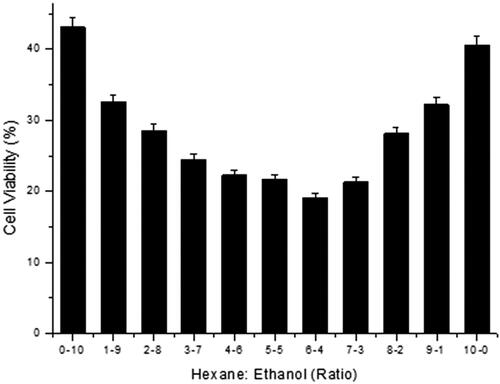
Phytocompounds from Naringi crenulata leaves were extracted with water, ethanol and hexane. The anti-cancer effect of extracts was tested in SK-BR3 cells. SK-BR3 cells were treated with different extracts, for 22–24 h, after which the cell viability was assessed in the MTT assay. The results showed a varied cytotoxicity pattern in SK-BR3 cells incubated with different solvent extracts (). The hexane and ethanol extracts showed maximal effect at 24 h of exposure. Akrout et al. [Citation25] reported that the ethanol extract of Artemisia campestris (wormwood) has revealed a higher cytotoxic effect. SK-BR3 cells exposure to both extracts showed a similar cell survival curve pattern, although with a distinct reduction in the cell viability when compared to the water extract. Due to the loss of cells, differences in the cell number could also be visualized in the culture dish following treatment with ethanol and hexane extracts for 24 h in comparison to the control dish (nontreated cells). These results showed that the extracts in various solvents exhibited a potent cytotoxic activity in the cancer cells (). In the next step, we applied a series of different ratios of hexane and ethanol-containing solvent systems in the extraction and evaluated the cell viability. Marwa et al. [Citation26] explored the anticancer effect of Orobanche crenata methanolic extract on cancer cell lines, where the study identified that most of the cells were diminished, and the remaining cells were reduced in size with obvious cell shrinkage and further monolayer destruction and chromatin clumping. Kanoh et al. [Citation27] used hexane and ethanol mixed solvent for the extraction of phenylahistin (anticancer compound). Compared to all the assayed proportions of Hexane: Ethanol, the ratio of 60:40 showed the highest cell growth inhibition activity (). It is inferred that the content of the cytotoxic compound in the hexane extract was higher than in the ethanol extract, which was reflected in the different ratios. Cell viability decreased with the gradual increase in hexane concentration up to 60%. The ethanol extract also showed cytotoxic activity, but some highly active compounds were probably missing in the extract.
Figure 1. Effects of Naringi crenulata extract on SK-BR3 cells in vitro. (a) Cell viability and cell morphology (b) following treatment with water, ethanol and hexane extracts compared with the control (nontreated). Images were captured with a 40x objective on an inverted light microscope. Note. (a) The cell viability in the nontreated control was taken as 100%. (b) The cell viability was varied thereby cells lost their adhesion property. Scale bars correspond to 50 μm.
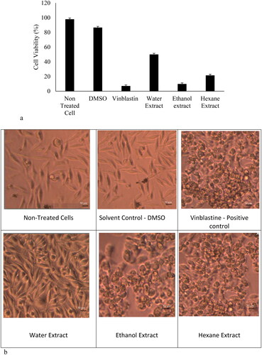
Figure 2. Effects of Hexane: Ethanol ratio for extraction of the cytotoxic compound(s) from the leaves of Naringi crenulata. Note. The cell viability in the nontreated control was taken as 100%.
Effect of extract fractions on the viability of SK-BR3 cells
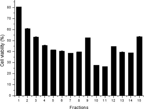
The obtained crude extract contains numerous phytocompounds; therefore, column fractionation was performed to screen for the effective cytotoxic compound. Brinda et al. [Citation28] investigated the antiproliferative activity of l-methioninase, where the crude l-methioninase was purified by acetone precipitation and diethylaminoethyl column methods. Upon column purification, the activity of l-methioninase in the 6th fraction of the column purified sample was 11.07-fold higher than in the crude extract. As compared to the present study, five different polar solvents were used in the elution, for each solvent 3 fractions were collected from the column [Citation29]. The polarity of hexane is 0, so the higher polarity solvents such as chloroform and ethyl acetate can purify compounds eluted by the hexane. Compounds with increased polarity have been indicated to possess good anticancer activity [Citation30,Citation31]. Thus, fractions 1 − 9 with less polarity cannot effectively interact with proteins involved in cancer cell signaling [Citation32,Citation33]. Fractions 10 − 12 contain the mid polar compounds and fractions 13 − 15 contain the highly polar compounds. Fraction 11 (i.e. eluted with ethanol) showed higher cytotoxic activity in SK-BR3 cells; indicating that the presence of active compounds in the fraction is higher. These compounds were separated by column fractionation (). Other compounds are eluted with respective polar solvent elution. Screened compounds were subjected to GC–MS analysis.
Figure 3. Cell viability following treatment with compounds in different fractions collected from the column fractionation process. Note. The cell viability in the nontreated control was taken as 100%.
Phytochemical profile of screened extract of Naringi cranulata
We used GC–MS to distinguish the compounds in the bioactive extract and characterize each component both qualitatively and quantitatively [Citation34–37]. In the present study, GC–MS analysis showed multiple peaks for a purified fraction (), and the compounds were matched against the database. Based on the GC–MS analysis 6,7-Dimethoxy-1-methyl-3,4-dihydro isoquinoline, 2,4-Dimethylbenzo[h] quinoline, 7,8-Dihydroneopterin, Naringetol, Hesperetin, Hesperetin 7-rhamnoglucoside, Methyl limonilate, Tanakamine, Tanariflavanone B, Chloroquine, 4a,8a-Methanophthalazine-1,4-dicarboxylic acid, Naringenin, and Estragole were identified in fraction 11. shows the structures of all of the compounds. Their structural properties were evaluated with the help of DruLito software and their structure was acquired utilizing PubChem ().
Figure 4. Identified phytocompounds in Naringi crenulata extract. GC–MS analysis (a) and chemical structures (b) of phytocompounds present in the higher activity of fraction 11 obtained from column fractionation. Note. Compound codes are listed in . The solvent ratio of 6:4 (hexane: ethanol) was used to obtain the extract analyzed subsequently.
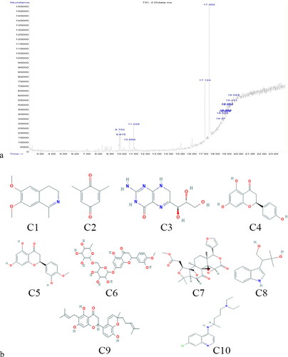
Table 1. Phytochemicals identified from the extract of Naringi crenulata by GC–MS.
Cytotoxicity assay
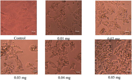
The ayurvedic medicine system suggests that the crude extract for any treatment contains compounds from the same plants having antioxidant and anti-inflammatory properties which help to avoid certain side effects [Citation38,Citation39]. Considering the adverse effects, toxicity and higher costs of common cancer treatment modalities, the use of novel herbal medicine-based therapies can provide promising health outcomes [Citation40]. Purified NCE has many active compounds; the drug-likeness property was analyzed by the DruLito, so NCE was taken for further analysis. The effect of NCE on SK-BR3 cells was analyzed by utilizing a cell viability assay. indicates that treated cells gradually grew relative to untreated cells, which explains that NCE (hexane to ethanol ratio of 6:4) has lower cytotoxicity to cancer cells with an anti-proliferative effect compared to other phytocompounds. Cells that undergo apoptosis exhibit several morphological and biochemical changes, such as aberrant chromatin condensation and/or fragmentation, and the formation of apoptotic bodies. This biochemical change hypothesis was examined using specific assays. Talib and Mahasneh [Citation41] obtained 27 μg/mL IC50 for Methanol fractions of Ononis hirta. At the concentration of 24.5 μg/mL (p < 0.01), approximately 50% cell growth inhibition was witnessed in NCE. From the outcomes of the present study, there was no significant difference between the solvent control and cells treated with NCE.
Figure 5. SK-BR3 cells kept for incubation at different concentrations of NCE (hexane to ethanol ratio of 6:4) for about 24 h and observed under an inverted light microscope. Note. Images were captured with a 40× objective on an inverted light microscope. Scale bars correspond to 50 μm.
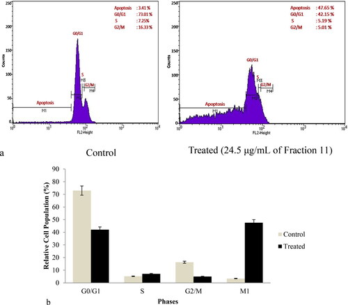
NCE compounds induced cell cycle arrest
Cancerous cells bypass several cell cycle safeguards as well as checkpoints, which could prevent the division of cells with aneuploidy and other defects, to allow unrestricted proliferation. This phenotype is the result of acquiring numerous genetic and epigenetic molecular modifications that hyper activate or inactivate the main cell cycle components. It imposes specific cellular dependencies upon cancer cells to sustain aberrant proliferation [Citation42,Citation43]. Şimşek et al. [Citation44] demonstrated that DNA-damage and cell cycle arrest initiated anti-cancer potency of super tiny carbon dots on MCF7 cell line, where the study identified cell cycle arrest in the G0/G1 phase and the induction of cell cycle arrest was reported to occur in a concentration-dependent manner. NCE compounds induce cell cycle arrest in SB RK3 cells, which is evaluated by cell cycle distribution by utilizing flow cytometry. Upon 24 h of the treatment period, NCE caused a significant accumulation of cells in the G1 phase followed by a logical decrease of the cells in G0/G1 phase and a parallel induction in the M1 phase (). The results indicate that NCE induces cellular apoptosis. shows the cell cycle distributions. These findings indicate that the NCE exercised its growth-suppressive activity by disrupting the progression of the cell cycle and triggering cell death by inducing apoptosis. Extracted compounds are majorly involved in the apoptotic induction pathway, so further, the apoptotic activity of the extract was studied in the further in-vitro experimental part.
Figure 6. Effects of NCE on cell cycle distribution. Representative results (a) and histograms (b). SK-BR3 human breast cancer cells were treated with 24.5 μmol/L NCE for 24 h and then harvested and stained with PI, and analyzed using flow cytometry.
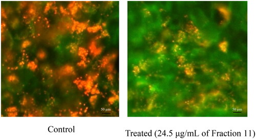
NCE effect on mitochondrial membrane potential (ΔΨm)
We explored another primary marker of apoptosis to further elucidate the mechanisms by which NCE induced cell death in cancerous cell lines. Mitochondria are instrumental in inducing apoptosis in mammalian cells. Ngoc et al. [Citation45] reported that compounds from Adenosma bracteosum induced significant increase in the production of reactive oxygen species accompanied by attenuation of mitochondrial membrane potential, thus inducing the activation of caspase-3 activity in human cancer cells. To determine whether treatment with NCE affects the permeabilization of the mitochondrial membrane and subsequently induces cell death, the MMP was evaluated before and after exposure to the IC50 concentration of NCE (). Caspase-3/7 activity was investigated to examine the possible effects of NCE on mitochondrial-mediated apoptosis. The mitochondrion is one of the main regulators in controlling cell death and survival mechanisms in cells. The JC-1 green fluorescence ratio signals measured in SK-BR3 cells with and without NCE treatment are illustrated in . Studies have reported that when JC-1 is introduced to living cells, it gets localized predominantly in the mitochondria [Citation46–48]. In such organelles, the JC-1 accumulation results in the production of J-aggregates (especially red fluorescence emission maximum at 590 nm), in addition to the unique green fluorescence of J-monomers (emission maximum of ∼529 nm). The lack of mitochondrial (ΔΨ) results in the reduction of J-aggregate formation as well as the depression of JC-1 mitochondrial accumulation. Thus, the ratio of red and green fluorescence of cells loaded with JC-1 is commonly utilized for detecting the mitochondrial membrane potential [Citation46–48]. The results showed that NCE treated cells exhibited a higher elevated reduction in the JC-1 fluorescence ratio by 76% relative to untreated cells (p < 0.05). The mitochondrial-mediated apoptotic intrinsic pathway leads to consequent cytochrome C release via depolarizing the membrane of the mitochondria, and this in turn leads to complex formation with caspase-9 and Apaf-1. Further, the caspase-9 contributes to the stimulation of the caspase-3/7 executioner. The results indicate that the NCE induced intrinsic apoptotic pathway by mitochondrial mediation.
Figure 7. JC-1 green fluorescence ratio signals in SK-BR3 cells with and without treatment with Fraction 11. Note: SK-BR3 human breast cancer cells were treated with 24.5 μmol/L of Fraction 11 for 24 h. Images were captured with a 40× objective on an inverted imaging system.
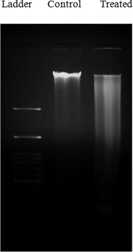
DNA fragmentation
The inter-nucleosomal fragmentation of genomic DNA is widely utilized as one of the biochemical indicators of apoptosis [Citation49]. A previous study identified the anticancer effect of piperine-free Piper nigrum extract and reported that the anticancer effect was associated with apoptosis, where the apoptosis was visualized using DNA fragmentation, thereby indicating the DNA cleavage by the activation of a nuclear endonuclease enzyme [Citation50]. Similarly, in the present study, a conventional agarose gel electrophoresis was performed to identify the presence of DNA fragmentation in SK-BR3 cells after treatment with NCE at the IC50 (24.5 μg/ml) for about 24 h. The untreated cells retained the intact genomic DNA without internucleosomal degradation (lane 1 and 2, ). The NCE treated SK-BR3 cells displayed a laddering pattern at IC50 (24.5 μg/mL) (lane 2). The results indicated the association with the enhancement in caspase-3/7 activity in SK-BR3 cells treated with the NCE. There is a clear expectation that the DNA fragmentation is strongly linked with the activation of caspase-3 as an apoptosis endpoint feature. A DNA smearing pattern was detected in the cells treated with NCE (lane 2). It, therefore, indicates that the apoptotic cells gain entry into the later stage of apoptosis due to a lack of cell remnant engulfment by the phagocytes. DNA fragmentation was observed in the cells treated with NCE. Activation of caspases results in apoptotic morphological changes and DNA fragmentation by selective cleavage of essential cellular substrates. Correspondingly, identification of DNA fragmentation in NCE treated cells indicates that NCE contributes to the activation of caspase 3. The cell activates DNase enzymes after the activation of caspase-3, resulting in DNA fragmentation and ultimately apoptosis.
Figure 8. DNA fragmentation in SK-BR3 cells after treatment with NCE. The cells are treated with the NCE at IC50 for 24 h. Lane 1: untreated SK-BR3 cells; Lane 2: SK-BR3 cells treated with 24.5 μg/mL.
Conclusions
The results of the present study indicate that Naringi crenulata extracts could be investigated further as promising anti-cancer phytochemicals against breast cancer. It is hypothesized that NCE could reduce cancer cell resistance by triggering cytotoxicity. NCE also deepens awareness about the potential of cellular models in studies on natural bioactive compounds, e.g. in this study NCE demonstrated anti-proliferative potential against SK-BR3 cells. More interestingly, SK-BR3 cells exposure to NCE induced cell cycle arrest (in S and G2/M phases; in G0/G1 phase). Thus, suggesting that NCE might exhibit anti-proliferative potential and pro-apoptotic potential. Naringi crenulata extract which exhibits various medicinal properties might serve as an outstanding candidate for anticancer treatment.
Disclosure statement
We have no conflicts of interest to disclose.
Data availability statement
The data that support this study are available from the corresponding author upon reasonable request.
References
- Chattopadhyay S, Hemminki O, F€Orsti A, et al. Impact of family history of cancer on risk and mortality of second cancers in patients with prostate cancer. Prostate Cancer Prostatic Dis. 2019;22(1):143–149.
- Gershon N, Berchenko Y, Hall PS, et al. Cost effectiveness and affordability of trastuzumab in sub-Saharan Africa for early stage HER2-positive breast cancer. Cost Eff Resour Alloc. 2019;17:5.
- Gutierrez C, Schiff R. HER2: biology, detection, and clinical implications. Arch Pathol Lab Med. 2011;135(1):55–62.
- Wang J, Xu B. Targeted therapeutic options and future perspectives for HER2-positive breast cancer. Signal Transduct Target Ther. 2019;4:34.
- Ryan Q, Ibrahim A, Cohen MH, et al. FDA drug approval summary: lapatinib in combination with capecitabine for previously treated metastatic breast cancer that overexpresses HER-2. Oncologist. 2008;13(10):1114–1119.
- Koch KM, Reddy NJ, Cohen RB, et al. Effects of food on the relative bioavailability of lapatinib in cancer patients. J Clin Oncol. 2009;27(8):1191–1196.
- Oun R, Moussa Y, Wheate N. The side effects of platinum-based chemotherapy drugs: a review for chemists. Dalton Trans. 2018;47(19):6645–6653.
- Kebebe D, Liu Y, Wu Y, et al. Tumor-targeting delivery of herb-based drugs with cell-penetrating/tumor-targeting peptide-modified nanocarriers. Int J Nanomed. 2018;13:1425–1442.
- Kuttan R, Bhanumathy P, Nirmala K, et al. Potential anticancer activity of turmeric (Curcuma longa). Cancer Lett. 1985;29(2):197–202.
- Sheikh E, Bhatt ML, Tripathi M. Bio-based synthesised and characterized monodispersed Curcuma longa silver nanoparticles induces targeted anticancer activity in breast cancer cells. Phcog Mag. 2018;14(57):340.
- Shishodia S, Aggarwal B. Nuclear factor-kappaB activation mediates cellular transformation, proliferation, invasion angiogenesis and metastasis of cancer. Cancer Treat Res. 2004;119:139–173.
- Hosseini A, Ghorbani A. Cancer therapy with phytochemicals: evidence from clinical studies. Avicenna J Phytomed. 2015;5(2):84–97.
- Wang H, Khor TO, Shu L, et al. Plants vs. cancer: a review on natural phytochemicals in preventing and treating cancers and their druggability. Anticancer Agents Med Chem. 2012a;12(10):1281–1305.
- McDermott M, Conlon N, Browne BC, et al. HER2-targeted tyrosine kinase inhibitors cause therapy-induced-senescence in breast cancer cells. Cancers. 2019;11(2):197.
- Sun SH, Huang HC, Huang C, et al. Cycle arrest and apoptosis in MDA-MB-231/Her2 cells induced by curcumin. Eur J Pharmacol. 2012;690(1–3):22–30.
- Thapliyal RC, Phartyal S. Dispersal and germination syndromes of tree seeds in a monsoonal forest in northern India. Seed Sci Res. 2005;15(1):29–42.
- Nicolson Sarada K, Margret RJ, Mohan VR. Anti-inflammatory activity of ethanol extracts of leaf and bark of Naringi crenulata (Roxb.). Int J Pharma Sci Res (IJPSR). 2012;3(11):4540–4544.
- Prasannaraj G, Perumal V. Green engineering of biomolecule-coated metallic silver nanoparticles and their potential cytotoxic activity against cancer cell lines. Adv Nat Sci Nanosci Nanotechnol. 2017;8:1–11.
- Ladole M, Nair R, Bhutada Y, et al. Synergistic effect of ultrasonication and co-immobilized enzymes on tomato peels for lycopene extraction. Ultrason Sonochem. 2018;48:453–462.
- Kar SS, Bhat VG, Shenoy VP, et al. Design, synthesis, and evaluation of novel diphenyl ether derivatives against drug-susceptible and drug-resistant strains of Mycobacterium tuberculosis. Chem Biol Drug Des. 2019;93(1):60–66.
- Forterre AV, Wang JH, Haraszti R, et al. Optimizing loading and expression of HChrR6 mRNA in extracellular vesicles (EVs) for side effect-free prodrug-mediated treatment of HER2 + ve breast cancer. J Extracell Vesic Suppl 1 (Abingdon). 2018;7(65-65)
- Pandey P, Sayyed U, Tiwari RK, et al. Hesperidin induces ROS-mediated apoptosis along with cell cycle arrest at G2/M phase in human gall bladder carcinoma. Nutr Cancer. 2019;71(4):676–687.
- Zouheira D, Agbor GA, Singh R, et al. In vitro antioxidant properties and inhibitory effect of extracts and fractions of Plectranthus glandulosus leaves on copper sulfate (CuSO4)-induced oxidation in human low-density lipoprotein. J Drug Delivery Ther. 2020;10(4):133–145.
- Sultan AS, Marie MA, Sheweita SA. Novel mechanism of cannabidiol-induced apoptosis in breast cancer cell lines. Breast. 2018;41:34–41.
- Akrout A, Gonzalez LA, El Jani H, et al. Antioxidant and antitumor activities of Artemisia campestris and Thymelaea hirsuta from Southern Tunisia. Food Chem Toxicol. 2011;49(2):342–347.
- Hegazy MG, Imam AM, Abdelghany BE. Evaluation of cytotoxic and anticancer effect of Orobanche crenata methanolic extract on cancer cell lines. Tumor Biology. 2020; 42(5):1-11.
- Kanoh K, Kohno S, Katada J, et al. Synthesis and biological activities of phenylahistin derivatives. Bioorg Med Chem. 1999;7(7):1451–1457.
- Brinda BKB, Diana ANK, Kohini B, et al. Purification, characterization, and antiproliferative activity of l-methioninase from a new isolate of Bacillus haynesii JUB2. J Appl Pharma Sci. 2020;10(10):054–061.
- Li S, Wang W, Tang H, et al. Comparison of counter-current chromatography and preparative high performance liquid chromatography applied to separating minor impurities in drug preparations. J Chromatogr A. 2014;1344:51–58.
- Fu Y, Lee SK, Min HY, et al. Synthesis and structure-activity studies of antofine analogues as potential anticancer agents. Bioorg Med Chem Lett. 2007;17(1):97–100.
- Wang Z, Wu M, Wang Y, et al. Synthesis and SAR studies of phenanthroindolizidine and phenanthroquinolizidine alkaloids as potent anti-tumor agents. Eur J Med Chem. 2012b;51:250–258.
- Alpert AJ. Hydrophilic-interaction chromatography for the separation of peptides, nucleic acids and other polar compounds. J Chromatogr. 1990;499:177–196.
- Boström J, Norrby PO, Liljefors T. Conformational energy penalties of protein-bound ligands. J Comput Aided Mol Des. 1998;12(4):383–396.
- Deepalakshmi K, Mirunalini S. Antiproliferative and apoptotic effect of Pleurotus ostreatus on human mammary carcinoma cell line (Michigan cancer foundation-7). Cancer Transl Med. 2016;2(4):95–104.
- Maruthupandian A, Mohan VR. GC–MS analysis of some bioactive constituents of Pterocarpus marsupium Roxb. Int J Chem Tech Res. 2011;3(3):1652–1657.
- Sermakkani M, Thangapandian V. GC–MS analysis of Cassia italic leaf methanol extract. Asian J Pharm Clin Res. 2012;5(2):90–94.
- Sujatha M, Karthika K, Sivakamasundari S, et al. GC–MS analysis of phytocomponents and total antioxidant activity of hexane extract of sinapisalba. Int J Pharm Chem Biol Sci. 2014;4(1):112–117.
- Kusumoto IT, Nakabayashi T, Kida H, et al. Screening of various plant extracts used in ayurvedic medicine for inhibitory effects on human immunodeficiency virus type 1 (HIV-1) protease. Phytother Res. 1995;9(3):180–184.
- Vani T, Rajani M, Sarkar S, et al. Antioxidant properties of the ayurvedic formulation Triphala and its constituents. Int J Pharmacogn. 1997;35(5):313–317.
- Abdulridha MK, Al-Marzoqi AH, Al-Awsi G, et al. Anticancer effects of herbal medicine compounds and novel formulations: a literature review. J Gastrointest Cancer. 2020;51(3):765–773.
- Talib WH, Mahasneh AM. Antiproliferative activity of plant extracts used against cancer in traditional medicine. Sci Pharm. 2010;78(1):33–45.
- Cancer Genome Atlas Network. Comprehensive molecular portraits of human breast tumours. Nature. 2012;490:61–70.
- Thu KL, Soria-Bretones I, Mak TW, et al. Targeting the cell cycle in breast cancer: towards the next phase. Cell Cycle. 2018;17(15):1871–1885.
- Şimşek S, Şüküroğlu AA, Yetkin D, et al. DNA-damage and cell cycle arrest initiated anti-cancer potency of super tiny carbon dots on MCF7 cell line. Sci Rep. 2020;10(1):13880.
- Ngoc HN, Qui THT, Quang TP, et al. Anticancer activity of novel plant extracts and compounds from Adenosma bracteosum (Bonati) in human lung and liver cancer cells. Molecules. 2020;25:2912.
- Elefantova K, Lakatos B, Kubickova J, et al. Detection of the mitochondrial membrane potential by the cationic dye JC-1 in L1210 cells with massive overexpression of the plasma membrane ABCB1 drug transporter. IJMS. 2018;19(7):1985.
- Ortiz-Lazareno PC, Bravo-Cuellar A, Lerma-Diaz JM, et al. Sensitization of u937 leukemia cells to doxorubicin by the mg132 proteasome inhibitor induces an increase in apoptosis by suppressing NF-kappa B and mitochondrial membrane potential loss. Cancer Cell Int. 2014;14(1):13.
- Seppet E, Gruno M, Peetsalu A, et al. Mitochondria and energetic depression in cell pathophysiology. IJMS. 2009;10(5):2252–2303.
- Elmore S. Apoptosis: a review of programmed cell death. Toxicol Pathol. 2007;35(4):495–516.
- Aman T, Araya K, Siribhorn M, et al. Anticancer effects of piperine-free Piper nigrum extract on cholangiocarcinoma cell lines. Phcog Mag. 2020;16(Suppl S1):28–38.