
Abstract
Background
Systemic AL amyloidosis arises from the misfolding of patient-specific immunoglobulin light chains (LCs). Potential drivers of LC amyloid formation are mutational changes and post-translational modifications (PTMs). However, little information is available on the exact primary structure of the AL proteins and their precursor LCs.
Objective
We analyse the exact primary structure of AL proteins extracted from 10 λ AL amyloidosis patients and their corresponding precursor LCs.
Materials and Methods
By cDNA sequencing of the precursor LC genes in combination with mass spectrometry of the AL proteins, the exact primary structure and PTMs were determined. This information was used to analyse their biochemical properties.
Results
All AL proteins comprise the VL and a small part of the CL with a common C-terminal truncation region. While all AL proteins retain the conserved native disulphide bond of the VL, we found no evidence for presence of other common PTMs. The analysis of the biochemical properties revealed that the isoelectric point of the VL is significantly increased due to introduced mutations.
Conclusion
Our data imply that mutational changes influence the surface charge properties of the VL and that common proteolytic processes are involved in the generation of the cleavage sites of AL proteins.
Introduction
The formation and deposition of fibrils derived from immunoglobulin light chains (LC) is a hallmark of AL amyloidosis [Citation1]. It is the most common form of systemic amyloidosis in industrialised countries and usually results from an underlying plasma cell dyscrasia in which malignant plasma cells overproduce a monoclonal LC [Citation2]. The annual incidence is approximately 10 new patients per 1 million population in the Western world [Citation2]. The clinical and pathological disease manifestations are diverse and AL amyloid deposits can be found in different tissues and organs, but frequently occur in the heart and kidneys [Citation2]. Many patients present with non-specific symptoms, leading to late diagnosis of the disease. For example, once symptoms of heart failure appear, the prognosis is poor and the median survival rate is < 7 months in patients with advanced cardiac involvement [Citation3].
AL amyloidosis is a patient-specific disease. Each individual patient overproduces an unique LC that is the precursor of the AL proteins deposited in the fibrils [Citation4]. The basis for this uniqueness is the great natural variability of LCs which is a consequence of the stochastic rearrangement of variable (V), joining (J), and constant (C) germ line (GL) gene segments (combinatorial diversity), the imprecise joining of V and J gene segment (junctional diversity) as well as the introduction of point mutations by somatic hypermutation [Citation5]. Furthermore, two LC isotypes with their own set of gene segments exist, dividing the LC repertoire into κ and λ LCs [Citation5]. In AL amyloidosis, the λ isotype is overrepresented in a ratio of 3:1 to the κ isotype, while in healthy individuals or patients with multiple myeloma the κ isotype predominates 1:2 [Citation6]. In addition, there is also a preferential use of certain GL gene families, with κ1, λ1, λ2, λ3 and λ6 being the most dominant [Citation6–8]. The GL gene segments IGLV1-44, IGLV2-14, IGLV3-1, IGLV3-21 and IGLV6-57 are preferentially associated with cardiac or renal involvement [Citation6–9].
Structurally, a LC is composed of an N-terminal variable domain (VL) and a C-terminal constant domain (CL). The special feature of the VL are three small regions, the complementarity determining regions (CDRs), which show the highest sequence variability and determine antigen specificity. The remaining parts of the VL and the CL have relatively conserved amino acid sequences and are referred to as framework regions (FRs) [Citation5]. The LC consists of an immunoglobulin fold that contains 9 β strands comprising the FRs, organised in two antiparallel β sheets that are connected via a disulphide bond [Citation5]. Three unstructured loops at the tip of the VL form the antigen binding region and contain the CDRs [Citation5].
Current evidence suggests that primary structural features of the LCs or AL proteins are important for triggering AL amyloidosis. Many studies suggest a lower stability of amyloidogenic LCs compared to their non-amyloidogenic counterparts caused by destabilising mutations in the FR of the VL [Citation10–14], the CDRs [Citation15,Citation16] or the linker region of VL and CL [Citation15]. Such mutations may lead to a decreased thermodynamic stability of LC domains [Citation10–12,Citation16], increased conformational dynamics [Citation12,Citation16,Citation17], the formation of aggregation-prone regions [Citation18] or altered interactions between LC domains [Citation10,Citation19]. Furthermore, post-translational modifications (PTMs) have also been discussed to be critical for fibril formation. AL fibrils usually do not contain the intact full-length LC, but truncated fragments of it [Citation20], suggesting that proteolytic processes may direct a LC towards aggregation through the loss of intra- or intermolecular stabilising interactions [Citation10,Citation21,Citation22]. Other PTMs like glycosylation [Citation4,Citation23], pyroglutamate formation [Citation24,Citation25] or a disulphide bridge [Citation25] may also influence the formation of AL fibrils. The great diversity of LC sequences makes it extremely difficult to identify specific protein properties that allow a better understanding of whether a given LC sequence will be amyloidogenic and why. A key limitation arises from the fact that hardly any AL protein is known by its exact primary structure. For example, the AL database lists more than 500 entries of LCs, with the vast majority based on DNA sequencing and only 27 entries confirmed at the protein level. Therefore, little information can be deduced about posttranslational modifications and the exact biochemical properties of the corresponding AL proteins.
In this study, we determine the exact primary structure of λ-AL proteins and their precursor LCs from 10 patients with dominant heart or kidney involvement. For this purpose, we have used a complementary approach of cDNA sequencing and tandem mass spectrometry (MS). Based on the obtained sequences we analysed the number and location of mutations, the presence of truncations and other PTMs and their effect on biochemical properties, such as hydrophobicity, aggregation propensity and isoelectric point (pI). Given that there are only few exact AL protein primary structures available that have also been confirmed at the protein level, our results drastically expand the current knowledge concerning the molecular nature of the AL fibril proteins and their proteolytic cleavage sites.
Methods
Patient population and clinical data
Ten patients with high quality cDNA sequence information and sufficient amount of amyloid in fat samples (n = 8) or heart tissue (n = 2) were included (). This study was approved by the Ethics Committee of the University of Heidelberg (S-123/2006, last time renewed 12.06.2018) and followed the Helsinki guidelines for research of human subjects. Clinical parameters were collected and analysed on the basis of the clinical patient reports (). The patients were sampled at time of diagnosis at the Amyloidosis Centre (bone marrow, fat aspirate and heart tissue).
Table 1. Clinical parameter and GL gene segments of the included AL amyloidosis patients and sample tissue of extractions.
cDNA sequencing of the precursor LC gene
The bone marrow sample preparation, isolation of RNA, reverse transcriptase as well as the amplification and sequencing of the precursor LC gene were performed as recently published [Citation9]. The corresponding primer are listed in the SI Tab. 1.
Identification of the GL gene segments
IGLV and IGLJ family assignment was performed using VBase2 [Citation26]. IGLJ1*01 (Gene ID: 28833) was defined as IGLJ1. IGLJ2*01 and IGLJ3*01 (Gene ID: 28832) display the same DNA and were therefore defined as IGLJ2. IGLJ3*02 (Gene ID: 28831) was defined as IGLJ3. IGLC family assignment was done using the BLAST/BLAT search on ENSEMBL [Citation27]. The GL gene segments with the highest similarity score to the patient specific cDNA sequence were considered the most likely GL gene segment. Since, with one exception (9.6%), no more than 4% (9 AS) are missing for complete coverage of IGLV, IGLJ and IGLC these sequences are referred to as full length.
Fibril extraction from abdominal fat tissue
Extraction procedure for heart tissue was described elsewhere [Citation28]. For fibril extractions from abdominal fat, a slightly adapted protocol with different amounts was used. Unless otherwise stated, all centrifugation steps were conducted at 3100 × g for 5 min at 4 °C. Depending on the contamination, the tissue was resuspended a total of 5–9 times with tris-calcium-buffer (TCB) containing 20 mM Tris pH 8.0, 138 mM NaCl, 2 mM CaCl2, 0.1% v/v NaN3 and then centrifuged to remove blood components. 200 µL TCB per 100 mg of initial fat tissue was used in each washing step. Afterwards, collagenase solution from Clostridium histolyticum (Sigma-Aldrich, St. Louis, Missouri) was added and the sample was incubated over night at 37 °C and 750 rpm shaking using an IKA® MTS 2/4 digital shaker. The collagenase solution was prepared as follows: 2 mg collagenase was dissolved in 400 µL TCB containing EDTA-free protease inhibitor (Roche, Basel, Switzerland) per 100 mg of initial fat tissue used. The next day, the sample was centrifuged for 30 min at 3100 × g at 4 °C. The supernatant was removed using a syringe and the solid fat phase that stuck to the side of the tube was removed with a spatula. The remaining pellet was resuspended a total of 3–4 times in tris-EDTA-buffer (TEB) containing 20 mM Tris pH 8.0, 140 mM NaCl, 10 mM EDTA, 0.1% NaN3 and centrifuged. The same volume was used as for TCB. Afterwards, to extract fibrils, the remaining pellet was resuspended a total of 5–7 times in icecold water and centrifuged. 100 µL water per 100 mg initial tissue was used. The supernatant containing the fibrils was kept and stored at 4 °C. Success of the water extraction was evaluated with denaturing protein gel electrophoresis and negative stain transmission electron microscopy.
Total mass analysis by MS
The total mass of the AL proteins was determined from non-reduced samples. For this purpose, fibril containing water extracts were pooled and lyophilised. 100 µg of freeze-dried powder was dissolved in 100 µL of 50 mM Tris (pH 8.0) containing 6 M guanidine hydrochloride and incubated at 4 °C overnight to disaggregate the fibrils. A sample containing about 1 µg of protein was diluted with 0.1% trifluoroacetic acid (TFA) to reach a final volume of 15 µL. The sample was applied to a U3000 RSLCnano (Thermo Fisher Scientific, Idstein, Germany) HPLC system operated with solvent A (0.1% (v/v) formic acid (FA)) and solvent B (86% (v/v) acetonitrile (ACN), 0.1% (v/v) FA) and online coupled to a LTQ Orbitrap Elite mass spectrometer (Thermo Fisher Scientific, Bremen, Germany). First, the sample was injecting on a C18 µ-precolumn (0.3 mm × 5 mm, PepMap, Dionex LC Packings) (Thermo Fisher Scientific, Bremen, Germany) which removes salts and other polar contaminants by washing with 0.1% (v/v) FA for 5 min at a flow rate of 30 µL/min into waste. The sample is directly eluted to an Acclaim® PepMapTM analytical column (75 µm × 500 mm, 2 µm, 100 Å pore size) (Thermo Fisher Scientific, Bremen, Germany) which was initially equilibrated with 5% (v/v) solvent B. Elution is carried out at a flow rate of 250 nl/min using a linear gradient from 5 to 40% (v/v) solvent B over 30 min. The same gradient applied also for the analytical column. Eluting fractions from the analytical column were directly injected into the electrospray module of the mass spectrometer for further mass analysis. Total mass was measured using an LTQ Orbitrap Elite system (Thermo Fisher Scientific, Bremen, Germany). The mass spectrometer was equipped with a nanoelectrospray ion source and distal coated SilicaTips (FS360-20-10-D) (New Objective, Woburn, Massachusetts). The instrument was externally calibrated using standard compounds (LTQ Velos ESI Positive Ion Calibration Solution) (Thermo Fisher Scientific, Rockford, Illinois) and operated using the following parameters: spray voltage: 1.5 kV; capillary temperature: 250 °C; S-Lens radio frequency level: 68.9%. The software XCalibur 2.2 SP1.48 (Thermo Fisher Scientific, Bremen, Germany) was used for data-dependent tandem MS analysis. Full scans ranging from mass to charge ratio (m/z) 370 to 1,700 were acquired in the Orbitrap at a resolution of 30,000 (at m/z 400) with automatic gain control enabled and set to 106 ions and a maximum fill time of 500 ms. The raw data was deconvoluted by the MASH Explorer [Citation29] using default settings and the ‘Quick Deconvolution’ feature. All calculated monoisotopic masses with a score equal to or above 94% resulting from initial m/z peaks with 5 charges or more were considered as correct. The deconvoluted mass spectra were further assigned with protein species by using the software mMass [Citation30] considering an error of 0.1 Da. The assignment of the total mass peaks was only possible if the presence of intact disulphide bonds was taken into account.
De-novo sequencing of proteins using MS
2 µg of extracted, lyophilised fibril protein was resuspended in 15 µL Lämmli buffer [280 mM Tris/HCl (pH 6.8), 9% (w/v) sodium dodecyl sulphate, 33.3% (w/v) glycerol, 100 mM dithiothreitol] and processed by denaturing protein gel electrophoresis. After Coomassie-staining, protein bands were excised and washed individually by alternating incubation in the respective protease buffer (described below) and a mixture of 50% (v/v) protease buffer and 50% (v/v) ACN for 10 min each. After washing three times, gel slices were vacuum dried. Subsequently, they were reduced with 5 mM dithiothreitol (AppliChem, Darmstadt, Germany) in 50 mM ammonium bicarbonate buffer (pH 8.0) for 20 min at room temperature followed by an alkylation with 55 mM iodoacetamide (Sigma-Aldrich, St. Louis, Missouri) in 10 mM ammonium bicarbonate buffer (pH 8.0) for 20 min at 37 °C. The gel slices were reconstituted in five different protease solutions (trypsin in 50 mM ammonium bicarbonate (pH 8.0); LysC in 50 mM ammonium bicarbonate (pH 8.0); elastase in 50 mM Tris/HCl (pH 9.0); chymotrypsin in 50 mM Tris/HCl buffer (pH 8.0), 10 mM CaCl2; pepsin in 40 mM HCl (pH 1.5)). Each protease was used at 0.33 ng/µL concentration and digestion was carried out overnight at 37 °C (except for chymotrypsin at 25 °C). Resulting peptides were washed out of the gel slices by adding 20 µL of 50% (v/v) ACN and 0.1% (v/v) TFA, followed by an incubation in an ultrasonic bath (Bandelin Sonorex Super10P) at 100% intensity for 10 min each. ACN was evaporated and samples were filled to 15 µL with 0.1% TFA (v/v). HPLC purification, settings of the mass spectrometer and the method for acquiring full scans was conducted according to the same protocol as described above. Per survey scan, ten ions were selected for fragmentation employing higher-energy collisional dissociation (HCD). Single charged ions were rejected and the m/z peaks of fragmented ions were excluded for further selection for 60 s. Automatic gain control was set to 50,000 ions and a maximum fill time of 100 ms was allowed. Following HCD activation for 100 ms, the resulting fragments were detected in the Orbitrap mass analyser at a resolution of 15,000. De novo sequencing of the peptides was accomplished by using the Peaks AB Software (Bioinformatics Solutions, Waterloo, Canada). For all analyses, the mass accuracy was set to 20 ppm on intact peptide masses and 0.05 Da for fragmented ions. For amino acid sequence determination, identification parameters in Peaks AB were kept at default values and amino acids were accepted at confidence levels of 85% and above. Deamidation of asparagine or glutamine residues, pyroglutamate modifications of glutamine, oxidation of methionine, as well as carbamidomethylated cysteine as a result of the alkylation were considered as PTMs. isoleucine/leucine as well as glutamine/lysine have the same molecular weights and could not be unequivocally determined. These residues were assigned based on the respective cDNA sequences.
Protein sequence analysis
In addition to the protein sequences generated in this study, two protein sequences from previously published LCs and their corresponding AL proteins were also included (FOR005, FOR006) [Citation28]. Protein sequences of the patient specific LCs were obtained by translating the determined cDNA sequences with the Translate tool from Expasy (https://web.expasy.org/translate) [Citation31]. In seven cases, the underlying cDNA sequence did not cover the last 6 to 21 C-terminal amino acids of the full-length LC. In these cases, the protein sequence was filled with the corresponding amino acids of the GL protein. At the undefined amino acid position 133 in the CL of FOR159 the corresponding amino acids of the GL protein was used for calculations. Mutations were defined as differences between the amino acid sequence of the patient-specific LC and the amino acid sequence of its respective GL protein. The GL proteins corresponding to the patient-specific LCs were constructed by linking the ‘gap free’ amino acid sequences of the functional GL reference genes for IGLV, IGLJ and IGLC gene segments listed in IMGT/GENE-DB (http://www.imgt.org/genedb, program version: 3.1.34 from 31 May 2021) [Citation32] according to the identified V-J-C family combination (SI Tab. 2). The GL protein does not necessarily imply a biological, non-amyloidogenic LC, but represents a genomically encoded amino acid sequence as reference for the respective gene segment at the respective gene locus [Citation33]. The Ensembl Variation database lists several commonly found sequence variations (incl. incomplete terminal codon variants and length polymorphisms). Those variants are not included in the analysis at hand but can be accessed via the Ensembl under the respective transcript ID. Amino acid positions were sequentially numbered and the position of the CDRs were determined after Kabat using Abysis [Citation34]. The grand average of hydropathy (GRAVY) value as an indicator of protein hydrophobicity and the pI value as an indicator of protein charge at a given pH were calculated using the ProtParam tool from Expasy (https://web.expasy.org/protparam/) [Citation35]. Total aggregation scores were calculated using the aggregation prediction tool TANGO (http://tango.crg.es/) [Citation36] where TANGO’s AGG value was normalised by dividing by the sum of amino acids of the corresponding sequence. ΔGRAVY, ΔpI and ΔAGG values were calculated by subtracting raw values of the GL sequence from the LC sequence. The prediction of N-glycosylation was carried out with the bioinformatic tool NetNGlyc 1.0 (https://services.healthtech.dtu.dk/service.php?NetNGlyc-1.0) [Citation37]. Default settings were applied and only glycosylation sites with the motif N-X-S or N-X-T (X ≠ P) were considered.
Statistics
Statistical significance was tested with a two-tailed t-test using Excel. Effect size of the t-test was calculated as Cohens d (dCohen).
Three-dimensional visualisation
Three-dimensional visualisation of native LC structures was performed with Chimaera 1.15 (https://www.rbvi.ucsf.edu/chimaera/) [Citation38]. PDB files corresponding to the respective VL GL were selected by using the Basic Local Alignment Search Tool BLAST (https://blast.ncbi.nlm.nih.gov/Blast.cgi). The PDB entry with the best total similarity score for the VL was chosen for further 3D investigations.
Results
Sequence determination and mutational analysis of the precursor LC protein
Our study includes 8 patients with dominant cardiac and 2 patients with dominant renal involvement (). The median age at diagnosis was 56.5 years (range: 34–80 years). Five patients had smouldering myeloma and one an overt multiple myeloma as underlying disease. The median dFLC was 374 mg/L (range: 96–1382 mg/mL). Seven of 8 patients with heart involvement showed very severe cardiac amyloidosis (cardiac stage IIIb) [Citation3]. The 2 patients with kidney involvement did not have advanced renal stage (both I) [Citation39]. All the fat aspirates used contained a large amount of amyloid (3+ or 4+) [Citation40]. All precursor LCs overproduced in the patients belong to the λ isotype but originate from different IGLV gene segments, 1 from IGLV1-40 (FOR138), 5 from IGLV1-44 (FOR102, FOR107, FOR128, FOR142, FOR006), 2 from IGLV2-14 (FOR101, FOR159) and 2 from IGLV3-19 (FOR103, FOR005) (). These IGLV families are often associated with AL amyloidosis [Citation6–9]. Due to the small total sample size no statement can be made about a preferential use of the IGLJ and IGLC gene segments.
In a first step, we determined the amino acid sequence of the precursor LCs of the different patients. From 2 patients (FOR005, FOR006), the protein sequence of the precursor LCs and their corresponding AL proteins had already been determined as part of a study on the cryo-EM structure of the corresponding fibril [Citation28]. The amino acid sequence of the remaining cases was determined mainly by translation of the cDNA sequence obtained from isolated CD138+ plasma cells. In three out of eight cases (FOR101, FOR142, FOR159) the cDNA sequence contained 1 to 2 uncertain base positions leading to two possible amino acids. With the help of additional MS data, the sequences could be confirmed and it was possible to accurately identify the ambiguous amino acids within one of the two possibilities that resulted from the cDNA sequencing. The only exception in this regard was the case FOR159, where amino acid position 133 in the CL could not be accurately identified because this position is not part of the fibril protein and thus MS could not provide information about this position. However, the DNA sequence at this position encodes for either a valine or an alanine. Since an alanine at this position corresponds to GL and there is no evidence of mutations in this region, the alanine was considered to be the corresponding amino acid.
In a next step, we performed a mutational analysis of the obtained LC protein sequences. The aim was to find mutations in comparison to the GL sequence in order to analyse their influence on the biochemical properties of LCs and their fragments. The total number of mutations per LC ranges between 6 and 14 and thus shows a relatively large variability (, SI Table 3). While mostly all mutations are found in the VL, the CL often contains no mutation or only rarely one (SI Table 3). As expected, the vast majority of mutations in the VL accumulate in the CDRs, in particular in the CDR3 (). Three positions accumulate mutations in more than 50% of all LCs, namely position 25 (CDR1), 54 (CDR2) and 101 (CDR3). However, no exchange in the direction of a particular amino acid or side chain property can be detected at these sites. Only one LC, FOR138, carries an additional amino acid at position 100 in CDR3, which can be explained by junctional diversity, while all other LCs do not carry an additional amino acid at this site with respect to the chosen reference (), suggesting that junctional diversity does not play a major role in the amyloidogenicity of the LCs in our cohort. Outside the CDRs most mutations are more or less unique to a specific LC. One exception is position 40 in the FR2 of LCs belonging to the IGLV1-44 subfamily. Here, in 3 of the 5 sequences (FOR102, FOR107, FOR142), a mutation from glutamine to histidine is found (), whereby a polar amino acid is replaced by a positively charged one. Whereas positions 40, 54, and 101 correspond to mutational hotspots discussed as relevant to λ-AL amyloidosis [Citation17], other positions such as 70 [Citation11] or 80 [Citation12] were not observed with high frequency in the LCs studied here. However, the limited number of LCs in this study is not sufficient to make clear statements about potentially significant mutations.
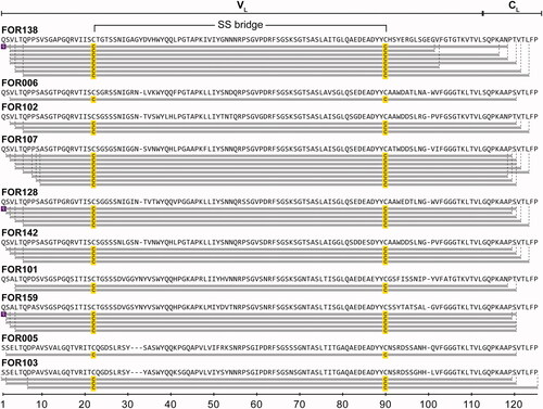
Figure 1. Location of mutational positions in the patient-specific LC proteins. Sequence alignment of the 10 LCs. Amino acid positions are numbered consecutively. Red: mutation, Green: non-encoded amino acid that arose from junctional diversity, Violet: uncertain position. Underlined amino acids at the C-terminus could not be resolved by cDNA sequencing and represent the amino acids of the respective GL. Above the sequence alignment, the secondary structure elements corresponding to the native structure of a LC (PDB: 6QB6-L) are shown. Arrows indicate β strands and continuous lines indicate ordered conformation. Below the sequence, number of mutations per amino acid position is plotted. Asterisks indicate a mutated position in more than 50% of all LCs. The sequences shown are composed of cDNA sequencing and MS results. Sequences corresponding to FOR005 and FOR006 were already published [Citation28].
![Figure 1. Location of mutational positions in the patient-specific LC proteins. Sequence alignment of the 10 LCs. Amino acid positions are numbered consecutively. Red: mutation, Green: non-encoded amino acid that arose from junctional diversity, Violet: uncertain position. Underlined amino acids at the C-terminus could not be resolved by cDNA sequencing and represent the amino acids of the respective GL. Above the sequence alignment, the secondary structure elements corresponding to the native structure of a LC (PDB: 6QB6-L) are shown. Arrows indicate β strands and continuous lines indicate ordered conformation. Below the sequence, number of mutations per amino acid position is plotted. Asterisks indicate a mutated position in more than 50% of all LCs. The sequences shown are composed of cDNA sequencing and MS results. Sequences corresponding to FOR005 and FOR006 were already published [Citation28].](/cms/asset/c2a659dc-9f1b-420d-81e5-809ceae26774/iamy_a_2095618_f0001_c.jpg)
Since the presence of glycosylation is discussed as a possible cause of aggregation of LCs [Citation4,Citation41], we bioinformatically analysed the protein sequences obtained for potential glycosylation sites. In none of the LC and GL sequences could we identify a canonical N-glycosylation site corresponding to the motif asparagine-X-serine/threonine (X ≠ proline). This shows that none of the mutations inserted during LC formation generated such a glycosylation site and indicates that N-glycosylation is not involved in the aggregation process of the LCs studied. In contrast to N-glycosylation, the prediction of O-glycosylation is challenging because no amino acid consensus has yet been identified. Therefore, we did not perform a computational prediction of O-glycosylation.
Analysis of the primary structure of the fibril proteins
In order to obtain the primary structure of the fibril proteins, we extracted AL amyloid fibrils from abdominal fat from the patients using an established protocol for the extraction of amyloid fibrils from human tissue [Citation28]. It was previously demonstrated that the fibrils from abdominal fat are representative for the fibrils deposited within other organs of the same patient [Citation28]. To identify the exact AL protein primary structure, we used the following workflow. The extracted AL fibrils were disaggregated and the monomerized fibril proteins applied to MS to either obtain total mass or to perform de-novo sequencing using tandem MS (, SI Table 7, SI Figure 2). The obtained masses were assigned to the protein sequence of the precursor LCs considering common PTMs such as deamidation, pyroglutamate modifications, oxidation or disulphide bridges. Exemplary MS results, including liquid chromatography chromatogram, raw MS spectrum and deconvoluted MS spectrum of FOR142 are presented in the supporting information (SI Figure 2). In all cases, we found the fibril protein species only in the mass range of 10,000–12,300 Da (SI Table 7), which is consistent with the molecular weight of the fibril protein found in previous studies [Citation25,Citation28] and indicates a truncation of the precursor LC. All analysed proteins could be assigned to the VL and a small part of the CL (), which is consistent with previous reports about λ LCs [Citation28,Citation42–44]. Most fibril proteins lack the first 1 to 4 N-terminal amino acids and only rarely is the N-terminus intact or further amino acids are missing (, SI Table 7). When the expected N-terminus is intact, we usually found a pyroglutamate at position 1 (, SI Table 7), which is a typical PTM of natural antibodies [Citation45]. Most fibril proteins end C-terminally in a region around amino acid 120 (according to our sequential numbering), but the exact position of the C-terminus is slightly heterogeneous (, SI Table 7). This region is part of the N-terminal β-strand of the natively folded CL (). In only one case, FOR138, we found additional protein species terminating at around amino acid 100), which corresponds to CDR3 of the VL (). Another striking feature of all fibril proteins is that the native disulphide bridge of the VL is preserved (, SI Table 7), indicating that AL fibril formation must generally occur around the intact disulphide bond. In addition to these features, we found no evidence of glycosylation, which is consistent with our analysis of the primary structure of the LCs (see above). Furthermore, we also found no evidence for other PTMs such as deamidation, oxidation or phosphorylation. Taken together, our data show that the fibril proteins share common features such as presence of the VL and a small part of the CL as well as common PTMs such as N- and C-terminal truncations and the presence of the native disulphide bridge of the VL.
Figure 2. Overview of the sequence coverage of the fibril proteins of the 10 AL patients found by MS. Disulphide bonded cysteine is highlighted in yellow and pyroglutamate is highlighted in violet. The dashed lines indicate the N- and C-terminal ends.
Analysis of the mutational effects on the protein properties of precursor LC and fibril protein
To investigate the effects of the mutations on the biochemical properties of the precursor LCs and the fibril proteins, we bioinformatically analysed their amino acid sequence and compared them with their respective GL sequence. All analyses were performed comparatively for the identified fibril proteins, the full-length LCs and the VL and CL separately, as the vast majority of mutations occur in the VL and therefore mainly alter the properties of this domain separately and not necessarily those of the full-length LC. We focussed on properties that are discussed to be key determinants of aggregation, such as hydrophobicity, aggregation propensity and protein net charge. Hydrophobicity was analysed by calculation of the grand average of hydropathy (GRAVY), which is defined by the sum of hydropathy values of all amino acids divided by the protein length. The difference values of the patient-specific proteins to the GL proteins, calculated from the GRAVY of the respective protein sequences and expressed as ΔGRAVY, scatter around the value 0 (, SI Table 5). This shows that the mutations alter hydrophobicity to a small extent, but do not lead to a clear trend in any direction, neither for VL, CL, nor the full-length LC. The same pattern could be observed for the global aggregation propensity predicted with TANGO [Citation36], which also shows only subtle changes with no clear trend (, SI Table 4). In summary, mutations alter the hydrophobicity and aggregation propensity, but these properties do not provide a conclusive explanation for the fibril formation of the LCs and fibril proteins studied.
Figure 3. Mutational effects on the biochemical and structural properties of the LC proteins. (A) Analysis of the hydrophobicity (GRAVY) and aggregation propensity (AGG). Shown are the difference values ΔGRAVY and ΔAGG between the LC proteins and their respective GL proteins separately for the VL and CL. (B) Analysis of the pI values. Shown are the individual pI values and the mean pI values of the LC proteins and their respective GL proteins separately for the VL and CL. Asterisks indicate a significant difference. (C) Location of mutations in a natively folded LC (PDB: 6QB6). Colour coding as indicated.
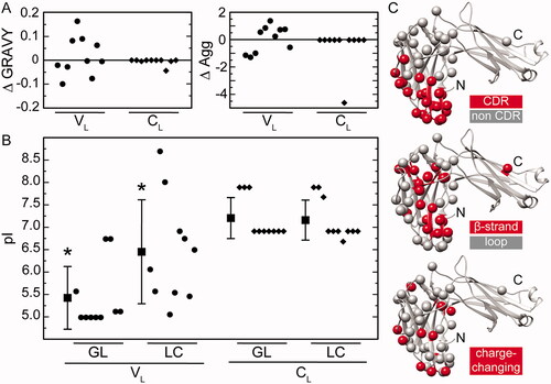
We also investigated the effects of the mutations on the net charge of the protein, which depends on its pI and the pH of the environment. It is well established that the reduction of the net charge of a protein increases its propensity to aggregate [Citation46]. The comparison of the GL protein sequence and the LC protein sequence revealed a significant increase of the mean pI of the VL by about 1 pH unit from 5.5 ± 0.7 to 6.5 ± 1.2 (mean ± SD, p = .045, effect size dCohen = 0.986, , SI Table 6), suggesting that the mutations render this domain less soluble under physiological pH conditions. In contrast, the mean pI values of the CL do not differ at all, as mutations occur only rarely (7.2 ± 0.5 to 7.2 ± 0.5, mean ± SD, p = .854, effect size dCohen = 0.084, , SI Table 6). Given this, it is not surprising that the mean pI values of the full-length GL protein (6.6 ± 0.6) and LCs (7.0 ± 0.8) are not significantly different (p = .185, effect size dCohen = 0.619, SI Table 6). In agreement with this, the mean pI difference of the fibril proteins (7.4 ± 1.0) to the corresponding GL sequence (6.7 ± 0.7) is 0.7 pH units (p = .086, effect size dCohen = 0.818, SI Table 8), since they consist of the VL and a part of the CL. We conclude from our data, that the main effect caused by the mutations is a significant increase in the net protein charge of the VL.
Since mutations can also influence the native state of a LC, we next analysed the location and type of mutations using a native structure of a LC from the protein data base (PDB). For this purpose, PDB files corresponding to the respective VL GL were selected based on sequence similarity. The PDB entry with the best total similarity score for the VL was used for further analysis. Since the native fold of LCs in different GL families is very similar, the mutation sites of all cases examined were exemplified in the crystal structure of an IGLV1-44 derived LC (PDB:6QB6). However, we also provide a figure showing mutations of cases with the same GL V segment in a natively folded LC crystal structure of the same family (SI Figure 1). We find that the mutations in the CDRs and mutations outside the CDRs cluster into 2 distinct regions in the VL (). The mutations within the CDRs (red) are located at the lower part of the VL, that is involved in antigen binding and spatially distant from the CL, while mutations outside the CDRs (grey) are concentrated in the upper part of the VL, that is close to the CL. These 2 structural regions correlate with previously described mutational clustering in amyloidogenic LCs [Citation25]. In contrast, the distribution of mutations with respect to secondary structure elements shows no obvious preference for loops or β strands (). Based on the findings about the net charge of the VL, we also analysed the distribution of charge changes in the structure caused by mutations (). Positions with charge changes (red) also show no clustering in a specific region, but are rather distributed throughout the structure. However, they are mainly located on the VL surface, suggesting that these mutations may affect the surface charge of the VL and alter its inter- and intramolecular interactions.
Discussion
In this study, we provide a systematic overview of the exact primary structure of the fibril proteins of AL amyloidosis patients and their respective precursor LCs and analysed their biochemical properties. Major findings of our study are a significant increase of the mean pI of the VL of the patient specific LCs due to the mutational changes, the presence of common N- and C-terminal truncations in the fibril proteins and the preservation of the native disulphide bridge in the fibril proteins.
By combining MS and cDNA sequencing it was possible to successfully resolve three out of four uncertain positions within the cDNA sequences. These uncertainties resulted at the cDNA level from overlapping signals for individual nucleotides most probably caused by oligoclonal differences of the patient’s plasma cell population. The concept of intra-clonal heterogeneity and clonal evolution has been described in the closely related plasma cell disease multiple myeloma [Citation47,Citation48]. In all stages of the disease – from monoclonal gammopathy of undetermined significance to plasma cell leukaemia – sub clonal heterogeneity can be found [Citation48]. Such clonal differences can be picked up by cDNA sequencing. However, the combination of cDNA sequencing and MS allowed the identification of fibril-forming clones.
Our analysis of the mutational effects on the biochemical properties of the λ precursor LCs and the fibril proteins revealed an increase of the pI of the VL of 1.0 pH unit from 5.5 ± 0.7 to 6.5 ± 1.2. Previous biochemical studies have determined the pI values of some AL associated λ LCs and derived fragments from different sources [Citation44,Citation49–52]. Reported values range from 5.0 ± 1.3 for urinary Bence Jones proteins [Citation49], 7.2 to 8.0 for free monoclonal LCs from serum [Citation50], 5.2 to 8.8 for AL proteins purified from tissue [Citation51], and 5.5 and 5.4 for full-length LCs, respectively [Citation44,Citation52]. Together with our data, these data show a great variability in pI values and suggest that the charge properties are very different for different LCs and AL proteins. As we have only studied a relatively small cohort, one has to be careful when generalising our data and its implications. Therefore, we can only speculate at this point what impact the changed pI may have. The solubility of globular proteins depends on the pI and strictly correlates with fibril formation [Citation53]. It is determined by the amino acid content, the pK values of the ionisable groups and environmental factors such as pH [Citation54]. The strong increase in the pI of the VL is caused by the relative increase in positive charges and indicates a significant reduction in the net surface charge of this domain at the physiological pH of human blood. This may lead to changes in inter- and intramolecular interactions and reduced solubility of the VL. Furthermore, inter- and intramolecular interactions are important for the intrinsic stability of VL and CL and destabilisation of one domain can lead to fibril formation of the entire LC or increased proteolytic susceptibility [Citation21,Citation55,Citation56]. Taken together, we conclude that the increase of the pI of the VL might lead to an increased destabilisation and aggregation tendency of the LC and/or the fibril protein.
Information about truncations and other PTMs of the fibril proteins could be deduced from our MS investigations of highly purified AL fibrils from tissue. In all cases, we found that the major fibril protein species consist of the VL and a small part of the CL which show slight heterogeneity at the N- and C-terminus. The heterogeneity of the N-terminus we found is consistent with recent MS studies [Citation57] and cryo-EM studies of λ1 and λ3 AL fibrils [Citation25,Citation58,Citation59] showing solvent-exposed and conformationally disordered N-termini. Only in the case of a λ6-AL fibril does the N-terminus show no heterogeneity [Citation24,Citation57] and is buried in the fibril core [Citation52]. λ6 proteins were not part of our study and it is unclear whether this is a feature of the specific protein or the λ6 GL. The C-termini of AL proteins in all available cryo-EM structures are disordered and solvent-exposed, and the ordered fibril core terminates well before the C-terminal region we and others identified [Citation25,Citation52,Citation58,Citation59]. The presence of a ubiquitous truncation region, also reported by other studies [Citation24,Citation44,Citation57], suggests that a common proteolytic event is involved in the formation of LC fragments. But when this event takes place in terms of time cannot be deduced from it and it is a very controversial discussion. Amyloidogenic full-length LCs are less kinetically stable than non-amyloidogenic full-length LCs, but often do not form fibrils under physiological conditions, suggesting that an initial proteolytic event may initiate aggregation by releasing an amyloidogenic fragment containing the less stable VL from its highly stable and protective CL [Citation12,Citation15,Citation21,Citation55,Citation60]. On the other hand, studies have found full-length LC in amyloid deposits, suggesting that deposition occurs first, followed by truncation [Citation24,Citation44,Citation61]. However, it is reasonable to assume that the small N- and C-terminal heterogeneity of the fibril proteins reflects the activity of exoproteases that process the deposited polypeptide chain [Citation24].
The strength and at the same time the possible limitation of our conclusions regarding the truncation sites of AL proteins is the extraction procedure. The prerequisite for our analysis is the extraction of amyloid fibrils that are as pure as possible. Only with a defined and highly pure fibril sample can it be ensured that the analysed proteins are actually part of the fibrils and have not been co-purified by the extraction procedure. The water extraction protocol, in which the connective tissue around the AL fibrils is digested by incubation with collagenase, enables precisely this restrictive extraction of AL fibrils. On the other hand, the enzymatic digestion raises the question of possible artefactual degradation of the fibrils, which is a limitation for determining the exact primary structure of the AL proteins. Therefore, the analysis of corresponding non-digested samples would be desirable to exclude the possibility that artifactual fibril degradation has occurred. However, without an overnight enzymatic digestion, we were not able to obtain pure AL fibrils.
Previous studies on AL deposits found in addition to truncations further PTMs like disulphide bonds, deamidation, oxidation, phosphorylation, glycosylation and pyroglutamate formation [Citation4,Citation24,Citation25,Citation41,Citation44]. Here, we only found evidence that the major fibril protein species contains the conserved disulphide bond of the VL and sometimes a pyroglutamylation at the intact N-terminus. Further PTMs could not be detected. While glycosylation has been reported as an important feature for amyloidogenicity of κ LCs [Citation23], it occurs rather rarely in amyloidogenic λ LCs and thus seems to play a minor role. [Citation25]. Pyroglutamylation is a typical PTM of natural antibodies [Citation45]. Although we cannot exclude that pyroglutamylation influences the aggregation of LCs. Based on our data, it is not very likely that it has a major impact on fibril formation. The disulphide bond in the VL is not only a general feature of natively folded LCs but also consistently present in all AL proteins described here and AL proteins found in cryo EM structures so far [Citation25,Citation28,Citation52,Citation58,Citation59]. This suggests a central mechanism of fibril formation by LCs in which the native disulphide bond of the VL is preserved and unfolding and misfolding must occur around the intact disulphide bond. Whether correlation with respect to the C-terminal truncation site, PTMs and pI represent common pathogenic principles in AL amyloidosis will become apparent as more AL protein sequences become available.
| Abbreviations | ||
| ACN | = | Acetonitrile |
| AL | = | Amyloid light chain |
| C | = | Constant |
| CDR | = | Complementarity determining region |
| CL | = | Constant light domain |
| dCohen | = | Cohen’s d |
| dFLC | = | Difference between involved and uninvolved free light chains |
| f | = | Female |
| FA | = | Formic acid |
| FR | = | Framework region |
| GL | = | Germ line |
| GRAVY | = | Grand average of hydropathy |
| HCD | = | Higher-energy collisional dissociation |
| IGLC | = | Immunoglobulin lambda constant |
| IGLJ | = | Immunoglobulin lambda joining |
| IGLV | = | Immunoglobulin lambda variable |
| J | = | Joining |
| LC | = | Light chain |
| m | = | Male |
| MGCS | = | Monoclonal gammopathy of clinical significance |
| MM | = | Multiple myeloma |
| MS | = | mass spectrometry |
| PTM | = | Post-translational modification |
| SD | = | Standard deviation |
| SMM | = | Smouldering multiple myeloma |
| TCB | = | Tris-calcium-buffer |
| TEB | = | Tris-EDTA-buffer |
| TFA | = | Trifluoroacetic acid |
| UMC | = | Underlying medical condition |
| V | = | Variable |
| VL | = | Variable light domain |
Supplemental Material
Download MS Word (258.9 KB)Acknowledgements
We thank the Deutsche Forschungsgemeinschaft for funding (FOR2969; grant numbers: HA 7138/3 to C.H., HE 8472/1-1 to U.H., HU 2400/1-1 to S.H., SCHO 1364/2-1 to S. O. S.).
Disclosure statement
No potential conflict of interest was reported by the author(s).
Data availability statement
The data that support the findings of this study are available from the corresponding author, C. H., upon reasonable request. In addition, the MS data are accessible in the MassIVE database under the dataset identifier MassIVE MSV000089772.
Additional information
Funding
References
- Gertz MA. Immunoglobulin light chain amyloidosis: 2016 update on diagnosis, prognosis, and treatment. Am J Hematol. 2016;91(9):947–956.
- Blancas-Mejía LM, Ramirez-Alvarado M. Systemic amyloidoses. Annu Rev Biochem. 2013;82:745–774.
- Wechalekar A, Schönland S, Kastritis E, et al. A European collaborative study of treatment outcomes in 346 patients with cardiac stage III AL amyloidosis. Blood. 2013;121(17):3420–3427.
- Bellotti V, Mangione P, Merlini G. Review: immunoglobulin light chain amyloidosis – the archetype of structural and pathogenic variability. J Struct Biol. 2000;130(2–3):280–289.
- Murphy K, Travers P, Walport M. Janeway immunologie, 7th ed. Heidelberg: Spektrum Akademischer Verlag; 2009.
- Gertz RA, Kyle MA. Primary systemic amyloidosis: clinical and laboratory features in 474 cases. Semin Hematol. 1995;32(1):45–59.
- Comenzo RL, Zhang Y, Martinez C, et al. The tropism of organ involvement in primary systemic amyloidosis: contributions of Ig V L germ line gene use and clonal plasma cell burden. Blood. 2001;98(3):714–720.
- Abraham RS, Geyer SM, Price-Troska TL, et al. Immunoglobulin light chain variable (V) region genes influence clinical presentation and outcome in light chain-associated amyloidosis (AL). Blood. 2003;101(10):3801–3808.
- Berghaus N, Schreiner S, Granzow M, et al. Analysis of the complete lambda light chain germline usage in patients with AL amyloidosis and dominant heart or kidney involvement. PLoS One. 2022;17(2):e0264407.
- Baden EM, Randles EG, Aboagye AK, et al. Structural insights into the role of mutations in amyloidogenesis. J Biol Chem. 2008;283(45):30950–30956.
- Hurle MR, Helms LR, Li L, et al. A role for destabilizing amino acid replacements in light-chain amyloidosis. Proc Natl Acad Sci USA. 1994;91(12):5446–5450.
- Kazman P, Vielberg MT, Pulido Cendales MD, et al. Fatal amyloid formation in a patient’s antibody light chain is caused by a single point mutation. Elife. 2020;9:e52300.
- Del Pozo-Yauner L, Wall JS, González Andrade M, et al. The N-terminal strand modulates immunoglobulin light chain fibrillogenesis. Biochem Biophys Res Commun. 2014;443(2):495–499.
- Rennella E, Morgan GJ, Yan N, et al. The role of protein thermodynamics and primary structure in fibrillogenesis of variable domains from immunoglobulin light chains. J Am Chem Soc. 2019;141(34):13562–13571.
- Weber B, Hora M, Kazman P, et al. The antibody light-chain linker regulates domain orientation and amyloidogenicity. J Mol Biol. 2018;430(24):4925–4940.
- Rottenaicher GJ, Weber B, Rührnößl F, et al. Molecular mechanism of amyloidogenic mutations in hypervariable regions of antibody light chains. J Biol Chem. 2021;296:100334.
- Poshusta TL, Sikkink LA, Leung N, et al. Mutations in specific structural regions of immunoglobulin light chains are associated with free light chain levels in patients with AL amyloidosis. PLoS One. 2009;4(4):e5169.
- van der Kant R, Karow-Zwick AR, Van Durme J, et al. Prediction and reduction of the aggregation of monoclonal antibodies. J Mol Biol. 2017;429(8):1244–1261.
- Garofalo M, Piccoli L, Romeo M, et al. Machine learning analyses of antibody somatic mutations predict immunoglobulin light chain toxicity. Nat Commun. 2021;12(1):10.
- Buxbaum JN, Chuba JV, Hellman GC, et al. Monoclonal immunoglobulin deposition disease: light chain and light and heavy chain deposition diseases and their relation to light chain amyloidosis. Clinical features, immunopathology, and molecular analysis. Ann Intern Med. 1990;112(6):455–464.
- Weber B, Hora M, Kazman P, et al. Domain interactions determine the amyloidogenicity of antibody light chain mutants. J Mol Biol. 2020;432(23):6187–6199.
- Wall JS, Gupta V, Wilkerson M, et al. Structural basis of light chain amyloidogenicity: comparison of the thermodynamic properties, fibrillogenic potential and tertiary structural features of four Vλ6 proteins. J Mol Recognit. 2004;17(4):323–331.
- Stevens FJ. Four structural risk factors identify most fibril-forming kappa light chains. Amyloid. 2000;7(3):200–211.
- Mazzini G, Ricagno S, Caminito S, et al. Protease-sensitive regions in amyloid light chains: what a common pattern of fragmentation across organs suggests about aggregation. Febs J. 2022;289(2):494–506.
- Radamaker L, Karimi-Farsijani S, Andreotti G, et al. Role of mutations and post-translational modifications in systemic AL amyloidosis studied by cryo-EM. Nat Commun. 2021;12(1):6434.
- Retter I, Althaus HH, Münch R, et al. VBASE2, an integrative V gene database. Nucleic Acids Res. 2005;33(Database issue):D671–D674.
- Howe KL, Achuthan P, Allen J, et al. Ensembl 2021. Nucleic Acids Res. 2021;49(D1):D884–D891.
- Annamalai K, Liberta F, Vielberg MT, et al. Common fibril structures imply systemically conserved protein misfolding pathways in vivo. Angew Chem Int Ed Engl. 2017;56(26):7510–7514.
- Wu Z, Roberts DS, Melby JA, et al. MASH explorer: a universal software environment for top-down proteomics. J Proteome Res. 2020;19(9):3867–3876.
- Niedermeyer THJ, Strohalm M. mMass as a software tool for the annotation of cyclic peptide tandem mass spectra. PLoS One. 2012;7(9):e44913.
- Gasteiger E, Gattiker A, Hoogland C, et al. ExPASy: the proteomics server for in-depth protein knowledge and analysis. Nucleic Acids Res. 2003;31(13):3784–3788.
- Giudicelli V, Chaume D, Lefranc MP. IMGT/GENE-DB: a comprehensive database for human and mouse immunoglobulin and T cell receptor genes. Nucleic Acids Res. 2005;33(Database issue):D256–D261.
- Kawasaki K, Minoshima S, Nakato E, et al. One-megabase sequence analysis of the human immunoglobulin lambda gene locus. Genome Res. 1997;7(3):250–261.
- Swindells MB, Porter CT, Couch M, et al. abYsis: integrated antibody sequence and structure-management, analysis, and prediction. J Mol Biol. 2017;429(3):356–364.
- Gasteiger E, Hoogland C, Gattiker A, et al. Protein identification and Analysis Tools on the ExPASy Server. Proteomics Protoc Handb. 2005;2005:571–607.
- Fernandez-Escamilla AM, Rousseau F, Schymkowitz J, et al. Prediction of sequence-dependent and mutational effects on the aggregation of peptides and proteins. Nat Biotechnol. 2004;22(10):1302–1306.
- Gupta R, Brunak S. Prediction of glycosylation across the human proteome and the correlation to protein function. Authors to correct!. 2002;2002:310–322.
- Pettersen EF, Goddard TD, Huang CC, et al. UCSF Chimera – a visualization system for exploratory research and analysis. J Comput Chem. 2004;25(13):1605–1612.
- Palladini G, Hegenbart U, Milani P, et al. A staging system for renal outcome and early markers of renal response to chemotherapy in AL amyloidosis. Blood. 2014;124(15):2325–2332.
- Van Gameren II, Hazenberg BPC, Bijzet J, et al. Amyloid load in fat tissue reflects disease severity and predicts survival in amyloidosis. Arthritis Care Res. 2010;62(3):296–301.
- Dispenzieri A, Larson DR, Rajkumar SV, et al. N-glycosylation of monoclonal light chains on routine MASS-FIX testing is a risk factor for MGUS progression. Leukemia. 2020;34(10):2749–2753.
- Glenner GG. Amyloid deposits and amyloidosis: the beta-fibrilloses (second of two parts). N Engl J Med. 1980;302(24):1333–1343.
- Solomon A, Frangione B, Franklin EC. Bence Jones proteins and light chains of immunoglobulins. Preferential association of the V(λVI) subgroup of human light chains with amyloidosis AL(λ). J Clin Invest. 1982;70(2):453–460.
- Lavatelli F, Perlman DH, Spencer B, et al. Amyloidogenic and associated proteins in systemic amyloidosis proteome of adipose tissue. Mol Cell Proteomics. 2008;7(8):1570–1583.
- Chelius D, Jing K, Lueras A, et al. Formation of pyroglutamic acid from N-terminal glutamic acid in immunoglobulin gamma antibodies. Anal Chem. 2006;78(7):2370–2376.
- Chiti F, Dobson CM. Protein misfolding, functional amyloid, and human disease. Annu Rev Biochem. 2006;75:333–366.
- Walker BA, Wardell CP, Melchor L, et al. Intraclonal heterogeneity is a critical early event in the development of myeloma and precedes the development of clinical symptoms. Leukemia. 2014;28(2):384–390.
- Rasche L, Chavan SS, Stephens OW, et al. Spatial genomic heterogeneity in multiple myeloma revealed by multi-region sequencing. Nat Commun. 2017;8(1):268.
- Bellotti V, Merlini G, Bucciarelli E, et al. Relevance of class, molecular weight and isoelectric point in predicting human light chain amyloidogenicity. Br J Haematol. 1990;74(1):65–69.
- Kaplan B, Ramirez-Alvarado M, Dispenzieri A, et al. Isolation and biochemical characterization of plasma monoclonal free light chains in amyloidosis and multiple myeloma: a pilot study of intact and truncated forms of light chains and their charge properties. Clin Chem Lab Med. 2008;46(3):335–341.
- Kaplan B, Livneh A, Gallo G. Charge differences between in vivo deposits in immunoglobulin light chain amyloidosis and non-amyloid light chain deposition disease. Br J Haematol. 2007;136(5):723–728.
- Swuec P, Lavatelli F, Tasaki M, et al. Cryo-EM structure of cardiac amyloid fibrils from an immunoglobulin light chain AL amyloidosis patient. Nat Commun. 2019;10(1):1269.
- Schmittschmitt JP, Scholtz JM. The role of protein stability, solubility, and net charge in amyloid fibril formation. Protein Sci. 2003;12(10):2374–2378.
- Schein CH. Solubility as a function of protein structure and solvent components. Biotechnology. 1990;8(4):308–317.
- Rennella E, Morgan GJ, Kelly JW, et al. Role of domain interactions in the aggregation of full-length immunoglobulin light chains. Proc Natl Acad Sci USA. 2019;116(3):854–863.
- Klimtchuk ES, Gursky O, Patel RS, et al. The critical role of the constant region in thermal stability and aggregation of amyloidogenic immunoglobulin light chain. Biochemistry. 2010;49(45):9848–9857.
- Lavatelli F, Mazzini G, Ricagno S, et al. Mass spectrometry characterization of light chain fragmentation sites in cardiac AL amyloidosis: insights into the timing of proteolysis. J Biol Chem. 2020;295(49):16572–16584.
- Radamaker L, Lin YH, Annamalai K, et al. Cryo-EM structure of a light chain-derived amyloid fibril from a patient with systemic AL amyloidosis. Nat Commun. 2019;10(1):1103.
- Radamaker L, Baur J, Huhn S, et al. Cryo-EM reveals structural breaks in a patient-derived amyloid fibril from systemic AL amyloidosis. Nat Commun. 2021;12(1):875.
- Morgan GJ, Kelly JW. The kinetic stability of a full-length antibody light chain dimer determines whether endoproteolysis can release amyloidogenic variable domains. J Mol Biol. 2016;428(21):4280–4297.
- Buxbaum J. Mechanisms of disease: monoclonal immunoglobulin deposition. Amyloidosis, light chain deposition disease, and light and heavy chain deposition disease. Hematol Oncol Clin North Am. 1992;6(2):323–346.