
Abstract
Objectives: The aim of our study was to determine if redox imbalance caused by the activities of antioxidant enzymes existed in erythrocytes of type 1 myotonic dystrophy (DM1) patients.
Methods: The activities of erythrocyte superoxide dismutase, catalase, glutathione peroxidase, and glutathione reductase were measured in 30 DM1 patients and 15 healthy controls (HCs). The obtained values were correlated with the Muscular Impairment Rating Scale (MIRS) score and creatine kinase (CK).
Results: Superoxide dismutase and catalase activities were lower in DM1 patients compared to HCs. A positive correlation was found between disease duration and MIRS score as well as with glutathione reductase activity. In DM1 patients, there were positive correlations between catalase, glutathione peroxidase, and glutathione reductase activities. After sub-dividing DM1 patients according to CK levels, superoxide dismutase activity was still statistically different from HCs. However, catalase activity was significantly lower only in DM1 patients with increased CK.
Discussion: Undesirable alterations in antioxidant enzyme activities during DM1 disease progression may result in conditions favoring oxidative stress and changes in metabolism which together could contribute to muscle wasting.
Introduction
Type 1 myotonic dystrophy (DM1) is the most common form of muscular dystrophy in adults with a worldwide prevalence of 1–35 patients per 100 000 inhabitants.Citation1 DM1 is an autosomal-dominant disease caused by the expansion of Cytosin, Thymine, Guanine (CTG) trinucleotide repeats within the non-coding 3′-untranslated region of the gene encoding dystrophia myotonica-protein kinase on chromosome locus 19q13.3.Citation2 This mutation has a trans-dominant effect through the regulation of alternative splicing of pre-messenger RNA for various proteins, mRNA translation and mRNA stability, which contribute to the multiple features of DM1.Citation3 However, the alternative splicing of many different transcripts fails to fully explain muscle wasting in DM1 patients. DM1 is a multi-systemic disease that affects many organs and tissues besides muscle.Citation4 DM1 has been described as a disease characterized by premature aging, which involves oxidative stress in its pathogenesis.Citation5 Reduced or increased cellular antioxidant activity and increased oxidative stress parameters have been reported in DM1, which may be associated with muscular and extra-muscular signs of the disease.Citation5–Citation8 There is considerable evidence suggesting that reactive oxidative species (ROS) and reactive nitrogen species, such as superoxide (O2.−) and nitric oxide (NO.), can play roles in age-related loss of muscle mass and function.Citation9 The inability of aged muscle to respond to stress is explained by deteriorating redox signaling processes and redox-mediated adaptation.Citation10 Current knowledge supports the likelihood that interactions between the primary genetic defect and disruptions in the normal production of free radicals contribute to muscle atrophy in muscular dystrophies.Citation10 The inability of muscle regeneration to keep up with apoptotic and necrotic events following oxidative stress during normal muscular exercise may underlie muscle atrophy in DM1.Citation11 In sporadic and familiar amyotrophic lateral sclerosis, also characterized by muscle wasting, decreased levels of both copper–zinc superoxide dismutase (SOD1) and catalase (CAT) activities in erythrocytes have been described.Citation12 Thus, circulating oxidative stress could be related to skeletal muscle wasting in DM1.
The aim of our study was to analyze the levels of antioxidant defense enzymes in erythrocytes of patients with DM1, to compare them with those in healthy controls (HCs) and to correlate individual antioxidant enzyme activity with DM1 clinical data, Muscular Impairment Rating Scale (MIRS) scores and creatine kinase (CK) levels. Erythrocytes are a good model for studying systemic oxidative stress and have been exploited in other studies.Citation12
Methods
Patients selection
This cross-sectional study comprised 30 (14 males and 16 females) adult patients aged 41 ± 5 years primarily diagnosed with DM1, who were examined in the outpatient and inpatient units of the Neurology Clinic in Belgrade from 1 September until 31 December 2011 and 15 HCs matched for gender (seven males and eight females) age 36 ± 5 years. Patients with congenital and late adult form of DM1 were not included. In addition to standard clinical and electromyography data, genetic diagnosis of CTG repeat expansion was obtained from all the patients (mean was 691 ± 57). The study was approved by the Ethical Board of the Neurology Clinic, University of Medical Sciences, Belgrade, N° 29/X-5. All patients gave informed written consent to participate in the study.
The severity of DM1 was assessed by the MIRS (mean was 3.4 ± 0.2).Citation13 The MIRS is an ordinal five-point rating scale established in accordance with the clinically recognized distal to proximal progression of muscular involvement in DM1. The man level of serum CK in patients was 251 ± 50 IU/l.
Biochemical analysis
Each study participant donated a 5 ml blood sample obtained by venous puncture between 8 and 9 a.m. after an overnight fast and a period of tobacco abstinence (overnight 12 hours). Erythrocyte lysates were prepared as follows. After the heparin-treated blood samples were centrifuged at 2000 × g for 15 minutes at 4°C, the plasma was discarded. The separated erythrocytes were washed three times with 0.9%, w/v NaCl. Washed cells (0.5 ml) were then lysed by adding 3 ml of ice-cold distilled water followed by thorough mixing.
Hemoglobin (Hb) concentration
The total Hb content of the hemolysates was measured as cyanmethemoglobin using the Drabkin method.Citation14
Hemoglobin removement
To remove Hb 1.0 ml of an ethanol/chloroform (1:1, v/v) mixture was added to an aliquot (0.5 ml) of the hemolysate cooled on ice.Citation15 This mixture was stirred constantly for 15 minutes before being diluted with 0.5 ml of distilled water. After centrifugation for 10 minutes at 1600 × g, the pale-yellow supernatant was separated from the protein precipitate and was used to assay SOD enzyme activity spectrophotometrically and for native polyacrylamide gel electrophoresis (PAGE).
Spectrophotometric measurements
Antioxidant enzyme activities were measured as previously described.Citation12
SOD1 activity was determined by the epinephrine method, which is based on the capacity of SOD1 to inhibit autoxidation of adrenaline to adrenochrome. Reaction mixtures consisted of 3 × 10⁻⁴ M adrenaline, 1 × 10⁻⁴ M EDTA, and 0.05 M Na2CO3, pH 10.2. One unit of SOD is defined as the amount of protein causing 50% inhibition of the autoxidation of adrenaline.
CAT activity is defined as the amount of enzyme, which decomposes 1 mmol hydrogen peroxide (H2O2) in 1 minute at pH 8.0 in 1 M Tris–5 mM EDTA buffer measured at 230 nm.
GSH-Px—glutathione peroxidase activity was measured according to NADPH consumption (NADPH oxidation by glutathione reductase) monitored at 340 nm. The reaction mixture consisted of 50 mM potassium phosphate buffer (pH 7.0), 1 mM EDTA, 1 mM GSH, 1 mM sodium azide, 1 U/ml glutathione reductase, 0.2 mM NADPH, and 3 mM t-butyl hydroperoxide.
GR—glutathione reductase activity measurement is based on NADPH oxidation concomitant with GSH reduction. The reaction mixture consisted of 0.5 M sodium phosphate buffer (pH 7.5), 0.1 mM EDTA, 0.1 mM NADPH, and 0.1 mM GSSG. Enzyme activity is expressed in U/g of soluble protein. One unit of enzyme activity is defined as the amount of enzyme required to transform 1 µmol of substrate per minute under the above described assay conditions.
Native PAGE
Was performed according to Laemmli, 1970 using 12% acrylamide under non-denaturing conditions.Citation16 Prior to PAGE, aliquots of SOD1 from erythrocytes were incubated for 2 hours at 37°C with gentle stirring in the absence (control) and presence of 5 mM H2O2. Incubated SOD samples were diluted to 2 U/ml using a solution containing 12% glycerol, 0.5 mM Tris–HCl (pH 6.8), and 0.2 M EDTA before loading 50 µl per well. SOD1 bands (achromatic zones on an otherwise uniformly violet-blue gel) were visualized using the activity staining procedure described by Beauchamp and FridovichCitation17 requiring the reduction of nitroblue tetrazolium (NBT) with superoxide produced by photochemical reduction of riboflavin with N,N,N′,N′,-tetramethylethylenediamine. To quantify SOD activity, the gels were scanned using a densitometer (GS-700, Bio-Rad) and the tiff image files were analyzed using Multi-Analyst 1.0.2 software (Bio-Rad).
Statistical analysis
Statistical analyses were performed according to the protocols originally described by ManleyCitation18 and Blagojević et al.,Citation19 which have been subsequently applied to study other pathophysiological conditions.Citation12,Citation20 Differences in antioxidant enzyme activity between DM1 patients and controls were analyzed by one-way analysis of variance (ANOVA) followed by Tukey's Student's t-test. Spearman's coefficient was used for correlation of two variables. Since correlation analysis calculates relationships between individual components, we performed canonical discriminant analysis that calculated differences between groups, taking into consideration the complete correlation matrix (i.e. composition of antioxidant defense) of separate patient groups. In all analyses, P < 0.05 was considered as significant.
Results
The activities of SOD1 and CAT were lower in DM1 patients compared to HCs (Fig. ).
Figure 1. Antioxidant enzyme activities in erythrocytes from DM1 patients (n = 30) and HCs (n = 15). Results are expressed as mean ± SE and compared by t-test. ***P < 0.001, **P < 0.01.
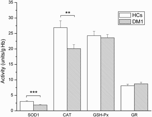
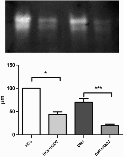
Other measured antioxidant enzymes (GSH-Px and GR) showed no differences. In-gel SOD activity showed the same result (lower activity of SOD1 in DM1 patients compared to HCs). Figure illustrates a representative SOD1 activity profile in non-denaturing PAGE conditions. In addition, SOD1 in HCs was more active (45% of activity remaining; P < 0.05) after partial inhibition with equimolar concentration of H2O2, compared to that in DM1 patients (25% activity remaining; P < 0.001).
Figure 2. Native PAGE gel stained for SOD1 enzyme activity with NBT. Lanes, from left to right: HCs; HCs + H2O2; DM1 and DM1 + H2O2. Histogram presents quantitative analysis of SOD activity. Graphs represent % from SOD activity HCs taken as 100% (±SE). Statistical analysis showed a more pronounced reduction in activity after H2O2 treatment of DM1 SOD compared with to HC SOD (***P < 0.001; n = 7, vs. *P < 0.05; n = 5). Note the difference between samples with respect to relative band intensity (in-gel SOD1 activity). For details, see ‘Methods’ section.
When DM1 patients were segregated according to MIRS grade score, SOD1 activity in erythrocytes of DM1 patients with MIRS grades III and IV was significantly lower than HCs (Table ). Differences in CAT activity were just above the threshold for a statistically acceptable ANOVA P value (therefore the post hoc test showed no statistical significance). The activities of GSH-Px and GR were not different compared to those found in HCs.
Table 1 The activity of antioxidant enzymes in DM1 patients with different severity of muscle impairment
After segregating the patients according to their CK levels, normal (60–174 IU/l) or increased (>180 IU/l), SOD1 was still statistically lower in both groups of DM1 patients compared to HCs. However, CAT activity was significantly lower only in DM1 patients with increased CK (Table ). GSH-Px and GR activities were not different compared to HCs.
Table 2 Comparison of antioxidant defense in HCs and DM1 patients with normal and increased CK values
Inter-correlations between the activities of different antioxidant enzymes and clinical parameters as well as MIRS score, CK levels, and CTG number in DM1 patients are shown in Table .
Table 3 Correlation analysis of different clinical parameters with activity of antioxidant enzymes in DM1 patients (n = 30)
There were no significant correlations between the level of measured antioxidant enzymes and clinical parameters (age of patients, disease duration, age of disease onset), clinical MIRS scores, CK levels, and CTK number, except for GR activity [it was significantly positively correlated with duration of the disease (σ = 0.42, P < 0.05). In contrast, significant positive correlations were found between antioxidant enzyme activities in DM1 patient erythrocytes: CAT versus GSH-Px (P < 0.001), CAT versus GR (P < 0.05), and GSH-Px versus GR (P < 0.05).
As antioxidant enzymes form complex inter-connecting physiological functions, we performed canonical discriminant analysis between different groups of patients and HCs. Canonical discriminant analysis significantly separated DM1 patients with MIRS III (P < 0.001) and MIRS IV (P < 0.01) scores from HCs. The composition of antioxidant enzymes in MIRS II and MIRS V groups was not significantly different from that in HCs (Fig. A).
Figure 3. Canonical discriminant analysis of the activity of antioxidant enzymes in different forms of DM1 according to (A) different severity of MIRS and (B) CK levels, presented as two-dimensional canonical space. Canonical analysis significantly separated DM1 patients with MIRS III and IV stages from HCs and the other two stages (P < 0.001). DM1 patients were significantly separated from HCs regardless if CK levels were increased (P < 0.001).
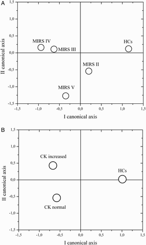
Furthermore, there were no differences in the composition of antioxidant enzymes in patients with different CK levels (CK normal and CK increased), but both groups of patients (CK normal and CK increased) had a different composition compared to HCs (Fig. B). For all comparisons SOD1 activity was most dominant (P < 0.001).
Discussion
Our results clearly showed decreased SOD1 activity in DM1 patient erythrocytes compared to HCs. The decrease in SOD1 activity may have been caused by H2O2-mediated inhibition,Citation21 as demonstrated by native PAGE (Fig. ). After partial inhibition with H2O2, SOD1 activity was more reduced in DM1 patients than in HCs. It is well known that H2O2 inhibits SOD1 activity. In addition, H2O2 directly degrades the enzyme itself.Citation20 It appears that SOD1 in DM1 patients is more susceptible to this process (Fig. ). Previous studies have shown increased apoptosis in myogenic cell lines possessing increased number of CTG repeats after administration of methylmercury, a known ROS producer.Citation22 Advanced oxidation protein products are significantly higher in sera of DM1 patients compared to HCs.Citation10 These findings are in line with our own results, which suggest that there is a certain degree of disturbance in the activity of antioxidant enzymes in DM1 patients. Decreased levels of antioxidant enzymes in blood samples from DM1 patients, including decreased SOD, have also been reported.Citation23 In contrast, earlier reports showed increased SOD1 activity.Citation5 These latter activities were measured in blood serum and could have been a reflection of erythrocyte lysis, or other interfering substances. In our current study, decreased SOD1 activity was measured in erythrocytes. This finding could represent a suppressive reaction to increased oxidative stress during a particular phase of the disease, since SOD1 activity from DM1 patients are more susceptible to H2O2-mediated inhibition. Additionally, in our cohort of patients, decreased SOD1 activity was particularly pronounced in patients with moderate muscular impairment severity (MIRS grades III and IV). This was confirmed by canonical discriminant analysis, as DM1 patients with MIRS III and IV scores were significantly separated from other groups when taking into account the composition of antioxidant enzymes in erythrocytes. These results also demonstrated that oxidative stress in DM1 patients is dynamic and changeable during the disease stages. Furthermore, the activity of antioxidant enzymes in erythrocytes did not correlate with MIRS score suggesting that oxidative stress and involvement of particular ROS is stage specific and does not linearly follow disease progression. Moreover, the activity of antioxidant enzymes in erythrocytes was not correlated with either CK level nor CTG number, although SOD1 and CAT activities were lower in DM1 patients regardless if CK levels were elevated (our results showed that CAT activity was significantly decreased only in patients with increased CK, but the significance was slightly below the confidence limit, P < 0.05). It has been shown that CK concentration is often, but not always, elevated in muscle disease.Citation24 Our results suggest that there is an association between muscle deterioration, increased CK levels and CAT inhibition in erythrocytes.
The effect of oxidative stress on muscle damage has been shown to be present in muscular diseases such as dystrophinopathy,Citation11 mitochondrial disordersCitation25 and in sarcopenia, which is age-related muscle loss.Citation5 Normal muscle tissue responds to the stress of contractions by up-regulating protective enzymes (including SOD1 and CAT) and stress proteins.Citation5,Citation26 The primary reactive species generated by muscle, both at rest and during contraction, are NO. and O2.−.Citation27 Small increases in reactive species are favorable and lead to increased force production. Greater production of reactive species contributes to the development of muscle fatigue.Citation28 The predominant effects of ROS and NO· on muscle force production are due to changes in calcium metabolism influenced by the ryanodine receptor and sarcoplasmic reticulum Ca2+ ATPase-SERCA.Citation29 Interestingly, both mRNAs of these proteins are mis-spliced in DM.Citation30
Since the above-mentioned mechanisms lead to aging in normal muscle tissue, they are also probably related to progeroid processes in DM1.Citation4 In line with this, there is evidence that increased cellular O2.− can cause muscle ageing since mice lacking SOD1 exhibited accelerated age-related loss of skeletal muscle mass and function.Citation31 At present treatment for DM1 is limited to symptomatic intervention and there is no therapeutic approach to prevent or reverse disease progression.Citation32 As oxidative stress plays a role in the progression of neurodegeneration and muscle wasting,Citation33 there is a possibility that supplemented antioxidant therapy could be beneficial, particularly at intermediate stages of DM1, when decreased SOD 1 is present.
In physiological settings erythrocytes exhibit a self-sustaining activity of antioxidant defense enzymes and their coordinated actions protect the erythrocytes' bio-macromolecules from free radical-mediated damage. This is the case in this current study, as positive correlation was found between CAT, GSH-Px, and GR activities in erythrocytes suggesting that erythrocytes adjust their antioxidant potential according to the presence of oxidative pressure especially by H2O2. However, we found that there was no significant correlation between the activity of the antioxidant enzymes and the clinical outcome. Therefore, the measurement of the activity of these enzymes cannot be exploited for diagnostics, but instead suggests that oxidative processes occur in DM1 patients providing new insight into the disease.
Disclaimer statements
Contributors All authors contributed equally.
Funding This work was supported by grants from the Ministry of Education, Science and Technological Development of the Republic of Serbia [Grants No. 175083 and 173014].
Conflicts of interest The authors state that there are no conflicts of interest regarding the publication of this article.
Ethics approval The study was approved by the Ethical Board of the Neurology Clinic, University of Medical Sciences, Belgrade, No29/X-5.
Acknowledgements
Special thanks for English language-editing by Dr David R. Jones from Paterson Institute for Cancer Research, The University of Manchester, United Kingdom.
References
- Emery AE. Population frequencies of inherited neuromuscular diseases—a world survey. Neuromusc Disord 1991;1:19–29. doi: 10.1016/0960-8966(91)90039-U
- Klein AF, Gasnier E, Furling D. Gain of RNA function in pathological cases: focus on myotonic dystrophy. Biochimie 2011;93:2006–12. doi: 10.1016/j.biochi.2011.06.028
- Lee JE, Cooper TA. Pathogenic mechanisms of myotonic dystrophy. Biochem Soc Trans 2009;37:1281–86. doi: 10.1042/BST0371281
- Harper PS. Myotonic dystrophy. 3rd ed. London, UK: Saunders WB; 2001.
- Toscano A, Messina S, Campo GM, Di Leo R, Musumeci O, Rodolico C, et al. Oxidative stress in myotonic dystrophy type 1. Free Radic Res 2005;39(7):771–76. doi: 10.1080/10715760500138932
- Ihara Y, Mori A, Hayabara T, Namba R, Nobukuni K, Sato K, et al. Free radicals, lipid peroxides and antioxidants in blood of patients with myotonic dystrophy. J Neurol 1995;242(3):119–22. doi: 10.1007/BF00936882
- Siciliano G, Pasquali L, Rocchi A, Falorni M, Galluzzi F, Rocco A, et al. Advanced oxidation protein products in serum of patients with myotonic disease type I: association with serum gamma-glutamyltransferase and disease severity. Clin Chem Lab Med 2005;43(7):745–47. doi: 10.1515/CCLM.2005.127
- Rodriguez MC, Tarnopolsky MA. Patients with dystrophinopathy show evidence of increased oxidative stress. Free Radic Biol Med 2003;34(9):1217–20. doi: 10.1016/S0891-5849(03)00141-2
- Vasilaki A, Mcardle F, Iwanejko LM, Mc Ardle A. Adaptive responses of mouse skeletal muscle to contractile activity: the effect of age. Mech Ageing Dev 2006;127(11):830–39. doi: 10.1016/j.mad.2006.08.004
- Jackson MJ. Control of reactive oxygen species production in contracting skeletal muscle. Antioxid Redox Signal 2011;15(9):2477–86. doi: 10.1089/ars.2011.3976
- Tidball JG, Wehling-Henricks M. The role of free radicals in the pathophysiology of muscular dystrophy. J Appl Physiol 2007;102(4):1677–86. doi: 10.1152/japplphysiol.01145.2006
- Nikolić-Kokić A, Stević Z, Blagojević D, Davidović B, Jones DR, Spasić MB. Alterations in anti-oxidative defense enzymes in erythrocytes from sporadic amyotrophic lateral sclerosis (SALS) and familial ALS patients. Clin Chem Lab Med 2006;44:589–93. doi: 10.1515/CCLM.2006.111
- Mathieu J, Boivin H, Meunier D, Gaudreault M, Bégin P. Assessment of a disease-specific muscular impairment rating scale in myotonic dystrophy. Neurology 2001;56(3):336–40. doi: 10.1212/WNL.56.3.336
- Tentori L, Salvati AM. Hemoglobinometry in human blood. Methods Enzymol 1981;76:707–15. doi: 10.1016/0076-6879(81)76152-4
- Tsuchihashi M. Zur Kenntnis der Blutkatalase. Biochemistry 1923;Z140:65–74.
- Laemmli UK. Cleavage of structural proteins during the assembly of the head of bacteriophage T4. Nature 1970;227:680–85. doi: 10.1038/227680a0
- Beauchamp C, Fridovich I. Superoxide dismutase: improved assays and an assay applicable to acrylamide gels. Anal Biochem 1971;44:276–87. doi: 10.1016/0003-2697(71)90370-8
- Manley BFJ. Multivariate statistical methods. A primer. London: Chapman and Hall; 1986.
- Blagojević D, Buzadžić B, Korać B, Saičić ZS, Radojičić R, Spasić MB, Petrović VM. Seasonal changes in the antioxidant defense in ground squirrel (Citellus citellus): possible role of GSH-Px. JEPTO 1998;17:241–50.
- Miljević Č, Nikolić-Kokić A, Saičić ZS, Milosavljević M, Blagojević D, Tosevski DL, et al. Correlation analysis confirms differences in antioxidant defence in the blood of types I and II schizophrenic male patients treated with anti-psychotic medication. Psych Res 2010;178(1):68–72. doi: 10.1016/j.psychres.2008.10.038
- Bray RC, Cockle SA, Fielden EM, Roberts PB, Rotilio G, Calabrese L. Reduction and inactivation of superoxide dismutase by hydrogen peroxide. Biochem J 1974;139(1):43–8. doi: 10.1042/bj1390043
- Usuki F, Takahashi N, Sasagawa N, Ishiura S. Differential signaling pathways following oxidative stress in mutant myotonin protein kinase cDNA-transfected C2C12 cell lines. Biochem Biophys Res Commun 2000;267(3):739–43. doi: 10.1006/bbrc.1999.2026
- Comim CM, Mathia GB, Hoepers A, Tuon L, Kapczinski F, Dal-Pizzol F, et al. Neurotrophins, cytokines, oxidative parameters and funcionality in Progressive Muscular Dystrophies. An Acad Bras Cienc 2015; 87(3):1809–18.
- Renard D. Serum CK as a guide to the diagnosis of muscle disease. Pract Neuro 2015; 15(2):121. doi:10.1136/practneurol-2014-001031.
- Peterson CM, Johannsen DL, Ravussin E. Skeletal muscle mitochondria and aging: a review. J Aging Res 2012;2012:194821. doi: 10.1155/2012/194821
- McArdle F, Spiers S, Aldemir H, Vasilaki A, Beaver A, Iwanejko L, et al. Preconditioning of skeletal muscle against contraction-induced damage: the role of adaptations to oxidants in mice. J Physiol 2004;561:233–44. doi: 10.1113/jphysiol.2004.069914
- Jackson MJ. Free radicals generated by contracting muscle: by-products of metabolism or key regulators of muscle function? Free Radic Biol Med 2008;44(2):132–41. doi: 10.1016/j.freeradbiomed.2007.06.003
- Reid MB, Khawli FA, Moody MR. Reactive oxygen in skeletal muscle. III. Contractility of unfatigued muscle. J Appl Physiol 1993;75:1081–87.
- Zima AV, Blatter LA. Redox regulation of cardiac calcium channels and transporters. Cardiovasc Res 2006;71:310–21. doi: 10.1016/j.cardiores.2006.02.019
- Kimura T, Nakamori M, Lueck JD, Pouliquin P, Aoike F, Fujimura H, et al. Altered mRNA splicing of the skeletal muscle ryanodine receptor and sarcoplasmic/endoplasmic reticulum Ca2+-ATPase in myotonic dystrophy type 1. Hum Mol Genet 2005;14(15):2189–200. doi: 10.1093/hmg/ddi223
- Muller FL, Song W, Liu Y, Chaudhuri A, Pieke-Dahl S, Strong R, et al. Absence of CuZn superoxide dismutase leads to elevated oxidative stress and acceleration of age-dependent skeletal muscle atrophy. Free Radic Biol Med 2006;40:1993–2004. doi: 10.1016/j.freeradbiomed.2006.01.036
- Magaña JJ, Cisneros B. Perspectives on gene therapy in myotonic dystrophy type 1. J Neurosci Res 2011;89(3):275–85. doi: 10.1002/jnr.22551
- Kumar A, Kumar V, Singh SK, Muthuswamy S, Agarwal S. Imbalanced oxidant and antioxidant ratio in myotonic dystrophy type 1. Free Radic Res 2014;48(4):503–10. doi: 10.3109/10715762.2014.887847