
Abstract
Introduction: Although the role of insulin in the development of benign prostatic hyperplasia (BPH) is well established, there are no studies regarding alteration in the gene expression of components of insulin-signaling pathway and their association with prostate size in BPH. Hence, the study was designed to analyze the gene and protein expression of insulin receptor and its related components in patients with BPH.
Materials and methods: Twenty-seven BPH patients aged between 55 and 75 years were recruited in the study and prostatic tissues were obtained after transurethral resection of the prostate. Gene expression levels of Insulin receptor (IR), insulin receptor substrate (IRS), insulin-like growth factor (IGF) and insulin-like growth factor-binding protein-3 (IGFBP-3) were assessed by q-PCR.
Results: Insulin receptor (IR-A and B) and insulin-like growth factors (IGF-1 and IGF-2) gene expression were significantly increased and IGFBP-3 gene expression was reduced in BPH patients with larger prostate size. Also, serum insulin was significantly increased and IGFBP-3 was significantly reduced in patients with larger prostate size.
Conclusion: Increased expression of IR-A, B and IGF-1, 2 genes and reduced IGFBP-3 gene expression was associated with larger prostate size in BPH.
Introduction
Benign prostatic hyperplasia (BPH) is the most common urological problem in aging men [Citation1]. Although BPH is highly prevalent, the pathogenesis involved in the development of prostate growth remains uncertain. Among hormones, androgens play a prominent and essential role in normal and hyperplastic prostate growth [Citation2]. Pradidarcheep has found an association between late onset hypogonadism and lower urinary tract symptoms (LUTS) indicating a link between low testosterone and symptoms suggestive of BPH [Citation3]. Previous studies have found an improvement in PSA, prostate volume and LUTS after testosterone replacement therapy in patients with hypogonadism [Citation4–7], where as Efesoy et al. and Meuleman et al. did not found any increase in prostate volume or PSA after treatment with testosterone [Citation8,Citation9]. Dutasteride, a 5-α reductase inhibitor used in the treatment of BPH was found to reduce PSA and prostate volume and it has been attributed to increase in testosterone levels [Citation10].
Previous studies have reported the role of other factors like insulin and insulin-like growth factors (IGF’s) in the development of BPH which might act through androgen independent mechanisms [Citation11–13]. A recent study has reported that androgen deprivation therapy increases fasting plasma insulin and decreases insulin sensitivity [Citation14]. Animal experiment studies and clinical studies have indicated the complications associated with insulin resistance in the pathogenesis of BPH [Citation13,Citation15,Citation16]. Yeh et al. has revealed an association of LUTS with metabolic syndrome in elderly male [Citation17]. Also earlier investigators have confirmed increased incidence of BPH in insulin resistant and diabetic population [Citation18].
Insulin is known to influence cell growth, differentiation and regulation of gene expression and alteration in insulin-signaling cascade has been demonstrated in many cancers [Citation19]. Insulin transduces its signaling through its cognate receptors, which consists of two isoforms, insulin receptor – A and B (IR-A and IR-B) [Citation20], which belong to family of receptor tyrosine kinases that includes the receptor for insulin-like growth factor 1 (IGF-1 R). Upregulated expression of IGF-1 R, IR and potentially hybrid IR/IGF-1Rs was observed in many tumor types that enable high activation of mitogenic, prosurvival and protein synthesis pathways with the increased levels of ligands like insulin, IGF-1, or IGF-2 [Citation21,Citation22].
Recent studies have demonstrated the significance of insulin receptor and its differential expression in prostate cancer [Citation23]. Over expression of insulin receptor and its higher activity of downstream signaling in malignant prostate tissues has been reported [Citation24,Citation25]. Differential expression of insulin receptor isoforms IR-A and IR-B and alteration in ratios of IR A/B were observed in prostate cancer and its adjacent tissues [Citation26]. The insulin-like growth factor-binding protein-3 (IGFBP-3) is known to exhibitproapoptotic and antiangiogenic properties. Previous studies have demonstrated the expression of IGFBP-3 in benign epithelial, stromal and tumor prostate cells [Citation27] and hypothesized that IGBP-3 gene suppresses metastasis in prostate cancer [Citation28]. But till date, there are no reports about the expression of IR, IGF and IGFBP-3 in BPH patients with different prostate size. Hence, the present study was designed to investigate gene expression of insulin receptor and associated factors in prostate tissues of BPH patients and their association with prostate size.
Materials and methods
Study subjects
The study was carried out July 2015 to August 2016 in the department of Biochemistry and Urology, JIPMER, Puducherry, India. Twenty-seven BPH patients, aged 50–75 years, who underwent transurethral resection of prostate were recruited in the study. BPH was diagnosed based on clinical findings, per rectal examination, ultrasound findings and confirmed by histopathological findings. Subjects with prostate cancer, other causes of lower urinary tract symptoms, diabetes, renal disease, cardiovascular disease and pancreatic disorders were excluded from the study. The study protocol was approved by scientific advisory committee and institute ethics committee. Written informed consent was obtained from all the subjects prior to the study.
Blood sample collection and estimation of biochemical parameters
Five milliliters of venous blood samples was collected after overnight fasting from the subjects. Serum was separated and the aliquoted samples stored at −80°C and used for further analysis of the test parameters namely insulin (CalbiotechInc, Spring Valley, California), PSA (CalbiotechInc, Spring Valley, California), IGF-1 (Raybiotech, Georgia, United States), IGFBP-3 (Raybiotech, Georgia, United States) by using ELISA kits.
Prostate tissue collection and processing
Prostate tissues was obtained from transition zone of the prostate after transurethral resection of prostate (TURP) and were transferred immediately to the laboratory in liquid nitrogen container, tissues were aliquoted and stored at −80 °C. BPH patients were divided into two groups based on their prostate size: Group A (n = 13) prostate size ≤30 ml, Group B (n = 14) prostate size by >30 ml.
RNA extraction, cDNA synthesis and real-time PCR
RNA was extracted from tissues using RNeasy Fibrous Tissue kit in combination with RNase-free DNase set (Qiagen) according to the manufacturer’s protocol. RNA concentration, purity were determined by measuring the absorbance at 260 and 280 nm in Nanodrop2000 (Thermo Scientific, MA, USA). One microgram of total RNA was used for single-strand complementary DNA synthesis by using the High Capacity cDNA Reverse Transcription Kit (Applied Biosystems, CA, USA) according to manufacturer’s protocol. Gene-specific oligonucleotide primers (IDT) were used as listed in . Expression levels of the genes were measured by real-time PCR using SYBR master mix (TaKaRa Bio Inc Japan). Real-time PCR was carried out in CFX96 real-time PCR detection system. Signals were normalized to the housekeeping genes β-actin or GAPDH as the endogenous internal control. The following real-time PCR protocol was used: Initial denaturation program (95 °C for 30 s), an amplification and quantification program repeated 40 cycles (95 °C for 5 s, 60 °C for 30 s). After the reaction complete, melting curve analysis performed in order to confirm the presence of a single PCR product. To determine the difference, mRNA levels derived from the group A (<30 ml) tissues were arbitrarily taken as control group. Fold change was calculated by using 2–ΔΔCt method. All real-time PCR expression data were presented as arbitrary units.
Table 1. Primers used for real-time PCR.
Western blot analysis
Prostate tissues were homogenized, and proteins were extracted using homogenizing buffer contains freshly added protease inhibitor cocktail (Mini complete protease inhibitor cocktail tablets, Roche, Germany) and incubated on ice for 20 min, followed by centrifugation at 12,000 rpm for 30 min at 4 °C. Thirty micrograms of proteins were resolved by SDS-PAGE (8–12%). After transferring to nitrocellulose membranes (Advantec, Japan), the membranes were then blocked with the skimmed milk solution, incubated overnight at 4 °C with appropriate primary antibodies (Rabbit monoclonal anti-IR, Cell Signaling Technology, MA, USA). And then membranes were washed with TBST thrice, incubated with horseradish peroxidase-conjugated secondary antibody at 25 °C for one hour, blots were visualized with an chemiluminescence blotting substrate (Roche, Germany) kit.
Statistical analysis
The normality of the data was tested by Kolmogorov–Smirnoff test. The data for the continuous variables were expressed as mean with standard deviation and median with range. Independent t-test was used to compare the differences between two groups. Statistical analysis was performed using SPSS, Graph Padsoftware and p values <.05 considered as statistically significant.
Results
shows the mean and standard deviation of age, insulin, PSA, IGF-1 and IGFBP-3 in BPH patients with prostate size less than 30 ml and more than 30 ml. Insulin and PSA levels are significantly increased in serum of larger prostate size group. IGFBP-3 was significantly reduced in larger prostate size group and there was no significant difference in IGF-1 levels.
Table 2. Mean and S.D or Median (range) of age, prostate size and biochemical parameters among BPH groups.
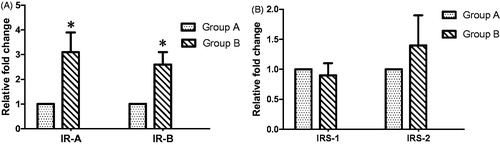
Gene expression of IR-a, IR-B, IRS-1 and IRS-2
We determined the gene expression levels of insulin receptor and its substrates in BPH patients with different prostate size. IR isoform A and B were significantly higher in BPH patients larger size prostate tissues when compared with lesser prostate size (). There was no significant difference of IRS-1 and IRS-2 expression between both the groups ().
Figure 1. (A) Relative fold change of IR A and IR B in prostate tissues of BPH patients (Group A: Prostate size <30 Ml, Group B: Prostate size >30 ml). Data were expressed in Mean ± SEM. *p = .007 for IR A, p = .01 for IR B. (B) Relative fold change of IRS-1 and IRS-2 in prostate tissues of BPH patients (Group A: Prostate size <30 Ml, Group B: Prostate size >30 ml). Data were expressed in Mean ± SEM. p value – .12 for IRS-1, p value – .25 for IRS-2.
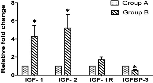
Gene expression of IGF-1, IGF-2, IGF-1 R and IGFBP-3
shows the gene expression of IGF-1, IGF-2, IGF-1 R and IGFBP-3 in BPH patients with different prostate size. IGF-1 and IGF-2 expression were significantly increased and IGFBP-3 expression was significantly reduced in patients with larger prostate size group when compared to small prostate size group. There was no significant difference in IGF-1 R expression among two groups.
Figure 2. Relative fold change of IGF-1, IGF-2, IGF-1 R and IGFBP-3 in prostate tissues of BPH patients (Group A: Prostate size <30 Ml, Group B: Prostate size >30 ml). Data were expressed in Mean ± SEM. *p = .001 for IGF-1, p = .003 for IGF-2, p = .003 for IGFBP-3.
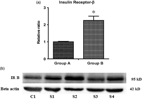
Protein expression of IR
shows the protein expression of insulin receptor beta subunit (IR-B) in prostate tissues. It was found that increase in IR-B expression was associated with increase in prostate size. Relative ratio of IR-B/Beta actin was higher in large prostate size group when compared with small prostate size group (p < .05).
Figure 3. (a) Relative ratio of IR-B/Beta actin protein expression in prostate tissues of BPH patients. Data were expressed in Mean ± SEM, *indicates statistically significant, p values – .05 for IR-B. (b) First line (C1) represent Group A (<30 ml prostate size), next four lines (S1, S2, S3, S4) represents Group B (>30 ml prostate size).
Discussion
Although several studies have reported IR expression in tissues that are classically insulin responsive, such as skeletal muscle and adipose tissue [Citation29], the data regarding IR expression and its physiological role in prostate tissues are limited. Cox et al. have confirmed the presence of insulin receptor in prostate tissue and demonstrated two isoforms of IR such as IR isoform A and IR isoform B [Citation23]. Overexpression of IR-A has been reported in several malignancies compared to IR-B [Citation26]. It has been reported that IR isoform A regulates cell proliferation and exhibits antiapoptotic effects, whereas IR isoform B is involved in the metabolic regulation [Citation30]. In the present study, we observed significantly increased gene expression of IR isoform A in BPH patients with prostate size more than 30 ml compared to those with less than 30 ml prostate size suggesting increased prostate growth is associated with over expression of IR-A. Also we found increased IR-B protein expression in BPH patients with larger prostate size.
Insulin-like growth factors (IGF-1 and IGF-2) are closely related and structurally similar to insulin. IGF-1 and IGF-2, through activation ofIGF-1 receptor (IGF-1 R) are involved in cell proliferation, migration, growth and apoptosis [Citation31,Citation32]. Previous studies have evaluated IGF-1, IGF-2 and IGF-1 R expression in prostate cancer and found significantly increased expression in prostate cancer when compared with normal prostate tissue [Citation33,Citation34]. These studies have concluded that the differential expression of components of IGF system are associated with aggressive tumor behavior and predicts the outcome of prostate cancer [Citation33]. In the present study IGF-1 and IGF-2 expression were greater in BPH patients with larger prostate size compared to those with smaller prostate size. Also IGF-1 R expression was higher in patients with prostate size more than 30 ml, but it was not statistically significant.
Insulin-like growth factor-binding protein-3 (IGFBP-3), the most abundant IGF binding proteins in circulation function as regulators of IGF-I and IGF-II bioavailability and inhibitors of IGF action by preventing IGFs from binding to their receptors [Citation35,Citation36]. Previous investigators have demonstrated significant local production of IGFBP-3 in the prostate and an association between higher nuclear IGFBP-3 expressions with recurrence of prostate cancer [Citation27,Citation37]. In the current study, we found that IGFBP-3 expression was significantly reduced in BPH patients with larger prostate size.
Binding of insulin and IGF to IR or IGF-IR, respectively, results in the activation of intracellular downstream signaling (e.g. insulin receptor substrate [IRS]) pathways with different biological responses. IRS-1 was reported to be associated with tumor growth and proliferation and IRS-2 was found to be associated with tumor motility and invasion [Citation38]. Increased expression of both IRS-1 and IRS-2 are demonstrated in hepatocellular, pancreatic and prostate cancers [Citation38]. In our study, there was no significant difference in IRS-1and IRS-2 gene expression in both the groups.
In the present study, alteration in the gene expression in BPH patients with different prostate size was supported by alteration in serum levels of insulin and IGFBP-3. When serum levels of insulin, IGF-1 and IGFBP-3 levels were compared between two groups, we found that insulin was significantly increased and IGFBP-3 was significantly reduced in BPH patients with larger prostate size. These findings were in accordance with earlier studies from several groups including our group, which linked hyperinsulinemia with prostate enlargement in BPH patients [Citation13,Citation39,Citation40].
Insulin resistance followed by secondary hyperinsulinemia is well established in BPH [Citation15]. Based on our findings, we speculate that binding of insulin with insulin receptor may activate downstream signaling pathways like IRS resulting in prostate enlargement. Also, hyperinsulinemia may reduce the serum levels of IGFBP-3, thereby increasing the expression of IGF-1 and IGF-2 which can lead to increase in prostate size in BPH patients.
The strength of our study is, this is the first study to report alteration in gene expression of IR isoforms, IGF-1 R and its ligands IGF-1, IGF-2, in prostate tissue in BPH patients. The main limitation of our study is that only few cases had larger prostate size (>50 ml) and noninclusion of malignant prostate tissue to assess the alteration between benign and malignant prostate enlargement.
The data from the study has got clinical implication, since it demonstrates an association between increased expressions of insulin receptor, insulin receptor substrate, insulin-like growth factor in the enlargement of prostate in BPH. Increase in prostate size can lead to acute urinary retention, one of the common complications of BPH. The findings of this study will form the basis of further prospective studies to see if the altered expression of insulin receptor and its related components have a causal effect on BPH and whether they can predict the rate of progression of BPH. These findings may also provide new prospects for developing novel drugs for the treatment of BPH.
Conclusions
The findings from the present study showed increased expression of Insulin receptor, insulin receptor substrate and insulin-like growth factor in BPH patients with larger prostate size indicating overexpression of components of insulin-signaling pathway might be associated with development of BPH.
Acknowledgements
This work was supported by a grant from JIPMER intramural fund sanctioned to the corresponding author.
Disclosure statement
No potential conflict of interest was reported by the authors.
Additional information
Funding
References
- Egan KB. The epidemiology of benign prostatic hyperplasia associated with lower urinary tract symptoms: prevalence and incident rates. Urol Clin North Am. 2016;43:289–297.
- Khvostova E, Otpuschennikov A, Pustylnyak V, et al. Gene expression of androgen metabolising enzymes in benign and malignant prostatic tissues. Horm Metab Res. 2014;47:119–124.
- Pradidarcheep W. Lower urinary tract symptoms and its potential relation with late onset hypogonadism. Aging Male. 2008;11:51–55.
- Yassin A, Nettleship JE, Talib RA, et al. Effects of testosterone replacement therapy withdrawal and re-treatment in hypogonadal elderlymen upon obesity, voiding function and prostate safety parameters. Aging Male. 2016;19:64–69.
- La Vignera S, Condorelli RA, Cimino L, et al. Late onset hypogonadism: the advantages of treatment with human chorionic gonadotropin rather than testosterone. Aging Male. 2016;19:34–39.
- Amano T, Imao T, Takemae K, et al. Testosterone replacement therapy by testosterone ointment relieves lower urinary tractsymptoms in late onset hypogonadism patients. Aging Male. 2010;13:242–246.
- Karazindiyanoğlu S, Cayan S. The effect of testosterone therapy on lower urinary tract symptoms/bladder and sexual functionsin men with symptomatic late-onset hypogonadism. Aging Male. 2008;11:146–149.
- Efesoy O, Apa D, Tek M, et al. The effect of testosterone treatment on prostate histology and apoptosis in men with late-onset hypogonadism. Aging Male. 2016;19:79–84.
- Meuleman EJ, Legros JJ, Bouloux PM, et al. Effects of long-term oral testosterone undecanoate therapy on urinary symptoms: data from a 1-year, placebo-controlled, dose-ranging trial in aging men with symptomatic hypogonadism. Aging Male. 2015;18:157–163.
- Wada N, Hashizume K, Matsumoto S, et al. Dutasteride improves bone mineral density in male patients with lower urinary tract symptomsand prostatic enlargement: a preliminary study. Aging Male. 2016;19:12–14.
- Lucia MS, Lambert JR. Growth factors in benign prostatic hyperplasia: basic science implications. Curr Urol Rep. 2008;9:272–278.
- Parsons JK, Carter HB, Partin AW, et al. Metabolic factors associated with benign prostatic hyperplasia. J Clin Endocrinol Metab. 2006;91:2562–2568.
- Nandeesha H, Koner BC, Dorairajan LN, et al. Hyperinsulinemia and dyslipidemia in non-diabetic benign prostatic hyperplasia. Clin Chim Acta. 2006;370:89–93.
- Urushima H, Inomata-Kurashiki Y, Nishimura K, et al. The effects of androgen deprivation therapy with weight management on serum aP2 and adiponectin levels in prostate cancer patients. Aging Male. 2015;18:72–76.
- Hammarsten J, Högstedt B. Hyperinsulinaemia as a risk factor for developing benign prostatic hyperplasia. Eur Urol. 2001;39:151–158.
- Vikram A, Jena GB, Ramarao P. Increased cell proliferation and contractility of prostate in insulin resistant rats: linking hyperinsulinemia with benign prostate hyperplasia. Prostate. 2010;70:79–89.
- Yeh HC, Liu CC, Lee YC, et al. Associations of the lower urinary tract symptoms with the lifestyle, prostate volume, and metabolic syndrome in the elderly males. Aging Male. 2012;15:166–172.
- Sarma AV, Kellogg Parsons J. Diabetes and benign prostatic hyperplasia: emerging clinical connections. Curr Urol Rep. 2009;10:267–275.
- Boucher J, Kleinridders A, Kahn CR. Insulin receptor signaling in normal and insulin-resistant states. Cold Spring Harb Perspect Bio. 2014;6:a009191.
- Belfiore A, Frasca F, Pandini G, et al. Insulin receptor isoforms and insulin receptor/insulin-like growth factor receptor hybrids in physiology and disease. Endocr Rev. 2009;30:586–623.
- Ulanet DB, Ludwig DL, Kahn CR, et al. Insulin receptor functionally enhances multistage tumor progression and conveys intrinsic resistance to IGF-1R targeted therapy. Proc Natl Acad Sci USA. 2010;107:10791–10798.
- Zhang H, Fagan DH, Zeng X, et al. Inhibition of cancer cell proliferation and metastasis by insulin receptor downregulation. Oncogene. 2010;29:2517–2527.
- Cox ME, Gleave ME, Zakikhani M, et al. Insulin receptor expression by human prostate cancers. Prostate. 2009;69:33–40.
- Merseburger AS, Hennenlotter J, Simon P, et al. Activation of the PKB/Akt pathway in histological benign prostatic tissue adjacent to the primary malignant lesions. Oncol Rep. 2006;16:79–83.
- Waalkes S, Simon P, Hennenlotter J, et al. Altered expression of Aktsignaling pathway parameters in prostate needle biopsies derived from benign, adjacent and cancerous tissue. Oncol Rep. 2010;23:1257–1260.
- Heni M, Hennenlotter J, Scharpf M, et al. Insulin receptor isoforms A and B as well as insulin receptor substrates-1 and -2 are differentially expressed in prostate cancer. PLoS One. 2012;7:e50953.
- Massoner P, Haag P, Seifarth C, et al. Insulin-like growth factor binding protein-3 (IGFBP-3) in the prostate and in prostate cancer: local production, distribution and secretion pattern indicate a role in stromal-epithelial interaction. Prostate. 2008;68:1165–1178.
- Mehta HH, Gao Q, Galet C, et al. IGFBP-3 is a metastasis suppression gene in prostate cancer. Cancer Res. 2011;71:5154–5163.
- Winkler G, Cseh K. Molecular mechanisms and correlations of insulin resistance, obesity, and type 2 diabetes mellitus. Orv Hetil. 2009;150:771–780.
- Frasca F, Pandini G, Sciacca L, et al. The role of insulin receptors and IGF-I receptors in cancer and other diseases. Arch Physiol Biochem. 2008;114:23–37.
- Christopoulos PF, Msaouel P, Koutsilieris M. The role of the insulin-like growth factor-1 system in breast cancer. Mol Cancer. 2015;14:43.
- Chao W, D'Amore PA. IGF2: epigenetic regulation and role in development and disease. Cytokine Growth Factor Rev. 2008;19:111–120.
- Cardillo MR, Monti S, Di Silverio F, et al. Insulin-like growth factor (IGF)-I, IGF-II and IGF type I receptor (IGFR-I) expression in prostatic cancer. Anticancer Res. 2003;23:3825–3835.
- Ryan CJ, Haqq CM, Simko J, et al. Expression of insulin-like growth factor-1 receptor in local and metastatic prostate cancer. Urol Oncol. 2007;25:134–140.
- Grimberg A. Mechanisms by which IGF-I may promote cancer. Cancer Biol Ther. 2003;2:630–635.
- Kelley KM, Oh Y, Gargosky SE, et al. Insulin-like growth factor-binding proteins (IGFBPs) and their regulatory dynamics. Int J Biochem Cell Biol. 1996;28:619–637.
- Seligson DB, Yu H, Tze S, et al. IGFBP-3 nuclear localization predicts human prostate cancer recurrence. Horm Canc. 2013;4:12–23.
- Mardilovich K, Pankratz SL, Shaw LM. Expression and function of the insulin receptor substrate proteins in cancer. Cell Commun Signal. 2009;7:14.
- Sreenivasulu K, Nandeesha H, Dorairajan LN, et al. Elevated insulin and reduced insulin like growth factor binding protein-3/prostate specific antigen ratio with increase in prostate size in Benign Prostatic Hyperplasia. Clin Chim Acta. 2017;469:37–41.
- Grosman H, Fabre B, Lopez M, et al. Complex relationship between sex hormones, insulin resistance and leptin in men with and without prostatic disease. Aging Male. 2016;19:40–45.