Abstract
Cranberry, the fresh or dried ripe fruit of Vaccinium macrocarpon Ait. (Ericaceae), is currently used as adjunct therapy for the prevention and symptomatic treatment of urinary tract infections. Data from clinical trials suggest that extracts of cranberry or cranberry juice reduce the bacterial load of E. coli and also suppress the inflammatory symptoms induced by E. coli infections. A methanol extract prepared from 10 kg of dehydrated cranberries did not directly inhibit the growth of E coli strains ATCC 700336 or ATCC 25922 in concentrations up to 256 μg/mL in vitro. However, the methanol extract (CR-ME) inhibited the activity of cyclooxygenase-2, with an IC50 of 12.8 μg/mL. Moreover, CR-ME also inhibited the NF-κβ transcriptional activation in human T lymphocytes with an IC50 of 19.4 μg/mL, and significantly (p < 0.01) inhibited the release of interleukin (IL)-1β, IL-6, IL-8 and tumor necrosis factor-α from E. coli lipopolysaccharide (LPS)-stimulated human peripheral blood mononuclear cells in vitro, at a concentration of 50 μg/mL. The extract had no effect on inducible nitric oxide synthase activity in the murine macrophage cell line RAW 264.7. The compounds responsible for this activity were identified using a novel LC-MS based assay as ursolic acid and ursolic acid derivatives. Taken together, these data suggest CR-ME and its constituent chemical compounds target specific pathways involved in E. coli-induced inflammation.
Introduction
Cranberry (CR), known scientifically as Vaccinium macrocarpon Ait., is a native North American plant and a member of the Ericaceae (heath family) (Mahady et al., 2001; Citation2008). Historically, the small, edible red-black berries were used by Native Americans as a dye, food, and for the treatment of wounds (CitationMahady et al., 2001). During the eighteenth century, cranberry juice was commonly employed throughout New England for the treatment of urinary tract infections (UTIs). Currently, cranberry juice and extracts are used as adjunct therapy for the prevention and symptomatic treatment of UTIs (CitationMahady et al., 2008). Numerous research and review articles have been published assessing the effects of cranberry on UTIs. However, conclusive data are still lacking (CitationAvorn et al., 1994; CitationJepson et al., 2001; CitationPapas et al., 1966; CitationRaz et al., 2004).
Limited data supporting the role of cranberry for the management of the symptoms of UTIs have been published (CitationAvorn et al., 1994; CitationPapas et al., 1966). In the 1960s, Papas et al. treated 60 symptomatic (frequency, dysuria, urgency, nocturia) patients with 16 oz. of cranberry juice daily for 21 days (CitationPapas et al., 1966). Following therapy, 53% of the patients had a reduction in urine bacterial counts and urogenital symptoms. Another 20% of treated patients exhibited a moderate response to the cranberry therapy. However, the therapeutic effects of cranberry were short-lived, with 61% of patients experiencing a relapse of symptoms and bacteriuria six weeks after discontinuing the cranberry juice (CitationPapas et al., 1966). Avorn et al. (2004) have also reported cranberry may reduce inflammatory symptoms associated with UTIs. In a randomized, double-blind, placebo-controlled trial involving 153 elderly women, a reduction in bacteriuria and pyuria following 6 months of therapy with 300 mL daily of cranberry juice cocktail was observed (Avorn et al., 2004). The incidence of urogenital symptoms in patients with bacteriuria and pyuria was 4% in the cranberry treated group versus 7% in the placebo group. While these studies are in no way conclusive, the data does suggest that cranberry products may have a potential role as adjunctive therapy for relief of the symptoms of UTIs. In addition, CitationMurphy et al. (2003) have shown that cranberry extracts also have antitumor activities in breast, cervical and prostate tumor cell lines, and that this activity was associated with ursolic acid derivatives and anti-inflammatory activity.
While many mechanisms of action have been proposed for cranberry, the potential anti-inflammatory activities of cranberry and related mechanisms have not been elucidated. To address these issues, we have assessed the antibacterial activity of methanol extracts of cranberry in two p-fimbriated E. coli strains, as well as the effect of cranberry extract on the activity of cyclooxygenase (COX-2), inducible nitric oxide synthase (iNOS) in murine macrophage cell line RAW 264.7, nuclear transcription factor κβ (NF-κβ) activation in human T lymphocytes, and the release of interleukin-1β (IL-1β), IL-6 and tumor necrosis factor alpha (TNF-α) from lipopolysaccharide (LPS)-stimulated human peripheral blood mononuclear cells in vitro. Using the novel PUF COX-2 assay (Citationvan Breemen et al., 1997) and other analytical methods, the active compounds were isolated and identified.
Materials and Methods
Plant material
The fresh cranberries (Vaccinium macrocarpon) were obtained from Ocean Spray Cranberries, Inc. (Lakeville, MA). The berries were lyophilized, milled, and kept frozen at 220ºC until used.
Reagents and chromatographic materials
All chemical reagents used in the study were analytical grade, CD3OD3, CDCl3, or HPLC grade (such as methanol, acetonitrile, acetone, ethyl acetate, and dichloromethane) and purchased from Fisher Scientific (Fair Lawn, NJ), silica gel 60 RP 18 for column chromatography 230–400 mesh, pre-coated silica gel 60 F254 TLC plates, and pre-coated RP-18 F254s TLC plates (EMD Chemicals Inc. Darmstadt, Germany). Fractions were monitored by TLC and spots were visualized by heating Si gel plates sprayed with 10% H2SO4 in EtOH and UV lamp. Octadecylsilane (ODS) 3 μm, 4.6 × 150 mm analytic HPLC column was obtained from Waters Corporation (Milford, MA). The 5 μm, 4.6 × 250 mm C18 semi-preparative HPLC column was purchased from Dionex Corporation (Sunnyvale, CA).
Instrumentation
HPLC analysis and purification were performed on a Dionex Summit System HPLC composed of P580 pump, Asi-100 automated sample injector, and PDA-100 photodiode array detector. LC-MS used in PUF-Assay for analysis of trapped ligands was carried out using a Hewlett-Packard (Palo Alto, CA), G1946A 1100 LC/MSD. IR spectra were run on a Jasco FT/IR-410 spectrometer, equipped with a Specac Silver Gate ATR system by applying a film on a germanium plate. NMR spectra were recorded on a Bruker DPX-300 or a Bruker DPX-400 NMR spectrometer. Chemical shifts (δ) were expressed in PPM with reference to TMS or the solvent signals. All NMR spectroscopic data were obtained by using standard pulse sequences supplied by the vendor. LREIMS were recorded on a Thermo Finnigan LCQ Mass Spectrometer. HRTOFMS spectra were recorded on a Micromass QTOF-2 spectrometer or a JEOL GCmate spectrometer.
Extraction and fractionation
Fresh cranberries (10 kg) were obtained from Ocean Spray Cranberries, Inc. The fruit was lyophilized, and 2 kg were milled and exhaustively extracted by percolation with 5 L MeOH three times by maceration. The extract was then dried in vacuo at 40°C. The concentrated extract was suspended in 90% MeOH and then partitioned with 1.5 L of petroleum ether which was also dried in vacuo. The aqueous methanol extract (CR-ME) was concentrated and 28 g was chromatographed on a reversed-phase C-18 column (Sigma, Saint Louis, MO) and eluted with a methanol-water gradient (from 1:1 to 100% methanol) as a mobile phase to afford 6 pooled fractions (F001-006). The crude extract and the fractions were tested in the primary COX-1 and COX-2 assays and the most active fractions subjected to PUF-LC-MS assay previously described (Citationvan Breemen et al., 1997). Of these fractions, F004 (156 mg) and F006 (208 mg) and the crude extract showed the most potent COX-2 inhibitory activity (65, 85, and 100% inhibition, respectively), at 50 μg/mL for the CRME extract and 10 μg/mL for each of the fractions. All solvents used for chromatographic separations were purchased from Fisher Scientific (Hanover Park, IL) and distilled prior to use.
PUF COX-2 assay and compound isolation
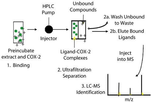
PUF-LC-MS (Citationvan Breemen et al., 1997) is a binding technique that can be used for rapid screening of complex mixtures and plant extracts for ligands that bind to a target protein, in this case cyclooxygenase (). During the PUF-LC-MS assay, a semi-permeable membrane is used to trap high molecular weight proteins but allow low molecular weight ligands to pass through (). Upon incubation of a mixture of ligands with the target protein, ligand-receptor complexes are formed. These complexes are then separated from the unbound ligands using ultrafiltration. Next, destabilizing condition such as organic solvent and/or low pH was used to release bound ligands for analysis by LC-MS. Control experiments in which active enzyme has been replaced with the denatured one were carried out to control for the non-specific protein binding as well as for non specific binding to the ultrafiltration membrane. This approach was enhanced by the use of the QqTOF mass spectrometer that provides an exact mass measurement for elemental composition determination of the ligands during the initial LC-MS screening assay (Citationvan Breemen et al., 1997).
Figure 1. Methods used for assessment of COX-2 activities in plant extracts using traditional enzyme assays and COX-2 PUF assay.
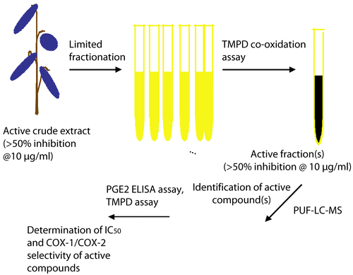
Figure 2. Schematic presentation of PUF-LC-MS assay.
Susceptibility testing
Two American Type Culture Collection (ATCC, Manassas, VA) strains of Escherichia coli were used in the susceptibility assay. The facultative anaerobic pyelonephritic strain E. coli ATCC 700336, a J96-like strain isolated from a patient with pyelonephritis, possesses P-fimbriae Classes I and III and is the prototypic uropathogenic strain from human urinary tract infections (CitationBates et al., 2004; CitationHütt et al., 2006). E. coli ATCC 25922, the NCCLS control strain for susceptibility studies, possesses Class II P-fimbriae typically found in pyelonephritis (CitationTurner et al., 2005). The organisms were stored at
280°C and underwent three subcultures on blood agar plates (Remel, Lenexa, KS) prior to MIC testing. The minimum inhibitory concentration (MIC) of the cranberry extract was determined per the National Committee for Clinical Laboratory Standards (CitationNCCLS, 2008) guidelines using the microbroth dilution method. Serial two-fold dilutions of the cranberry methanol extract (CME) were prepared in cation-adjusted Mueller-Hinton broth (caMHB, Difco, Sparks, MD), pH 7.3. CR-ME concentrations tested ranged from 0.25–256 μg/mL.
Each bacterial inoculum was prepared by direct suspension and adjusted with sterile saline until the turbidity matched a 0.5 McFarland standard using a spectrophotometer at 625 nm. The bacterial suspension was further diluted in caMHB to obtain a final inoculum of approximately 5 3 105 CFU/mL. The microtiter plates were incubated at 35°C (humidified air) and read at 20 h. All procedures were performed in duplicate. The MIC was defined as the lowest concentration at which there was no visible bacterial growth (CitationNCCLS, 2008).
Cyclooxygenase-2 (COX-2) assay
Functional COX-2 assays were performed by assessing human recombinant COX-2 catalytic activity as described previously by measuring prostaglandin E2 (PGE2) production (CitationKiemer et al., 2003; CitationWarner et al., 1999). Reaction mixtures were prepared in 100 mM Tris–HCl buffer (Sigma), pH 8.0, containing 1 μM heme, 500 μM phenol, 300 μM epinephrine, sufficient COX-2 enzyme (Cayman Chemical, Ann Arbor, MI) to generate 150 ng of PGE2/mL, and various concentrations of the cranberry extracts. The reaction was initiated by the addition of arachidonic acid (final concentration, 10 μM) and incubated for 10 min at room temperature (final volume, 200 μL). The reaction was terminated by adding 20 μL of the reaction mixture to 180 μL of indomethacin, and PGE2 was measured using an enzyme-linked immunosorbent (ELISA) kit. Samples were diluted to the desired concentration with 100 mM potassium phosphate buffer (Sigma), pH 7.4, containing 2.34% NaCl, 0.1% bovine serum albumin, 0.01% sodium azide, and 0.9 mM Na4EDTA. Following transfer to a 96-well plate coated with a goat anti-mouse IgG (Jackson Immuno Research Laboratories), the tracer (PGE2-acetylcholinesterase; Cayman Chemical) and primary antibody (mouse anti-PGE2; Monsanto, Saint Louis, MO) were added. The plates were then incubated at room temperature overnight, reaction mixtures were removed, and wells were washed with a solution of 10 mM potassium phosphate buffer (Sigma), pH 7.4 containing 0.01% sodium azide and 0.05% Tween 20. Ellman’s reagent (Sigma) (200 μL) was added to each well, and the plate was incubated at 37°C for 3–5 h, until the control wells yielded an optical density of 0.5–1.0 at 412 nm. A standard curve with PGE2 (Cayman Chemical) was generated from the same plate, which was used to quantify the PGE2 levels produced in the presence of the cranberry extracts. Results were expressed as a percentage relative to a control (solvent-treated samples). All determinations were performed in duplicate, and values generally agreed within 10%. Concentration-dependent curves were generated for the calculation of IC50 values.
NF-κβ reporter gene assay
To determine the effects of the cranberry extract on TNF-induced NF-κβ-dependent specific transcription, a Jurkat-derived clone (human T lymphocytes), 5.1-cell line (Pasteur Institute, Paris, France) was used and was maintained in complete RPMI medium (CitationSancho et al., 2002). These specific cells are stably transfected with a plasmid containing the firefly luciferase gene, driven by the HIV-1-LTR promoter. The promoter is dependent on NF-κβ activation induced by TNFα. Increased expression of luciferase activity reflects NF-κβ activation (CitationSancho et al., 2002). To determine NF-B dependent transcription, 5.1 cells were pre-incubated with the cranberry extract for 30 min followed by stimulation with TNF- (2 ng/mL) for 6 h. The cells were then lysed in 25 mM Tris-phosphate pH 7.8, 8 mM MgCl2, 1 mM DTT, 1% Triton X-100, and 7% glycerol. Luciferase activity was measured using a Sirius Luminometer (Fisher Scientific) following the instructions of the luciferase assay kit (Promega, Madison, WI) and protein concentrations were determined using the Bradford method. The background obtained with the lysis buffer was subtracted in each experimental value and the specific transactivation expressed as a fold induction. All the experiments were repeated at least three times. All cranberry extracts and pure compounds were tested at 6.25, 12.5, 25, 50, and 100 μg/mL.
Cytokine release
Modulation of cellular cytokine release was performed using the protocol of CitationWelker et al. (1996). Briefly, human peripheral blood mononuclear (PMN) cells (MDS, Bothell, WA) were treated with 25 ng/mL of E. coli LPS (Sigma) to stimulate the release of IL-1β and IL-6, and released cytokines in the cell supernatants were measured by ELISA. Cells (106/mL) with LPS were cultured for 16 h at 37°C in RPMI medium without serum, alone or with 0–100 μg/mL of CRME (CitationWelker et al., 1996).
For the TNF-α release assay, human PMN cells (5 3 105 cells, MDS, Bothell, WA) were pre-incubated at 37°C for 1 h in 0.5 mL of medium containing 50 μg/mL of the cranberry extracts. After three washes with phosphate-buffered saline (BPS), pH 7.4, the cells were further incubated at 37°C for 6 h in 0.5 mL of Eagle’s minimal essential medium (EMEM) containing 10% fetal bovine serum (FBS) (Sigma) in the presence of EC-LPS (25 ng/mL) and the various concentrations of each extract. After 6 h incubation, TNF-α levels in the conditioned medium were determined using a TNF-α ELISA kit according to the manufacturer’s instructions.
Nitric oxide synthase (iNOS assay)
RAW 264.7 mouse macrophage cells were obtained from RIKEN Gene Bank and cultured at 37°C with 5% CO2 in EMEM containing 10% FBS, penicillin G potassium (18 μg/mL) and streptomycin sulfate (50 μg/mL) (CitationIppoushi et al., 2003). The cells (5 3 105 cells) were pre-incubated at 37°C for 1 h in 0.5 mL of medium containing up to 100 μg/mL of the cranberry extracts or a non-specific inhibitor (NG-monomethyl-l-arginine acetate) (Sigma) of nitric oxide synthase (NOS). After three washes with PBS, pH 7.4, the cells were further incubated at 37°C for 12 h in 0.5 mL of EMEM containing 10% FBS in the presence of LPS (0.1 μg/mL) and the corresponding concentration of each extract. After incubation for 12 h, nitrite levels in the conditioned medium were determined spectrophotometrically by the quantitation of NO2 + NO3 (CitationIppoushi et al., 2003).
Statistical analysis
MIC and COX-2 assays were performed in duplicate and were the result of three experiments; the remaining experiments were performed in triplicate. Statistical analysis was performed by one way analysis of variance (ANOVA) (Graphpad Instat, San Diego, CA).
Results
In vitro susceptibility assay
The MIC of the cranberry methanol extract was >256 μg/mL for both of the P-fimbriated E. coli strains (). An acceptable (i.e., clinically relevant) concentration range for antimicrobial activity of a botanical extract is ≤50 μg/mL. Therefore, 256 μg/mL is well above the concentration that would be considered active, and thus the cranberry extract demonstrated no direct antibacterial activity against these E. coli strains.
Table 1. Minimum inhibitory concentration of a methanol extract of cranberry in two clinical E. coli strains. All experiments were performed in triplicate.
COX-2 inhibition
The CR-ME extract and 6 fractions were tested for COX activity in vitro (). At a 50 μg/mL concentration, the cranberry extract inhibited the activity of COX-2 by 100%. A median inhibitory concentration (IC50) for the extract was 12.8 μg/mL. At a concentration of 10 μg/mL, fraction 6 inhibited COX-2 activity by 85%, and fraction 4 by 65%. Performance of the COX-2 PUF-LC-MS on the extract and fraction 6 showed compounds with EI mass spectra of known compound 1 ursolic acid (MH+ m/z 456.71), compound 2 unidentified (MH+ m/z 504), and known compounds 3 and 4 cis- and trans-hydroxycinnamoyl ursolic acid (MH+ m/z 603.402 and 603.403) (). The compounds were isolated by reversed phase column chromatography and finally, structural information on the ligands was obtained by using 1H, 13C NMR, EI MS, LC-MS-MS. Compounds 1, 3, and 4 are known compounds, with 3 and 4 isolated from cranberry (). These two triterpene esters were isolated and identified previously by CitationMurphy et al. (2003) and were reported to have antitumor activities in vitro, although the mechanism was not elucidated. The EI mass spectrum and NMR spectral data were consistent with those previously described (CitationMurphy et al., 2003).
Table 2. Effect of various cranberry extracts on COX-2 activity in vitro. All experiments were performed in triplicate.
Figure 3. Structure of ursolic acid derivatives inhibiting COX-2 as determined using the COX-2 PUF-LC-MS assay.
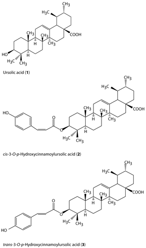
Ursolic acid (1) was obtained as colorless needles. The TOF MS ES-TIC gave an ion [M-H]- at 455.4, and the molecular formula of C30H48O3 was inferred from analysis of 1H, 13C NMR, and DEPT spectra. With the five high field 1H NMR singlet signals, there were five corner methyl groups in the molecular, and another two high field 1H NMR signals at δ 1.04 (d, J = 7.5 Hz), 0.98 (d, J = 7.2 Hz) typically indicated that there were two methyl groups substituted on ring. The signals at δ 139.8, 126.1 ppm of 13C NMR and the signal at δ 5.52 (br s) ppm of 1H NMR implied a C-C double bond in this molecular, one of the olefinic carbons was a quaternary carbon. The signal at δ 78.5 ppm of 13C NMR was correlated 1H NMR signal at δ 3.488 ppm (dd, J = 6.8,9.5 Hz) and indicated a ring carbon was substituted by a hydroxyl group (C-3). The 13C NMR signal at δ 180.3 ppm indicated an acid carbonyl carbon. Compound 1 was identified as ursolic acid.
Ursolic acid 3-O-p-hydroxy-cis-cinnamate (3) (cis-hydroxycinnamoyl ursolic acid) was obtained as a colorless powder. TOF MS ES-TIC gave the ion [M+H]- at 603.402, EI-MS gave a signal at m/z at 483[M-C8H7O]+. In addition, with the data of 1H, 13C NMR, the molecular formula was determined as C39H54O5. With 13C NMR data of compound 1, there existed a triterpenoid moiety, corresponding with ursolic acid.The additional nine carbon signals, which were all in low field, were read as aromatic carbons. The 1H NMR data supported the presence of the ursolic acid moiety. The down field chemical shift signals at δ 7.66 (d, J = 8.5 Hz, 2H), 6.82 (d, J = 8.5 Hz, 2H) showed a 1,4-disubstituted benzene fragment in this molecular. The signals at δ 6.85 (d, J = 12.8 Hz, 1H), 5.82 (d, J = 12.8 Hz, 1H) were much more down field than general olefinic protons, which implied they were protons on a double bond conjugated with the aromatic ring, and the same coupling constant of 12.8 Hz were typical J value of cis-olefinic protons. The 13C NMR signal of the only carbon connected to oxygen atom in triterpenoid moiety (C-3) was at δ 81.25 ppm, which down field shifted in comparing to δ 78.2 ppm of C-3 in compound 1. Therefore, the p-hydroxycinnamic acid was linked at C-3 in the O-C ester bond. Compound 3 was identified as cis-3-O-p-hydroxycinnamoyl ursolic acid previously reported by CitationMurphy et al. (2003).
Ursolic acid 3-O-p-hydroxy-trans-cinnamate (4) (trans-hydroxycinnamoyl ursolic acid) was obtained as a colorless powder. It had a TOF MS ES-TIC [M+H]+ of 603.403, and the similar chemical shifts recorded in 1H, 13C NMR to those of compound 3. The 13C NMR signal at δ 129.75 ppm was up field from the signals at δ 132.28 ppm for the corresponding carbons of compound 3. The 1H NMR signals at δ 7.58 (d, J = 8.2 Hz, 2H), 6.86 (d, J = 8.7 Hz, 2H) showed the same 1,4-disubstituted benzene fragment. But the signals at δ 7.58 (d, J = 15.8 Hz, 1H), 6.58 (d, J = 15.8 Hz, 1H) were down field shifted from the signals for the corresponding carbons of the cis isomer of compound 3. The same coupling constant of J = 15.8 Hz were characteristic J values of trans-olefinic protons. Compound 4 was identified as trans-3-O-p-hydroxycinnamoyl ursolic acid, also previously isolated by CitationMurphy et al. (2003).
Inhibition of TNF-induced NF-κβ-dependent specific transcription
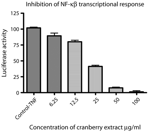
Since CR-ME extract significantly inhibited COX-2 activity, and inhibited NF-κβ activation in TNF-induced Jurkat clone 5.1, it was also tested in a non-radioactive NF-κβ reporter gene assay (). NF-κβ activity (indicated as relative luciferase activity) was increased after treatment of the cells with TNF for 6 h (positive control). When CRME was added, the extract inhibited NF-κβ transcriptional response with an IC50 of 19.4 μg/mL (). Fractions 6 and 8 also inhibited NF-κβ transcriptional response in concentrations of 10 μg/mL.
Figure 4. Cranberry extract (CRME) inhibits NF-κB-dependent transcriptional activities. CRME extract inhibited TNF-induced NF-κB activation in stably transfected human T lymphocytes 5.1 cells as assessed by a reporter gene assay. The luciferase activity was measured after 6 h and expressed as TNF induction = 100%. Values are means of ± SD of three independent experiments in triplicate.
iNOS
It is well known that nitric oxide has antimicrobial activity and that the transcription of the iNOS gene is regulated by NF-κβ (CitationIppoushi et al., 2003). Thus, we hypothesized that since CR-ME inhibited the NF-κβ transcriptional response it may also affect the activity of iNOS. However, the results showed that neither the cranberry extract nor any fractions inhibited the activity of this enzyme when tested in concentrations up to 100 μg/mL (data not shown).
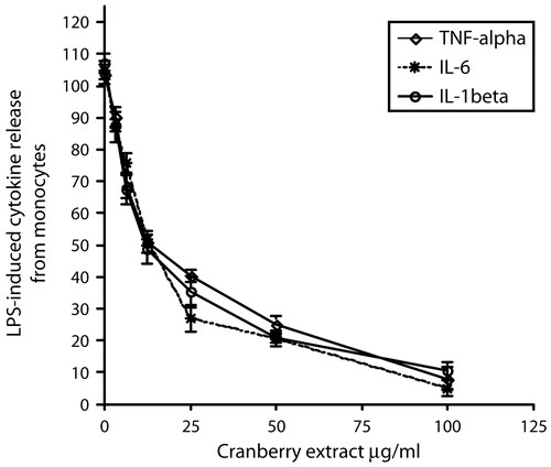
Cytokine release
The inhibition of LPS-induced cytokine release from PMNs by CRM extract is presented in . At 50 μg/mL, the CRM extract inhibited the release of TNF-α by 77% (p < 0.001), IL-1β by 85% (p < 0.001) and IL-6 by 83% (p < 0.001), with IC50 values of 12.5, 10.8, and 10.9 μg/mL, respectively.
Figure 5. Cranberry extract inhibited cytokine release in human peripheral blood mononuclear leukocytes (PMNs). Cells were stimulated with E. coli LPS (25 ng/mL) for 6–16 h in the presence or absence of cranberry extract (0-100 μg/mL). Data are expressed as the percentage of cytokines accumulated in the supernatant of LPS-induced PMNs (100%) and represent the means ± SEM of at least three experiments in triplicate. P < 0.001 represents a significant difference compared to the values seen in the LPS-activated cells
Discussion
Previously, the antimicrobial activity of cranberry juice was attributed to the urinary excretion of hippuric acid (HA), a bacteriostatic agent, believed to acidify the urine, although no minimum inhibitory concentrations (MICs) have been published. In this work, we attempted to establish MICs for cranberry extracts against two E. coli strains, one containing P-fimbriae classes I and III, and the other containing P-fimbriae class II, using modern standardized NCCLS methodology (CitationNCCLS, 2008). However, we were unable to demonstrate a direct bacteriostatic or bacteriocidal effect of a methanol extract of the berries against p-fimbriated strains of E. coli at concentrations up to, and including, 256 μg/mL. It is possible that cranberry has no direct antibacterial activity, but more likely functions through other mechanisms such as the reduction of E. coli adhesions (CitationOfek et al., 1996; CitationTurner et al., 2005). The lack of direct antibacterial activity led us to assess the effect of cranberry extracts on cellular mediators of inflammation.
It is well known that bacterial infections and bacterial lipopolysaccaride (LPS) activate several signal transduction pathways in host cells that induce an inflammatory response (CitationEjima et al., 2003; CitationFeezor et al., 2003; 1995; CitationMizgerd et al., 2002). In regard to urinary tract infections, CitationWheeler et al. (2002) reported increased urinary prostaglandin E2 production and COX-2 protein expression in patients with UTIs and other inflammatory diseases such as bladder cancer. Other investigations have shown that E. coli infections and bacterial LPS stimulate pro-inflammatory cytokine expression (CitationKohn & Kung, 1995) and COX-2 activity (CitationEjima et al., 2003), which are controlled by an increase the NF-κβ transcriptional response in the host cell (CitationKrull et al., 1998; CitationMarks-Knocezalik et al., 1998). NF-κβ comprises a family of inducible transcription factors that regulate gene expression involved in controlling cell growth and inflammation (CitationMarks-Knocezalik et al., 1998). Diverse signals, including inflammatory cytokines, pathogenic microorganisms, and bacterial LPS (CitationPistritto et al., 1999; CitationSamuelsson et al., 2004; CitationWang et al., 2000), stimulate the rapid phosphorylation of IκB, and subsequently allow NF-κβ to translocate to the nucleus, where it regulates multiple genes that encode pro-inflammatory cytokines and other chemokines (CitationD’Acquisto et al., 1997; Marks-Knocezalik et al., 1998; CitationPistritto et al., 1999; CitationSamuelsson et al., 2004; CitationWang et al., 2000). Therefore, NF-κβ transcriptional activation increases the transcription of TNF-α and other cytokines (IL-1β, IL-6) associated with E. coli infections (CitationMarks-Knocezalik et al., 1998). The data presented in this study demonstrate that treatment of human T lymphocytes with CR-ME extract led to a significant reduction in TNF-induced NF-κβ transcriptional activation in this cell line. In addition, LPS-induced IL-1β and IL-6 production in human peripheral blood mononuclear cells was also significantly reduced by treatment of the cell line with the cranberry extract.
Bacterial LPS and pro-inflammatory cytokines also induce the expression and activity of COX-2 and nitric oxide synthase (iNOS) (CitationSamuelsson et al., 2004, CitationMaloney et al., 1998). This activity is also under the control of several transcription factors, including NF-κβ (CitationWheeler et al., 2002). In fact, the 5’-promoter region of COX-2 contains two putative NF-κβ binding sites and NF-κβ can positively up-regulate COX-2 expression in murine macrophages and human colon adenocarcinoma cell lines exposed to bacterial LPS (CitationWheeler et al., 2002). Our data demonstrate the direct inhibition of COX-2 activity by cranberry extracts, and the active constituents were identified as ursolic acid and ursolic acid derivatives. Ursolic acid is a natural product well known for having anti-inflammatory activity. Thus, the results of this study support the role of ursolic acid as one of the anti-inflammatory constituents of cranberry extracts. Other mechanisms proposed for the effects of cranberry include the generation of nitric oxide (NO; 31). However, our cranberry extract had no effect on the activity of NO synthase activity in vitro.
Clinical studies suggest that cranberry juice and extracts may be effective for the management of UTIs and its symptoms (CitationAvorn et al., 1994; CitationJepson et al., 2001; CitationMahady et al., 2008; CitationPapas et al., 1966; CitationRaz et al., 2004; 2004). However, the mechanism of action has been somewhat elusive. Previous investigators have demonstrated that cranberry extracts have anti-adhesive activities (CitationGreenberg et al., 2005; CitationOfek et al., 1996; CitationTurner et al., 2005). In this study, a cranberry extract did not inhibit the in vitro growth of E. coli, but did inhibit the NF-κB transcriptional response in human T lymphocytes, LPS-stimulated release of IL-1β, IL-6, and TNF-α in PBMs, and the catalytic activity of COX-2. Taken together, these data suggest that cranberry may suppress elements in the signal transduction pathway that lead to the inflammatory response in response to bacterial-LPS. Since UTIs are characterized by a number of inflammatory symptoms, and the clinical trials suggest that cranberry extracts decrease these symptoms, we propose that in addition to its anti-adhesive activities, cranberry may reduce the symptoms of UTI through its anti-inflammatory effects.
Acknowledgements
This study was funded in part by NIH Grant AT001317 from the National Center for Complementary and Alternative Medicine. The contents are solely the responsibility of the authors and do not necessarily represent the views of the funding agency.
References
- Avorn J, Monane M, Gurwitz JH, Glynn RJ, Choodnovskiy I, Lipsitz LA (1994): Reduction of bacteriuria and pyuria after ingestion of cranberry juice. JAMA 271:751–754.
- Bates JM, Raffi HM, Prasadan K, Mascarenhas R, Laszik Z, Maeda N, Hultgren SJ, Kumar S (2004): Tamm-Horsfall protein knockout mice are more prone to urinary tract infection: Rapid communication. Kidney Int 65: 791–797.
- D’Acquisto F, Iuvone T, Rombola L, Sautebin L, Di Rosa M, Carnuccio R (1997): Involvement of NF-kappaB in the regulation of cyclooxygenase-2 protein expression in LPS-stimulated J774 macrophages. FEBS Lett 418: 175–178.
- Ejima K, Layne MD, Carvajal IM, Kritek PA, Baron RM, Chen YH, Vom Saal J, Levy BD, Yet SF, Perrella MA (2003): Cyclooxygenase-2-deficient mice are resistant to endotoxin-induced inflammation and death. FASEB J 17: 1325–1327.
- Feezor RJ, Oberholzer C, Baker HV, Novick D, Rubinstein M, Moldawer LL, Pribble J, Souza S, Dinarello CA, Ertel W, Oberholzer A (2003): Molecular characterization of the acute inflammatory response to infections with Gram-negative versus Gram-positive bacteria. Infect Immun 71: 803–5813.
- Greenberg JA, Newmann SJ, Howell AB (2005): Consumption of sweetened dried cranberries versus unsweetened raisins for inhibition of uropathogenic Escherichia coli adhesion in human urine: A pilot study. J Alter Complement Med 11: 875–878.
- Hütt P, Shchepetova1 J, Lõivukene K, Kullisaar T, Mikelsaar M (2006) Antagonistic activity of probiotic lactobacilli and bifidobacteria against entero- and uropathogens. J Applied Micobiol 100: 1324–1329.
- Ippoushi K, Azuma K, Ito H, Horie H, Higashio H (2003): [6]-Gingerol inhibits nitric oxide synthesis in activated J774.1 mouse macrophages and prevents peroxynitrite-induced oxidation and nitration reactions. Life Sci 73: 3427–3437.
- Jepson R, Mihaljevic L, Craig J (2001): Cranberries for preventing urinary tract infections. Cochrane Database Syst Rev 2: CD001321.
- Kiemer AK, Hartung T, Huber C, Vollmar AM (2003): Phyllanthus amarus has anti-inflammatory potential by inhibition of iNOS, COX-2, and cytokines via the NF-kappaB pathway. J Hepatol 38: 289–297.
- Kohn FR, Kung AH (1995): Role of endotoxin in acute inflammation induced by Gram-negative bacteria: Specific inhibition of lipopolysaccharide-mediated responses with an amino-terminal fragment of bactericidal/permeability-increasing protein. Infect Immun 63: 333–339.
- Krull M, Klucken AC, Wuppermann FN, Fuhrmann O, Magerl C, Seybold J, Hippenstiel S, Hegemann JH, Jantos CA, Suttorp N (1999): Signal transduction pathways activated in endothelial cells following infection with Chlamydia pneumoniae. J Immunol 162: 4834–4841.
- Mahady GB, Fong HHS, Farnsworth NR (2001): Cranberry, in: Botanical Dietary Supplements: Quality, Safety and Efficacy. Lisse, Netherlands, Swets Publishing, pp. 23–35.
- Mahady GB, Fong HHS, Farnsworth NR (2008):Fructus Macrocarponii in : WHO Monographs on Selected Medicinal Plants, Volume 4, Geneva, Switzerland, WHO Publications, pp.132–147.
- Maloney CG, Kutchera WA, Albertine KH, McIntyre TM, Prescott SM, Zimmerman GA (1998): Inflammatory agonists induce cyclooxygenase type 2 expression by human neutrophils. J Immunol 160: 1402–1410.
- Marks-Knocezalik J, Chu SC, Moss J (1998): Cytokine-mediated transcriptional induction of the human inducible nitric oxide synthase gene requires both activator protein 1 and nuclear factor kappaB-binding sites. J Biol Chem 273: 22201–22208.
- Mizgerd JP, Scott ML, Spieker MR, Doerschuk CM (2002): Functions of IkappaB proteins in inflammatory responses to Escherichia coli LPS in mouse lungs. Am J Respir Cell Mol Biol 27: 575–582.
- Murphy BT, MacKinnon SL, Yan XJ, Hammond GB, Vaisberg AJ, Neto CC (2003): Identification of triterpene hydrocinnamates with in vitro antitumor activity from whole cranberry fruit (Vaccinium macrocarpon). J Agric Food Chem 51: 3541–3545.
- NCCLS (National Committee for Clinical Laboratory Standards) (2008): Methods for dilution antimicrobial susceptibility tests for bacteria that grow aerobically. Approved Standard M7-A5, Wayne, PA, NCCLS.
- Ofek I, Goldhar J, Sharon N (1996): Anti-Escherichia coli adhesion activity of cranberry and blueberry juices. Adv Exp Med Biol 408: 179–183.
- Pahl HL (1999): Activators and target genes of Rel/NF-κB transcription factors. Oncogene 18: 6853–6854.
- Papas PN, Brusch CA, Ceresia GC (1966): Cranberry juice in the treatment of urinary tract infections. Southwestern Med 47: 17–20.
- Pistritto G, Franzese O, Pozzoli G, Mancuso C, Tringali G, Preziosi P, Navarra P (1999): Bacterial lipopolysaccharide increases prostaglandin production by rat astrocytes via inducible cyclo-oxygenase: Evidence for the involvement of nuclear factor kappaB. Biochem Biophys Res Commun 263: 570–574.
- Raz R, Chazan B, Dan M (2004): Cranberry juice and urinary tract infection. Clin Infect Dis 38: 1413–1419.
- Rhee KY, MacArthur C (2004): Antimicrobial mechanisms of cranberry juice. Clin Infect Dis 39: 877–879.
- Samuelsson P, Hang L, Wullt B, Irjala H, Svanborg C (2004): Toll-like receptor 4 expression and cytokine responses in the human urinary tract mucosa. Infect Innun 72: 3179–3186.
- Sancho R, Lucena C, Macho A, Calzado MA, Blanco-Molina M, Minassi A, Appendino G, Munoz E (2002): Immunosuppressive activity of capsaicinoids: Capsiate derived from sweet peppers inhibits NF-kB activation and is a potent anti-inflammatory compound in vivo. Eur J Immunol 32: 1753–1763.
- Turner AChen SN, Joike MK, Pendland SL, Pauli GF, Farnsworth NR (2005): Inhibition of uropathogenic Escherichia coli by cranberry juice: A new antiadherence assay. J Agric Food Chem 53: 8940–8947.
- van Breemen RB, Huang CR, Nikolic D, Woodbury CP, Zhao YZ, Venton DL (1997): Pulsed ultrafiltration mass spectrometry: A new method for screening combinatorial libraries. Anal Chem 9: 2159–2164.
- Wang X-C, Saban R, Kaysen JH, Saban MR, Allen PL, Benes EN, Hammond TG (2000): Nuclear factor kappa B mediates lipopolysacchardie-induced inflammation in the urinary bladder. J Urol 163: 993–998.
- Warner TD, Giuliano F, Vojnovic I, Bukasa A, Mitchell JA, Vane JR (1999): Nonsteroid drug selectivities for cyclo-oxygenase-1 rather than cyclo-oxygenase-2 are associated with human gastrointestinal toxicity: A full in vitro analysis. Proc Natl Acad Sci USA 96: 7563–7568.
- Welker P, Lippert U, Nurnberg W, Kruger-Krasagakes S, Moller A, Czarnetzki BM. (1996):Glucocorticoid-induced modulation of cytokine secretion from normal and leukemic human myelomonocytic cells. Int Arch Allergy Immunol 109: 110–115.
- Wheeler MA, Hausladen DA, Yoon JH, Weiss RM (2002): Prostaglandin E2 production and cyclooxygenase-2 induction in human urinary tract infections and bladder cancer. J Urol 168:1568–1573.