
Abstract
Context
The leech and centipede granules have good curative effects on many diabetic vascular diseases, including diabetes-induced erectile dysfunction (DIED).
Objective
To explore the effect of leech and centipede on erectile function in rats with diabetes-induced erectile dysfunction and its possible mechanism.
Materials and methods
Thirty male Sprague–Dawley DIED rats were randomly divided into the model group (Group M), low-dose group (Group DD), high-dose group (Group DG) and tadalafil group (Group T) (n = 6); diabetic rats were induced by streptozotocin. Apomorphine was used to induce diabetic erectile dysfunction. The ‘leech-centipede’ granules (0.15 and 0.6 g/kg) were intragastrically administered in the DD and DG groups for 8 weeks. Blood glucose, serum insulin, testosterone, cGMP levels and protein expression changes were measured in each group.
Results
After 8 weeks, the erectile function of rats in the DG group significantly improved (1.26 ± 0.73). Penis tissue cGMP levels were higher in the DG group (1.48 ± 0.11) than in the M group (0.58 ± 0.15). Protein and mRNA expression levels of NOS were significantly higher (0.77 ± 0.05; 0.61 ± 0.02) but those of PDE5 (0.43 ± 0.05; 0.61 ± 0.03) were lower in the DG group than in the M group (0.37 ± 0.06; 0.51 ± 0.01; 0.78 ± 0.06; 0.81 ± 0.04).
Conclusion
The leech-centipede can improve erectile dysfunction in DIED rats by regulating the expression of cGMP, NOS, and PDE5-related molecules in the PDE5 pathway. This study provides a potential mechanism for the treatment of DIED with leech-centipede.
Introduction
Erectile dysfunction (ED) is the consistent or recurrent inability to achieve and/or maintain penile erection sufficient for satisfactory sexual performance (Irwin Citation2019). ED has a serious effect on the quality of life of both male patients and their sexual partners. It is estimated that 322 million men worldwide will suffer from ED by 2025 (Shamloul and Ghanem Citation2013), which can seriously harm human health, spousal relationships, and family life. Diabetes mellitus (DM) is a chronic metabolic disease characterised by hyperglycaemia, with approximately 451 million people worldwide diagnosed with diabetes or at risk of early diabetes (Cho et al. Citation2018). Studies have shown that the probability of ED in patients with DM is 1.9–4 times higher than that in patients with normal blood glucose (Ryan and Gajraj Citation2012); it is estimated that up to 75% of patients with ED will also have DM (Castela and Costa Citation2016). The oral drug phosphodiesterase type 5 (PDE5) inhibitor is currently used as a first-line therapy for ED. However, some studies have shown that this class of drugs does not work effectively for some patients with severe diabetes-induced ED (DIED) (Assaly-Kaddoum et al. Citation2016); additionally, this drug sometimes produces side effects such as headache, backache, or flushing during use (Li et al. Citation2018).
ED is closely related to the nitric oxide (NO)-cyclic guanosine monophosphate (cGMP)-PDE5 pathway. NO synthase (NOS) produces NO when phosphorylated by arginine as a neurotransmitter that regulates penile smooth muscle relaxation, resulting in a normal penile erection after sexual stimulation. DM can reduce the activity of NOS and the levels of NO and cGMP in the corpus cavernosum of penis, which may be one of the most important mechanisms of DIED (Akingba and Burnett Citation2001; Podlasek et al. Citation2001; Sullivan et al. Citation2002). The advantages of Chinese medicine in the treatment of DIED has been gradually revealed in recent years. Chinese medicine scholars believe that ‘blood stasis blocking collaterals’ is the main pathological feature of DIED. A combination of leech and centipede is a key drug for promoting blood circulation and removing blood stasis in traditional Chinese medicine, and they belong to the family entomology; the ‘leech-centipede’ drug pair has been used in many prescriptions to treat diabetic vascular disease, including DIED (Zhang et al. Citation2014; Ma et al. Citation2017; Xu and Sheng Citation2018). However, its specific mechanism is not fully understood and requires deeper study.
In this study, leech (pinyin name Shui Zhi; Latin scientific name Hirudo; Hirudinea; Hirudinidae)-centipede (pinyin name Wu Gong; Latin scientific name Scolopendridae; Chilopoda; Scolopendridae), a common clinical drug pair for promoting blood circulation, was used as an intervention measure to observe its effect on the expression of PDE5 signalling pathway related molecules NOS, cGMP, and PDE5 in the penile cavernous body of DIED rats. We aimed to explore the possible mechanism of leech-centipede on improving erectile function of DIED rats and further explore the potential mechanism of DIED.
Materials and methods
Laboratory animals
Sixty male specific-pathogen free grade Sprague-Dawley (SD) rats, 12 weeks old, with an average body weight of 200 ± 20 g, were purchased from Beijing Huafukang Biotechnology Co., Ltd [Animal Licence No: SCXK (Jing) 2014-0004]. The rats were raised and tested in the clean animal laboratory of the Experimental Animal Centre, Dongzhimen Hospital, Beijing University of Traditional Chinese Medicine. After purchase, normal sexual function in the rats was proven by mating tests. Then, the animals were allowed access to deionized water and solid feed freely for 1 week. They were exposed to light for 10–12 h every day; indoor humidity was 55–80%, and the temperature was 18–25 °C. All experimental protocols were in accordance with the guidelines approved by the animal ethics committee of Beijing University of Traditional Chinese Medicine (authorization number: 17–27).
Drugs and reagents
All of the leech-centipede formula granules and PDE5 inhibitors used in the experiment were purchased by the Pharmaceutical Department of Dongzhimen Hospital, Beijing University of Traditional Chinese Medicine. The treatment drug was the traditional Chinese medicine formula granules, purchased from Beijing Kangrentang Pharmaceutical Co., Ltd. (Kang Rentang, Beijing, China). The specific drugs were as follows: leech 10 g (17016911, Kang Rentang, Beijing, China) and centipede 5 g (17016901, Kang Rentang, Beijing, China). The formula granules of 10 g leech and 5 g centipede were mixed and ground into fine powder. 3 g mixed fine powder was dissolved in 200 mL deionized water as low dose, 12 g was dissolved in 200 mL deionized water as high dose. All suspensions were stored in a 4 °C refrigerator. The suspension was mixed thoroughly before administration to the rats.
PDE5 inhibitor was purchased from Lilly company (batch number: H20170285, Eli Lilly Inc., IN, USA); Streptozotocin (STZ) was purchased from Sigma Company (S0130, St. Louis, MO, USA); apomorphine (APO) injection was purchased from Sigma Company (a4393, St. Louis, MO, USA); anti-NOS antibody was purchased from Bioss (bs-20609R, Woburn, MA, USA); anti-PDE5 antibody was purchased from Bioss (bs-1142R, Woburn, MA, USA); methanol was purchased from Sinopharm Chemical Reagent Co., Ltd. (batch No: 10014118, Shanghai, China); Trizma base was purchased from Sigma company (T1503, Saint Louis, MO, USA); protease inhibitors were purchased from Roche (11697498001, Basel, Basel-Stadt, Switzerland); Trizol reagent was purchased from Invitrogen Life Technologies (15596-026, Carlsbad, CA, USA); RevertAid First Strand cDNA Synthesis Kit was purchased from Thermo company (#K1622, Waltham, MA, USA); goat anti-rabbit IgG (H + L)-HRP purchased from Bioss Company (bs-0295G, Woburn, MA, USA); beta-actin was purchased from Immunoway Company (YM3028, Beijing, China); and enzyme-linked immunosorbent assay (ELISA) kits were purchased from Beijing Jipu Biotechnology (Beijing Jipu Biotechnology Co., Ltd, Beijing, China).
Modelling of diabetic erectile dysfunction rats
Sixty SD rats without abnormality in the first week of adaptive feeding were numbered, and their weights were recorded. According to the method of random number table, the rats were randomly divided into a blank control group with 10 rats and the DIED pre-built model group with 50 rats.
Construction of diabetes model
Rats in each group were fasted for 12 h. The 10 rats in the blank control group were injected with sodium citrate buffer, and the 50 rats in the pre-built model group were injected with streptozotocin (STZ) solution once in the right lower abdominal cavity according to the standard of 55 mg/kg to establish the type 2 diabetes model (Othman et al. Citation2020). Blood samples were taken from the tail vein of rats in the pre-built model separately 72 h later and 1 week after administration; their blood glucose was measured using Roche’s blood glucose metre and blood glucose test paper. One week later, rats with more than 16.7 mmol/L blood glucose and increased urine volume and diet were established as DM model rats. Four rats died 72 h later. The blood glucose of two rats were below 16.7 mmol/L after the second measure 1 week later. All the above rats were removed. After STZ injection, the rats were fed for 8 weeks. Blood glucose was tested in the remaining 44 rats once a week, and those with blood glucose lower than 16.7 mmol/L were removed. During the feeding period, another rat died, while the remaining 43 rats whose blood glucose level continued to be higher than 16.7 mmol/L, showed a significant decrease in weight, the hair was stained yellow, and the urine volume increased; these changes indicated successful establishment of the type 2 DM model. The rats in the blank control group were also in good condition.
Screening diabetic erectile function model
According to the method of Heaton et al. (Citation1991), 40 DM rats were weighed and placed in a quiet, dark observation box for 10 min. Then, APO was injected subcutaneously into the neck of the rats at a dose of 100 µg/kg. The state of the rats in the above environment was photographed using a video camera, and the penile erection of the rats was recorded for 30 min. The enlargement or growth of the penis, with the end of the penis exposed was counted as one time. The DM rats that did not have penile erection were regarded as DIED rats and included in the model group. Finally, 30 rats without erection were identified as DIED model rats.
Grouping and administration
After the successful establishment of the above animal model, 30 rats were randomly divided into four groups for drug intervention: model group (n = 6, group M), low-dose group (n = 6, group DD), high-dose group (n = 6, group DG), tadalafil group (n = 6, group T), and blank control group comprising 6 randomly selected rats from the original 10 rats (n = 6, group Z).
The intervention methods were as follows: rats in the Z and M groups were administered deionized water by gavage; rats in the DD and DG groups were administered leech-centipede granules at 0.15 g/kg (3 times of human dosage) and 0.6 g/kg (12 times of human dosage), respectively; and rats in the T group were administered 0.5 mg/kg PDE5 inhibitor solution by gavage. Additionally, all the experimental rats were fed conventional feed once a day at 10:00, according to the standard of 1 mL/100g body weight, and the drugs were administered by gavage for 8 weeks.
Draw materials
At the end of the experiment, rats in each group were fasted for 12 h and then anaesthetized with 1% pentobarbital sodium (30 mg/kg body weight). The sampling time was determined to be 1 h after the last administration. Blood (2 mL) was collected from the abdominal aorta, and serum tubes with EDTA anticoagulant were used. The tubes were centrifuged at 2,200 g for 20 min. Plasma and serum were separated for the detection of blood glucose, insulin, glucagon, serum endothelial NOS (eNOS), and sex hormones-follicle stimulating hormone (FSH) and testosterone. The penile cavernous tissue of rats was dissected and analyzed by western blotting (WB), polymerase chain reaction (PCR), hematoxylin-eosin (HE) staining, and transmission electron microscopy.
Observations
Evaluation of erectile function
After 8 weeks of treatment, each group of rats was placed in a transparent cage, with dim light and a quiet environment, which was good for observation. APO was injected into the neck of rats according the dosage of 100 μg/kg body weight. The number of erections within 30 min was observed and recorded.
Measurement of body weight, blood glucose, insulin, glucagon, serum eNOS and sex hormones (FSH and testosterone)
The rats in each group were weighed using an electronic scale, and the blood glucose was measured using an automatic biochemical analyzer (Basel, Basel-Stadt, Switzerland). Serum insulin, glucagon, serum eNOS, and sex hormones (FSH and testosterone) were determined by ELISA strictly according to the kit instructions. The absorbance value (optical density, OD) of each pore was measured at 450 nm by using a MultiSkan3 enzyme labelling instrument (Thermo, MA, USA). The unit concentration of the standard after multiple dilutions with buffer solution was used as the abscissa, and the measured absorbance OD value was used as the ordinate.
Histological analysis
The central part of the newly dissected penis tissue was fixed in 4% paraformaldehyde. Then, the samples were dehydrated by ethanol gradient and embedded in paraffin. According to the standard scheme of histological examination, approximately 4 μm-thick sections were prepared for HE staining. Automatic chemiluminescence imaging system (Tanon, Shanghai, China) was used to obtain photographs and analyze the pathological morphology of the penile cavernous tissue. At the same time, 1 mm long penile cavernous tissue was fixed in 1% osmium acid, dehydrated using a Lycra TP tissue processor, placed in the embedding capsule, and sliced and stained with saturated uranium acetate and lead citrate. The ultrastructure and endothelial cells of the penile cavernous tissue were observed under a transmission electron microscope (H-7500, Waltham, MA, USA).
Detection of cGMP in penile tissue by ELISA
To detect the expression of cGMP, ELISA kits (Beijing Huaying Biotechnology Research Institute, Beijing, China) were used according to the manufacturer’s instructions. Absorbance at 570 nm was detected using a STAT FAX 2100 Microplate Spectrophotometer (Awareness Technology Inc., Palm City, FL, USA).
Western blot to detect the expression of NOS and PDE5 proteins in the cavernous tissues
The cavernous tissues of each rat were dissected and placed in RIPA lysate (including phenylmethylsulfonyl fluoride 1 mmol/L) on ice for 30 min. Afterward, the tissues were centrifuged at 12,000 rpm for 15 min. The protein concentration of the supernatant was determined using a BCA protein detection kit (Solarbio, Beijing, China). After normalizing the protein content, 5× protein buffer was added, and the samples were denatured in a 100 °C metal salt bath for 15 min. The proteins were separated by 10% sodium dodecyl sulphate polyacrylamide gel electrophoresis; the gel strip containing the target protein band was cut and transferred to polyvinylidene difluoride membrane by wet rotation method. The membrane was blocked for 2 h at room temperature with 5% skimmed milk powder, and TBST was used to wash the membrane three times on a shaking table. Then membranes were incubated with a blocking solution containing antibodies against NOS and PDE5 at 4 °C overnight. Odyssey dual colour infra-red laser imaging system was used to scan and Alpha software was used for semi-quantitative analysis of protein bands. β-Actin was used as the internal control to calculate the relative expression of NOS and PDE5 proteins.
Detection of NOS and PDE5 mRNA expression by real-time reverse-transcription (RT) PCR
Tissue (100 mg) was fully ground using a tissue homogenizer. The homogenate was then centrifuged, and 250 μL of trichloromethane was added to the supernatant. The sample was allowed to stand for 3 min and then was centrifuged again. The supernatant was collected and incubated at −20 °C for 15 min, followed by centrifugation at 4 °C for 10 min. The white precipitate at the bottom of the tube is deemed RNA. Next, 20 μL of water without RNA enzyme was added to the precipitate to dissolve the RNA. Reverse transcription was carried out according to the instructions of the cDNA reverse transcription kit. The PCR amplification conditions were as follows: predenaturation at 95 °C for 2 min, denaturation at 95 °C for 15 s, extension at 55–68 °C for 30 s, 35–45 cycles, and annealing at 45 °C for 20 s. The primer sequence of real-time PCR was as follows ():
Table 1. The primer sequences for RT-PCR.
The changes of NOS and PDE5 expression were evaluated based on Ct values of the real-time RT-qPCR raw data using the 2–ΔΔCt method.
Statistical analysis
SPSS v20.0 software (SPSS Inc., Chicago, IL, USA) was used for all statistical analyses. Data were expressed as means ± standard deviation. If the data were normally distributed and conformed to the homogeneity of the variance, a t-test was used for pairwise comparison, but if the data were not normally distributed, the utilized statistical method depended on the variance. If the data were homogeneous with the variance, a one-way analysis of variance was used; otherwise, a nonparametric test was used. p < 0.05 was considered statistically significant.
Results
Erectile times of rats in each group after drug intervention
After 8 weeks of drug intervention, the number of erections within 30 min after APO injection was statistically analyzed. After APO induction, the number of erections in the M group was 0, which was significantly different from that in the Z group (2.67 ± 0.52) (p < 0.05). The number of erections in the T, DD, and DG groups (1.33 ± 0.51; 1.5 ± 0.49; 1.26 ± 0.73) was different from that in the M group (p < 0.05), and the number of erections in the DG group was better than that in the DD and T groups, but there was no significant difference between the groups (p > 0.05) ().
Table 2. Erectile times of rats in each group after drug intervention.
Body weight, blood glucose, insulin, glucagon and serum eNOS levels of rats in each group after drug intervention
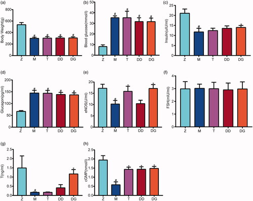
After 8 weeks of drug intervention, there was no significant difference in body weight between the T, DD, and DG groups (303.8 ± 25.3; 305.4 ± 24.4; 307.1 ± 19.3) and the M group (299.8 ± 19.0) (p > 0.05); the blood glucose level of the DD and DG groups (31.63 ± 3.60; 31.60 ± 3.35) was better than that of the M and T groups (35.28 ± 1.73; 35.25 ± 6.10), but there was no significant difference among the DIED groups. The serum insulin level of the DG group (13.99 ± 1.00) was significantly higher than that of the M group (11.75 ± 1.83) (p < 0.05), but there was no significant difference in insulin level between the T and DD groups (12.43 ± 1.20; 13.52 ± 1.23) and the M group (p > 0.05). There was also no significant difference in glucagon level between the T, DD, and DG groups (143.74 ± 11.98; 138.46 ± 8.88; 136.98 ± 8.46) and the M group (144.68 ± 11.47) (p > 0.05). The level of serum eNOS in the T and DG groups (15.85 ± 2.25; 17.13 ± 1.73) was significantly higher than that in the M group (p < 0.05), but there was no significant difference in the level of serum eNOS between the DD and M groups (10.40 ± 1.54; 10.24 ± 1.56) (p > 0.05) ()).
Figure 1. (a) The body weight in rats from the Z, M, T, DD, DG groups. (b) The blood glucose in rats from the Z, M, T, DD, DG groups. (c) The insulin in rats from the Z, M, T, DD, DG groups. (d) The glucagon in rats from the Z, M, T, DD, DG groups. (e) The blood eNOS in rats from the Z, M, T, DD, DG groups. (f) The blood FSH in rats from the Z, M, T, DD, DG groups. (g) The blood T in rats from the Z, M, T, DD, DG groups. (h) The cGMP protein in rats from the Z, M, T, DD, DG groups. Data are expressed as the mean ± SEM. Multiple comparisons analysis were conducted using ANOVA followed by t-testing.Differences with p < 0.05 were considered statistcally significant. #p < 0.05, the M group versus the Z group; *p < 0.05, the T, DD, DG groups vs the M group.
Serum FSH and testosterone levels of rats in each group after drug intervention
The testosterone level in the M group (0.16 ± 0.02) was significantly lower than that in the Z group (1.49 ± 0.65) (p < 0.05); however, the testosterone level in the DG group (1.16 ± 0.24) was significantly higher than that in the M group (p < 0.05). There was no significant difference in the testosterone level between the T and DD groups (0.17 ± 0.02; 0.41 ± 0.17) (p > 0.05). Additionally, there was no significant difference in the serum FSH level between the groups before and after drug intervention (p > 0.05) ().
Penile tissue structure and ultrastructure of rats in each group after drug intervention
HE staining results revealed that the trabeculae and blood sinuses were well distributed in the cavernous body of penises in group Z; some red blood cells were observed in the sinuses, and the inner walls of the sinuses were covered with flat endothelial cells. There were many smooth muscle cells, collagen fibres, and some small blood vessels in the blood sinus trabecula, and no hyperplasia in the interstitial tissue. In the M group, the number of blood sinuses in the cavernous body of the penis was significantly lower, the number of small blood vessels was significantly lower with disordered distribution of blood sinuses, the density of endothelial cells and smooth muscle cells were significantly lower, and the number of collagen fibres were significantly higher than the Z group. The distribution of blood sinuses in the T, DD, and DG groups was more regular than that in the M group, with higher density of endothelial cells, lower number of collagen fibres decreased, and some red blood cells in the blood sinuses ().
Figure 2. (a) HE staining of testicular tissues of rats from the Z, M, T, DD, DG groups (×200). In Group Z: (i) There were some red blood cells in the sinus space, and the inner wall of the blood sinus was covered with flat endothelial cells. There was no proliferation of interstitial tissue (Arrow ①). In Group M: (i) The number of blood sinuses decreased significantly, the number of small blood vessels decreased, and the distribution of blood sinuses was disordered (Arrow ②); (ii)The density of endothelial cells and smooth muscle cells decreased significantly, and the number of collagen fibres increased (Arrow ③). In Group T: (i) The distribution of blood sinuses was relatively uniform, and red blood cells could be seen locally in the blood sinus space (Arrow ④). In Group DD: (i) The distribution of blood sinus was more regular than that of group M (Arrow ⑤). In Group DG: (i) The density of endothelial cells increased and the number of collagen fibres decreased (Arrow ⑥). (b) Ultrastructure of penile tissue of rats from the Z, M, T, DD, DG groups (×5000). In Group Z: (i) Smooth muscle cells had normal morphology and structure, regular nucleus, abundant mitochondria, normal structure, clear inner ridge and tight arrangement of myofilaments (Arrow ①). In Group M: (i) some smooth muscle cells were swollen, the inner ridges of mitochondria in muscle cells were clear, and the myofilaments in some swelling cells were closely arranged and slightly loose (Arrow ②); In Group T, DD, DG: (i) Smooth muscle cells had normal morphological structure, regular nucleus, clear inner ridge of mitochondria and tight arrangement of myofilaments.(Arrow ③④⑤⑥).
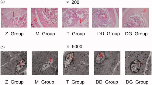
Under the transmission electron microscope, in the Z group, smooth muscle cells had normal morphology and structure, regular nucleus, abundant mitochondria, normal structure, clear inner ridge and tight arrangement of myofilaments. In the M group, some smooth muscle cells were swollen, the inner ridges of mitochondria in muscle cells were clear, and the myofilaments in some swelling cells were closely arranged and slightly loose. After 8 weeks of drug intervention, smooth muscle cells had normal morphological structure in the T, DD, and DG groups; we also observed that the nucleus is regular, the inner ridge of mitochondria is clear and the myofilaments are arranged closely ().
Expression of cGMP in penile tissue
The penile tissue level of cGMP in the M group (0.58 ± 0.15) was significantly lower than that in the Z group (1.94 ± 0.24) (p < 0.05) after 8 weeks of drug intervention. The cGMP level in the T, DD, and DG groups (1.42 ± 0.12; 1.43 ± 0.13; 1.48 ± 0.11) was significantly higher than that in the M group (p < 0.05), among which the DG group showed the most significant improvement. However, there was no significant difference between the T, DD, and DG groups (p > 0.05) ().
Expression of NOS and PDE5 protein in penile cavernous tissue of rats
The level of NOS protein expression in the M group (0.37 ± 0.06) was significantly lower than that in the Z group (0.97 ± 0.08) (p < 0.05). After 8 weeks of drug intervention, the expression of NOS protein in the T, DD, and DG groups (0.63 ± 0.08; 0.51 ± 0.06; 0.77 ± 0.05) was significantly higher than that in the M group (p < 0.05), and the increase in the DG group was more obvious. The expression of PDE5 protein in the M group (0.78 ± 0.06) was significantly higher than that in the Z group (0.33 ± 0.06) (p < 0.01). The expression of PDE5 protein in the T, DD, and DG groups (0.55 ± 0.07; 0.62 ± 0.08; 0.43 ± 0.05) was significantly lower than that in the M group (p < 0.01) ().
Figure 3. (a) The expression levels of two proteins (NOS, PDE5) in rats from the Z, M, T, DD, DG groups groups determined using western blotting. (b) Electrophoreto gram of two proteins (NOS, PDE5) in rats from the Z, M, T, DD, DG groups. (c) The mRNA expression levels of two proteins (NOS, PDE5) in rats from the Z, M, T, DD, DG groups determined using RT-qPCR. NOS, PDE5 Data are expressed as the mean ± SEM. Multiple comparisons analysis were conducted using ANOVA followed by t-testing. Differences with P < 0.05 were considered statistically significant. #p < 0.05 and ##p < 0.01, the M group versus the Z group; *p < 0.05 and **p < 0.01, the DD, DG groups vs the M group.
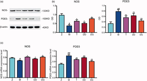
Real-time RT-qPCR was used to detect the expression of NOS and PDE5 mRNA in penile cavernous tissue of rats in each group
The level of NOS mRNA expression in the M group (0.51 ± 0.01) was significantly lower than that the Z group (0.58 ± 0.02) (p < 0.05). After 8 weeks of drug intervention, the expression of NOS mRNA in the T, DD, and DG groups (0.61 ± 0.01; 0.55 ± 0.02; 0.61 ± 0.02) was significantly higher than that in the M group (p < 0.05), and the increase in the DG group was more obvious. The expression of PDE5 mRNA in the M group (0.81 ± 0.04) was significantly higher than that in the Z group (0.51 ± 0.03) (p < 0.01). The expression of PDE5 mRNA in the T, DD, and DG (0.67 ± 0.02; 0.74 ± 0.03; 0.61 ± 0.03) groups was significantly lower than that in the M group (p < 0.01) ().
Discussion
The leech-centipede granules are common combinations of drugs for the treatment of blood stasis syndromes in clinical practice; they are also the core component of many prescriptions for promoting blood circulation and dredging collaterals. They have good curative effects on some vascular complications (Li et al. Citation2013). In this study, we observed that leech-centipede drugs can also significantly improve the erectile function of DIED rats. After 8 weeks of drug intervention, the erectile function of rats in the DD and DG groups significantly improved compared with that of rats in the M group, which showed that leech-centipede drugs can improve the erectile function of DIED rats. Their specific mechanism may be to improve the testosterone level of the rats and regulate PDE5 pathway-related protein and mRNA expression. The erectile process is dependent on androgen (Zhang et al. Citation2005); diabetes may cause testicular tissue damage, gonadal secretion axis function damage, and affect the synthesis and secretion of testosterone (Saad Citation2009), thus, leading to testosterone deficiency in patients. Testosterone can maintain normal cell morphology and function by acting on the androgen receptor of penile cavernous smooth muscle cells (Pu et al. Citation2008), to maintain the integrity of penile erectile structure. However, insufficient androgen secretion can lead to apoptosis of penile cavernous smooth muscle cells, which then cannot maintain the integrity of the erect penile structure. At the same time, testosterone can stimulate erection and maintenance of the penis by increasing male sexual desire (Wang et al. Citation2004), activating mononitrates (Chamness et al. Citation1995), and reducing penile cavernous vasoconstriction (Reilly et al. Citation1997). The results of this study showed that testosterone level and erectile function of rats in the M group were significantly lower than those in the Z group. After 8 weeks of drug intervention, the testosterone level and erectile function of rats in the DD and DG groups were significantly higher than those of rats in the M group. The above results showed a positive correlation between serum testosterone level and erectile function, which confirmed the role of testosterone on erectile function, and also showed that leech-centipede granules might improve erectile function by improving the testosterone level in DIED rats.
In recent years, studies have shown that the NO-cGMP-PDE5 pathway plays a key role in the physiological response to penile erection (Xin et al. Citation2001). NO is an important neurotransmitter in the corpus cavernosum mediating penile erection. During sexual stimulation, the parasympathetic nerve, non-adrenergic and non-cholinergic nerve endings, and vascular endothelial cells release NO under the action of NOS. NO activates guanylate cyclase and converts GDP into cGMP, which reduces intracellular calcium concentration, relaxes the cavernous smooth muscle, and increases penile blood flow to induce an erection. PDE5 promotes the degradation of cGMP into inactive GUMP and stops the process of penile erection. At present, PDE5 inhibitors, the first-line drugs in clinical ED treatment, are based on the above physiological processes: they selectively block the degradation process of cGMP and promote the increase in cGMP level to continue working. Therefore, it is an effective way to treat ED by improving the activity of NOS or inhibit the effect of PDE5, to improve the level of NO in the penile cavernous body. Our results showed that after 8 weeks of drug intervention, the expression of NOS protein in the DD and DG groups was significantly higher than that in the M group (p < 0.05), and this increase was more obvious in the DG group. The PDE5 mRNA expression level in the DD and DG groups was significantly lower than that in the M group (p < 0.01). Thus, our results show that leech-centipede could improve erectile function of DIED rats by increasing the expression of NOS protein and inhibiting the expression of PDE5 protein.
Studies have shown a close relationship between the severity of DIED and blood glucose control (Roth et al. Citation2003). The treatment of DIED is based on blood glucose control to prevent further damage of blood vessels and nerves caused by diabetes. In this study, we showed that after 8 weeks of leech-centipede treatment, the insulin level of rats in the DG group was significantly higher than that of rats in the M group, and the blood glucose control was relatively ideal. Thus, leech-centipede can improve erectile function and reduce the hyperglycaemia state of DIED rats. Our study still has many limitations; for example, we did not measure the concentration of CA + in penile tissues and the cells with functional gain and loss. IC50 ED50, LD50, MIC, etc. were also not measured. Therefore, we will continue to explore the potential mechanism of leech-centipede in improving diabetic ED from a cellular aspect.
Conclusions
The leech-centipede granules may improve the erectile function of DIED rats by improving blood glucose level, increasing testosterone secretion level, repairing pathological and ultrastructural damage of the penis cavernous body, and influencing the expression of cGMP, NOS, and PDE5-related molecules in the PDE5 pathway. This study provides a potential mechanism for the treatment of DIED with leech-centipede. It has increased the scientific basis for clinical application.
Disclosure statement
No potential conflict of interest was reported by the author(s).
Additional information
Funding
References
- Akingba AG, Burnett AL. 2001. Endothelial nitric oxide synthase protein expression, localization, and activity in the penis of the alloxan-induced diabetic rat. Mol Urol. 5(4):189–197.
- Assaly-Kaddoum R, Giuliano F, Laurin M, Gorny D, Kergoat M, Bernabé J, Vardi Y, Alexandre L, Behr-Roussel D. 2016. Low intensity extracorporeal shock wave therapy improves erectile function in a model of type II diabetes independently of NO/cGMP pathway. J Urol. 196(3):950–956.
- Castela Â, Costa C. 2016. Molecular mechanisms associated with diabetic endothelial-erectile dysfunction. Nat Rev Urol. 13(5):266–274.
- Chamness SL, Ricker DD, Crone JK, Dembeck CL, Maguire MP, Burnett AL, Chang TSK. 1995. The effect of androgen onnitric oxide synthase in the male reproductive tract of the rat. Fertil Steril. 63(5):1101–1107.
- Cho NH, Shaw JE, Karuranga S, Huang Y, da Rocha Fernandes JD, Ohlrogge AW, Malanda B. 2018. IDF Diabetes Atlas: Global estimates of diabetes prevalence for 2017 and projections for 2045. Diabetes Res Clin Pract. 138:271–281.
- Heaton JP, Varrin SJ, Morales A. 1991. The characterization of a bio-assay of erectile function in a rat model. J Urol. 145(5):1099–1102.
- Irwin GM. 2019. Erectile dysfunction. Prim Care. 46(2):249–255.
- Li JB, Liu WQ, Tang XH, Li ZF, Di PT, Liu WC, Peng JY. 2013. Analysis on clinical application of insect drugs. Shanghai J Trad Chinese Med. 47:66–68.
- Li X, Zhao Q, Wang J, Wang J, Dai H, Li H, Wang B. 2018. Efficacy and safety of PDE5 inhibitors in the treatment of diabetes mellitus erectile dysfunction: protocol for a systematic review. Medicine. 97(40):e12559.
- Ma HF, Liu Y, Wang B, Dang J, Ma JX, Zhu YT, Li HS. 2017. Clinical randomized controlled study for the treatment of erectile dysfunction by Tong Luo Xi Feng Qi Wei soup. China J Human Sex. 26:78–81.
- Othman MS, Hafez MM, Abdel Moneim AE. 2020. The potential role of zinc oxide nanoparticles in microRNAs dysregulation in STZ-induced type 2 diabetes in rats. Biol Trace Elem Res. 197(2):606–618.
- Podlasek CA, Zelner DJ, Bervig TR, Gonzalez CM, McKenna KE, McVary KT. 2001. Characterization and localization of nitric oxide synthase isoforms in the BB/WOR diabetic rat. J Urol. 166(2):746–755.
- Pu XY, Wang XH, Gao WC, Yang ZH, Li SL, Wang HP, Wu YL. 2008. Insulin-like growth factor-1 restores erectile function in aged rats: modulation the integrity of smooth muscle and nitric oxide-cyclic guanosine monophosphate signaling activity. J Sex Med. 5(6):1345–1354.
- Reilly CM, Stopper VS, Mills TM. 1997. Androgens modulate the alpha-adrenergic responsiveness of vascular smooth muscle in the corpus cavernosum. J Androl. 18(1):26–31.
- Roth A, Kalter-Lftbovhx O, Kerbis Y, Tekenbaum-Koren E, Chen J, Sobol T, Raz I. 2003. Prevalence and risk factors for erectile dysfunction in men with diabetes, hypertension, or both diseases: a community survey among 1,412 Israeli men. Clin Cardiol. 26(1):25–30.
- Ryan JG, Gajraj J. 2012. Erectile dysfunction and its association with metabolic syndrome and endothelial function among patients with type 2 diabetes mellitus. J Diabetes Complications. 26(2):141–147.
- Saad F. 2009. The role of testosterone in type 2 diabetes and metabolic syndrome in men. Arq Bras Endocrinol Metabol. 53(8):901–907.
- Shamloul R, Ghanem H. 2013. Erectile dysfunction. Lancet. 381(9861):153–165.
- Sullivan ME, Mumtaz FH, Dashwood MR, Thompson CS, Naseem KM, Bruckdorfer KR, Mikhailidis DP, Morgan RJ. 2002. Enhanced relaxation of diabetic rabbit cavernosal smooth muscle in response to nitric oxide: potential relevance to erectile dysfunction. Int J Impot Res. 14(6):523–532.
- Wang C, Cunningham G, Dobs A, Iranmanesh A, Matsumoto AM, Snyder PJ, Weber T, Berman N, Hull L, Swerdloff RS. 2004. Long-term testosterone gel (AndroGel) treatment maintains beneficial effects on sexual function and mood, lean and fat mass, and bone mineral density in hypogonadal men. J Clin Endocrinol Metab. 89(5):2085–2098.
- Xin ZC, Lin GT, Zhang ZW, Guo YL. 2001. Advances in the research of penile erection and erection dysfunction. Pro Phys Sci. 32:129–134.
- Xu M, Sheng Z. 2018. Research progress on the mechanism of traditional Chinese insects' medicine treatment of diabetic neurological complications. China J Clin Pharm Ther. 23:597–600.
- Zhang JJ, Liu QF, Wu CY. 2014. Study on activities of hyperglycemia and hyperlipemia of polysaccharides from Grateloupia filicina. J Fisheries Res. 36:21–28.
- Zhang XH, Morelli A, Luconi M, Vignozzi L, Filippi S, Marini M, Vannelli GB, Mancina R, Forti G, Maggi M. 2005. Testosterone regulates PDE5 expression and in vivo responsiveness to tadalafil in rat corpus cavernosum. Eur Urol. 47(3):409–416.