
Abstract
Context
Danshen, the dried root and rhizome of Salvia miltiorrhiza Bunge (Labiatae) and honghua, the dried flower of Carthamus tinctorius L. (Compositae) as the herb pair was used to treat cardiovascular diseases (CVD).
Objective
To study the effects of DHHP on MIRI and mechanisms based on apoptosis and mitochondria.
Materials and Methods
36 SD rats (n = 6) were randomly divided into control group (Con), the ischaemia-reperfusion group (IR), positive control (Xinning tablets, XNT, 1 g/kg/d) and DHHP (1.2, 2.4, and 4.8 g/kg/d). Except for Con, the other groups were intragastrically administrated for 5 d, the rat hearts were isolated to establish the MIRI model in vitro for evaluating the effects of DHHP on MIRI. 24 SD rats (n = 6) were randomly divided into Con, IR, DPPH2.4 (2.4 g/kg/d) and DPPH 2.4 + Atractyloside (ATR) (2.4 + 5 mg/kg/d), administered intragastrically for 5 d, then treated with ATR (5 mg/kg/d) by intraperitoneal injection in DPPH2.4 + ATR group, took rat hearts to establish MIRI model in vitro for revealing mechanism.
Results
Myocardial infarct sizes were, respectively, 0.35%, 40.09%, 15.84%, 30.13%, concentrations of NAD+ (nmol/gw/w) were 144, 83, 119, and 88, respectively, in Con, IR, DHHP2.4, DHHP2.4 + ATR group. Cleaved caspase-3 were 0.3, 1.6, 0.5 and 1.3% and cleaved caspase-9 were 0.2, 1.1, 0.4 and 0.8%, respectively, in Con, IR, DHHP2.4 and DHHP2.4 + ATR group. The beneficial effects of DHHP on MIRI were reversed by ATR.
Conclusions
The improvement of MIRI by DHHP may be involved in inhibiting MPTP opening, decreasing oxidative damage, alleviating ischaemic injury and inhibiting cardiomyocyte apoptosis.
Introduction
Cardiovascular diseases (CVD) are chronic diseases that seriously endanger human health, including myocardial infarction (MI), hypertension, atherosclerosis, angina pectoris, coronary heart disease, arrhythmia, heart failure and cardiomyopathy and so on. The morbidity and mortality of CVD rank first in the global, and continue increasing trend (Guo et al. Citation2016; Benjamin et al. Citation2017; Yan et al. Citation2017; Wang et al. Citation2019). It was forecasted that there were 2.5 million myocardial infarctions (MI) patients in China (Benjamin et al. Citation2017). Myocardial infarction occurs during ischaemia and anoxia of the coronary artery. While early reperfusion can prevent further deterioration of heart disease and even ischaemic necrosis induced by myocardial ischaemia and hypoxia. Reperfusion is one of the common therapy programs for ischaemic heart disease (IHD), however, the secondary damage of the myocardium such as myocardial haemorrhage, myocardial oedema, cell membrane rupture, mitochondrial swelling, excessive sarcomere contraction occurs in the process of reperfusion, which is called MIRI (Liu et al. Citation2018; Li et al. Citation2019). Although ischaemic pre-conditioning, ischaemic post-conditioning and remote ischaemic pre-conditioning, remote ischaemic post-conditioning can alleviate MIRI (Donato et al. Citation2017; Yang et al. Citation2019). However, ischaemic pre-conditioning or post-processing may cause damage to local tissues and organs, which is not accepted by medical ethics, so the above several methods are limited in clinical practice. Therefore, finding effective drugs and opening up new therapeutic ways are essential for alleviating MIRI (Jiang et al. Citation2014; Yuan et al. Citation2014; Wei et al. Citation2016; Li et al. Citation2018; Xie et al. Citation2018; Chen et al. Citation2019; Huang et al. Citation2019; Xiao et al. Citation2019).
Traditional Chinese medicines have good curative effects and broad application prospects in alleviating MIRI (Wang et al. Citation2014; Deng et al. Citation2017; Shi et al. Citation2017; Tan et al. Citation2017; Zhang et al. Citation2017; Koo et al. Citation2018; Yang et al. Citation2021). DHHP is widely used in the treatment of CVD for hundreds of years, with definite effects (Qian et al. Citation2018; Chen et al. Citation2019). However, the efficacy and mechanisms of DHHP in mitigating MIRI were still unclear, and further research was needed. It has been shown in the literature that MIRI was closely related to mitochondria (He et al. Citation2017; Pell et al. Citation2018; Miao et al. Citation2019; Sun et al. Citation2019). The aim of this research was to study the protective effects of DHHP on MIRI and potential mechanisms based on apoptosis and mitochondria.
Materials and methods
Ethics and experimental animals
All animal experiments were carried out in keeping with the State Council of the People’s Republic of China, Regulations for the Administration of Affairs Concerning Experimental Animals (2017 Revision) and the Animal Ethics Committee of Shaanxi University of Chinese Medicine (TCM-2018-030-E01). We made all efforts to minimize the pain and suffering of experimental animals throughout animal experiments.
Adult Specific pathogen free male Sprague–Dawley (SD) rats (body weight was 250 ± 20 g) were bought from the Experimental Animal Centre of Xi’an JiaoTong University Health Science Centre. (laboratory animal licence number SYXK (shaan) 2018-001; Xi’an, China). The rats were housed in a room with 25 ± 5 °C temperature, 45 ± 5% relative humidity, and 12 h light/dark cycle for 1 week to adapt to the experimental environment. All groups of rats were given free access to water and food. The rats fasted for 12 h before the experiment but freely received water.
Preparation of DHHP extractive
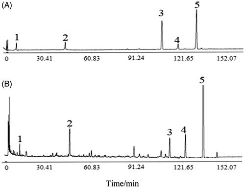
The dried pieces of danshen and honghua were used that complied with the standards of the Chinese Pharmacopoeia (ChP) 2015 edition from Shunwangcheng Chinese Medicinal Materials Market in Juancheng County of Shandong Province, China. A certificate specimen was kept in the Chinese Medicine Herbarium of College of Pharmacy, Shaanxi University of Chinese Medicine, Xianyang, China. The dried decoction pieces of danshen (3 kg) were soaked with water for 0.5 h, heated and boiled for 0.5 h, then cooled to room temperature, filtrated, added 1 kg of honghua, soaked in water (70–80 °C) for1 h, then boiled for 0.5 h, removed and cooled to room temperature, filtrated, combine the filtrate and concentrated under reduced pressure, freeze-dry to make powder. DHHP extractive was harvested. DHHP extractives were analyzed by HPLC, the results are shown in . Contents of danshensu, hydroxysafflor-yellow-A, rosmarinic acid, lithospermic acid and salvianolic acid B were 4.5, 16.7, 1.9, 4.6 and 55.8 mg/g, respectively, in DHHP extractives.
Figure 1. The HPLC chromatograms of standards and DHHP extracts. (A) Mix standards. (B) DHHP extracts. 1: Danshensu. 2: Hydroxysafflor-yellow-A. 3: Rosmarinic acid. 4: Lithospermic acid. 5: Salvianolic acid B.
Positive drug
Xinning tablets (positive drug, XNT), Xinning tablets consisting of including danshen, huaihua [the dried flowers and buds of Sophora japonica L. (Family Leguminosae)], chuanxiong [the dried rhizome of Ligusticum chuanxiong Hort. (Family Umbelliferae)], sanqi [the dried root and rhizome of Panax notoginseng (Burk.) F. H. Chen (Family Araliaceae)], jiangxiang [the dried heartwood of root and trunk of Dalbergia odorifera T. Chen (Family Leguminosae)], chishao [the dried root of Paeonia lactiflora Pall. or Paeonia veitchii Lynch (Ranunculaceae)] and honghua, were purchased from Jun Bisazza Pharmaceutical (Lot: 20181225). XNT are a common clinically used Chinese medicine for CVD, and are used in the treatment of CVD such as angina pectoris, coronary heart disease and myocardial ischaemia reperfusion, so XNT were selected as positive drugs.
Preparation of drug solution
An appropriate amount (12, 72, 48 g) of DHHP extractive was dissolved in water to make an aqueous solution (0.12, 0.24, 0.48 g/mL). Twenty tablets of XNT, with the coating removed, were ground, of which 10 g were dissolved in water to make an aqueous solution (0.10 g/mL). This aqueous solution was used for the animal experiments (The above dosages were the equivalent dose of laboratory animals based on the clinical dosage).
DHHP extractive was added to an appropriate amount of nutrient mixture F-12 (DMEM/F12), ultrasonicated for 30 min with China Ultrasonic Cleaner (Guangdong GT Ultrasonic Co., Ltd), made to dissolve, then centrifuged at 10,000 rpm for 10 min with a centrifuge (Weiya Technology Co., Ltd), and filtered with 0.22 µm sterile filter. A small amount was then dried in order to determine a precise weight of the dissolved DHHP extractive, and adjusted to a solution concentration according to the above results. The solution (400 µg/mL) with DHHP extractive was collected. Positive drug (Xinning tablets, XNT) solution (400 µg/mL) was prepared by the same method. The above aqueous solution was used for cell experiments. During cell experiments, sterile DHHP solution or sterile positive drug solution was added 20 µL per well, 200 µL total volume per well (reference blood concentration of rats).
Other materials
Radio-immunoprecipitation assay (RIPA), phenylmethyl sulphonyl fluoride (PMSF), enhanced BCA protein assay kit, SDS-PAGE gel electrophoresis buffer, beyoecl star reagent and terminal deoxynucleotidyl transferase dUTP nick end labelling (TUNEL) apoptosis assay kit bought from Beyotime Institute of Biotechnology (Shanghai, China). 2,3,5-Triphenyltetrazolium chloride (TTC) was bought from Wanleibio (Wuhan, China). Atractyloside (ATR; purity: ≥98%) were purchased from Shang Hai Pure One Biotechnology (Shanghai, China). ATR powder was dissolved in normal saline to inject intraperitoneally for the animal experiment. ATR powder was dissolved in DMEM/F12 medium (BOSTER Biological Technology Co. Ltd) for the cell experiments. The material for the Western blots that Cytochrome C antibody (WL02410), Bax antibody (WL01637), Bcl-2 antibody (WL01556), caspase-3/cleaved-caspase-3 antibody (WL02117), caspase-9/cleaved-caspase-9 antibody (WL03421), the secondary antibody goat anti-rabbit IgG-HRP (WLA023) and internal reference antibody GAPDH (WL01114) were bought from Wanleibio (Wuhan, China). XNT were purchased from Junbisazza Pharmaceutical Limited Company (Lot: 20181225).
Drug administration with experimental animals
In the first stage, 36 rats were randomly divided into the control group (Con), the ischaemia-reperfusion group (IR), Positive control group (XNT) and DHHP groups (1.2, 2.4 and 4.8 g/kg), 6 rats per group. Rats in Con and IR groups received 1 mL/100 g volume of water. Rats in XNT, DHHP groups were treated with corresponding drugs, XNT group at the dose of 1 g/kg/d, DHHP group 1.2, 2.4 and 4.8 g/kg/d, respectively. All groups were intragastrically administrated for 5 d, and rat hearts were isolated 1 h after corresponding drugs pre-treatment on the 5th day to establish the MIRI model in vitro for detecting the area of heart infarction and myocardial histopathology.
Based on the above research results, 2.4 g/kg DPPH was used for the research mechanism. Twenty-four rats were randomly divided into the control group (Con), the ischaemia-reperfusion group (IR), 2.4 g/kg DPPH dose group (DPPH2.4) and 2.4 g/kg DPPH combined with ATR group (DPPH 2.4 + ATR), 6 rats per group. Rats in Con, IR and DHHP2.4 were administrated according to the first stage. Rats in DPPH2.4 + ATR were administered intragastrically with DHHP (2.4 g/kg/d) for 5 d, then additionally treated with ATR (5 mg/kg/d) by intraperitoneal injection 1 h after the last DPPH pre-treatment, then anaesthetized and took out rat hearts 1 h after ATR pre-treatment to establish MIRI model in vitro for detecting cardiomyocyte apoptosis, myocardial mitochondria and related proteins, and analyzed potential mechanisms.
Establishment of MIRI model in vitro
The MIRI model in vitro was established according to methods in the literature (Wu et al. Citation2014; Liao et al. Citation2015; Xiao et al. Citation2018). In short, rats were anaesthetized with urethane at the dose of 1.2 g/kg by injecting subcutaneously; meanwhile, in order to prevent thrombus formation in the intracoronary, rats were administered intravenously heparin (1500 IU/kg), then opened the thoracic cavity, rapidly took out hearts of rat, then immediately placed them into 4 °C Krebs–Henseleit (K-H) solution (Liquid A: NaCl 6.9212 g; KCL 0.3517 g; KH2PO4 0.1621 g; MgSO4·7H2O2 0.2919 g; NaHCO3 2.1101 g, glucose 2.0112 g added double distilled water and stirred to dissolve; Liquid B: CaCl2 0.1664 g, added double-distilled water, stirred well to dissolve. Added liquid B to liquid A, stirred well, added 1 mmo1/L hydrochloric acid to adjust the pH to 7.30–7.40, and added water to make the volume to 1 L. Filtered with a 0.45 µm filter, and continuously inject a mixture of 95% O2 and 5% CO2 into the K-H perfusion solution for about 20 min before perfusion) with heparin and 100% oxygen, quickly removed the connective tissue, then swiftly hung heart on Isolated Heart Perfusion Apparatus, continuously perfused for 15 min using K-H solution containing 5% CO2 and 95% O2 at 37 °C, and stopped for 30 min for inducing ischaemia, then re-perfused 60 min for inducing MIRI. Hearts from the control group were placed in the K-H solution containing 5% CO2 and 95% O2 at 37 °C.
Myocardial histopathology
HE staining according to literature (Wang et al. Citation2019): After the end of ischaemia-reperfusion, removed the heart tissue block (generally no more than 0.5 cm thick), and immersed in 10% formalin solution for fixation at 25 °C for 36 h, then, with flowing water, washed the heart tissue block for 4 h. Using alcohol from low to high concentration as a dehydrating agent, gradually removed the water in the tissue mass (the heart tissue was immersed in 70% ethanol for 2 h, 80% ethanol overnight, 90% ethanol for 2 h, anhydrous ethanol I for 1 h, and anhydrous ethanol II for 1 h at room temperature). Then put the tissue block in xylene for 30 min to replace the alcohol in the tissue block with xylene, the dehydrated tissues were moved into the melted paraffin and placed in an incubator with 60 °C for 2 h, to allow paraffin to penetrate completely into the heart tissues, then the paraffin- penetrated heart tissues were buried in the centre of the metal frame with the dissolved paraffin, and cooled down, and became hard wax blocks, and fixed the embedded wax block on a microtome, and made into 5 µm slices. The cut slices were ironed in heated water, and then pasted on the glass slides, and placed in pathological slicing shelfs, dried in an incubator at 45 °C. then taken out and placed in xylene I 15 min and xylene II for 15 min; taken out and removed the residual liquid, and put into absolute ethanol I for 5 min and absolute ethanol II for 5 min; taken out and removed the residual liquid, and put into 95% ethanol for 2 min, 85% ethanol for 2 min, and 75% ethanol for 2 min; taken out and removed the residual liquid, and finally immersed in distilled water for 2 min.
Take out the section for HE staining: took out and removed residual distilled water, and placed into haematoxylin solution for 2 min, took out and removed the residual liquid, and placed into distilled water for 2 min, took out and removed the residual liquid, and placed into 1% hydrochloric acid alcohol for 3 s, took out and washed with flowing water for 20 min, took out and removed the residual liquid, and placed into distilled water for 2 min, took out and removed the residual liquid, and placed into eosin solution for 3 min: The stained paraffin sections were immersed in 75% ethanol for 2 min, 85% ethanol for 2 min, and 95% ethanol for 2 min, drained the residual liquid, immersed in absolute ethanol I for 5 min, absolute ethanol II for 5 min. Drained the residual liquid, immersed in xylene I for 10 min and xylene II for 10 min, took out and drained the surrounding liquid, added neutral gum, covered with a cover glass, the sections were fixed with neutral gum and dried at room temperature. Finally, all slices were observed under a microscope to evaluate the pathological changes of the heart tissue and captured images of magnification (200×).
Area of heart infarction
TTC staining according to literature (Li et al. Citation2018; Miao et al. Citation2019; Wang et al. Citation2019): In short, after the end of reperfusion, hearts were frozen it at −80 °C for 30 min. Cut the rat atrium, and quickly cut the ventricular part perpendicular to the long axis of the heart, made into 1 mm slices, placed the myocardial slice in 1% TTC at room temperature for 25 min, and fixed with 10% formalin solution. The un-infarcted myocardium is stained red and the infarcted myocardium is unstained. Photographs were taken with a digital camera image J software was used to analyze the images of the stained sections.
Myocardial apoptosis
After the end of reperfusion, molten paraffin was used for embedding heart tissue, and sliced after cooling (Wang et al. Citation2019), the flakes were ironed in heated water, and then pasted on the glass slides, dried in an incubator at 60 °C, placed in xylene I 15 min and xylene II for 15 min to remove the paraffin from the slices, and immersed absolute ethanol I for 5 min, shake off the remaining liquid, put it in absolute ethanol II, and soak for 5 min, shake off the remaining liquid, soaked in 95%, 85%, and 75% ethanol in sequence, and let each ethanol stay for 2 min, shake off the remaining liquid, and finally immersed in distilled water for 2 min, then rinsed 3 times with PBS, 5 min each time, and permeabilizated with 50 µL 0.1% triton X-100 at 37 °C for 8 min, and rinsed 3 times with PBS, 5 min each time, and closed endogenous peroxidase with 50 µL 3% H2O2 at 37 °C for 10 min, and then rinsed with PBS, and incubated with 50 µL TUNEL reaction solution at 37 °C for 60 min, and rinsed with PBS, and added converter-POD 50 µL, then incubated with at 37 °C for 30 min, and rinsed with PBS, wiped off excess solution around and. Stained using DAB: added 50 µL of DAB substrate, and immediately put it in water to stop the reaction when the colour just darkens, and rinsed with PBS, counterstained using haematoxylin, then dehydrated with 75% to 100% ethanol in sequence, and let each ethanol stay for 2 min, removed the remaining liquid, immerse it in absolute ethanol I, II, and xylene I, II for 10 min, respectively, take out the slices in turn, dried the surrounding liquid, added half a drop of neutral gum, and covered with a cover glass and dried at 37 °C. Finally, a microscope was used for observing all sections to assess apoptosis cells in heart tissues and images were captured at magnification (400×). The apoptosis rate is calculated by the number of apoptosis cells to the total number of cells × 100%.
Western blotting analysis
After the end of reperfusion, the ventricle parts of the heart were divided into pieces by cutting according to the method of literature (Quan et al. Citation2020; Xu et al. Citation2020), proteins were extracted from heart tissue using Whole Cell Lysis Assay (WLA019, Wanleibio, Wuhan, China) on the basis of the kit’s instructions. The enhanced bicinchoninic acid (BCA) Protein Assay Kit (WLA004, Wanleibio, Wuhan, China) was used for the measurement of protein concentration. Sodium dodecyl sulphate-polyacrylamide gel electrophoresis (SDS-PAGE) (10%) was used for separating proteins, and then proteins were transferred onto polyvinylidene difluoride (PVDF) membranes, 5% skim milk powder was used for blocking membranes at room temperature for 2 h, then incubated with primary antibody Bax (1:500), Bcl-2 (1:1000), cytochrome C (1:500), Cleaved caspase-9 (1:500), cleaved caspase 3 (1:500) overnight at 4 °C, incubated using the secondary antibody (1:5000) that conjugated HRP at 37 °C for 1 h. Enhanced chemiluminescence (ECL) reagent was used for detecting immunolabelled proteins. GAPDH was used as an inside reference. Analyze the optical density value of the target band with Gel-Pro-Analyzer software (Wanleibio, Wuhan, China). This part of the experiment was completed by Wanlei Biotechnology Co., Ltd.
Myocardial mitochondria
After the end of reperfusion, according to literature (Jia Citation2011; Jia et al. Citation2018), the left ventricle was quickly separated on ice, took a small piece of tissue, fixed with 2.5% glutaraldehyde (GA) and 1% acetic acid solution for 1 h, and quickly cut into a volume of about 1 mm3, dehydrated, infiltrated, embedded, and made into sections. Uranyl acetate and lead citrate were used for staining of sections, and a transmission electron microscope (JEM-1200EX, JEOL, Japan) was used for observing the mitochondria and photographed.
Concentrations of NAD+
At the end of reperfusion, according to a method described in the literature (Wu et al. Citation2014; Jia et al. Citation2018), extracted NAD+ from left ventricle tissues. Briefly, took left ventricle tissues, added 10 times the volume of the acidic extraction solution (mL) according to the tissue mass (g), ground in the ice bath, and kept at 60 °C for 1 h (extracted NAD+ and destroyed NADH at the same time), centrifuged at 10,000 rpm for 5 min, took the supernatant and placed it on an ice bath to cool, added alkaline extract, and mixed, preheated in 37 °C water bath for 2 min, accurately kept in 37 °C water bath for 10 min, centrifuged at 12,000 g for 10 min at 4 °C, took the supernatant for testing Concentrations of NAD+ using a spectrophotometer (DU 640, Beckman Coulter, Fullerton, CA, USA).
Sensitivity of MPTP to calcium
According to the literature (Morciano et al. Citation2017), the mitochondria in the heart tissues were extracted using a Mitochondrial Extract kit (Best Bio Co., Shanghai, China) according to the instructions. Briefly, myocardial tissue was chopped-up as much as possible, and added 0.25% trypsin and kept for 20 min in an ice bath, the trypsin was discarded, separating medium was used for washing the tissue and discarded the supernatant, added separating medium on ice, centrifugation at 1500 g for 10 min at 4 °C, took the supernatant, centrifugated at 15,000 g for 20 min at 4 °C, discarded the supernatant, mitochondria were obtained, and resuspend the precipitate with saving medium. The purified mitochondrial permeability transition pore (MPTP) Assay kit (Shanghai Genmed Pharmaceutical Technology Co., Ltd., Shanghai, China) was used for determining the sensitivity of MPTP to calcium according to the kit’s instructions. The value of optical density (OD) was determined at 520 nm by an enzyme-labeled instrument (Biotek, USA), the OD was determined at each minute for 10 min. The size of the OD value is related to the degree of opening of MPTP, and the decrease of the OD value indicates the opening of MPTP.
Mitochondrial membrane potential
The JC-1 mitochondrial membrane potential assay kit (Beyotime Institute of Biotechnology, Haimen, China) was used for determining the change of membrane potential in the isolated mitochondria according to the kit’s protocol. Fluorescence spectrophotometer (Lengguang Tech Co., Shanghai, China) was used for detecting fluorescence values before and after the addition of CCCP, and the difference of fluorescence (arbitrary units of fluorescence, AUF) meant the difference in the potential (measured as ΔAUF).
Cell culture
H9c2 cardiomyocytes were bought from Shanghai Meixuan Biological Science and technology LTD (Shanghai, China). DMEM/F12 was purchased from Wuhan Bude Biological Engineering Co., Ltd. (Wuhan, China). H9c2 cardiomyocytes were cultured in DMEM/F12 including penicillin (100 U/mL), 10% foetal bovine serum (FBS), streptomycin (100 µg/mL) in an incubator with 5% CO2 at 37 °C.
The establishment of H/R models
H9c2 cells were dominated by hypoxia/reoxygenation (H/R) to imitate MIRI model in vitro according to literature (Li et al. Citation2019). when the cells grew up to about 80% confluence in DMEM/F12 medium containing penicillin (100 U/mL), 10% FBS, streptomycin (100 µg/mL), washed with Phosphate Buffered Saline (PBS). DMEM/F12 medium containing 95% N2 and 5% CO2 without FBS was added to replace the previous medium, and cultured in a tri-gas incubator with 90% N2, 5% CO2 and 5% O2 at 37 °C for 2 h to induce hypoxia. Then, removed DMEM/F12 medium without FBS, cultured at DMEM/F12medium with penicillin (100 U/mL), 10% FBS, streptomycin (100 µg/mL) for 4 h in incubators with 5% CO2 and 95% O2 at 37 °C for reoxygenation. Models of MIRI of H9c2 cells in vitro were obtained.
Cell toxicity
The Cell Counting Kit-8 (CCK-8, Dojindo Molecular Technologies, Kumamoto, Japan) was used for checking up the cell toxicity of DHHP. Cells suspension (100 µL) per well was seeded into 96-well cultivation plates (number of cells 1500 per well) and were incubated for 24 h. Cells were divided into the control group (Con) and DHHP (40, 80, and 160 µg/mL). Cells in Con were cultured in an incubator with normal conditions; cells in DHHP groups were pre-treated with DHHP at a dose of 40, 80, and 160 µg/mL respectively, and cultured for 4, 8, 24 h, respectively, added 10 µL of CCK-8 reagent per well and incubated for 3 h at 37 °C. Microplate reader (Biotek Instruments, Inc., Winooski, UT, USA) was used for determining the absorbance value at 450 nm, percentage of viable cells in each group and Con group was used for evaluating cell toxicity of DHHP.
Cell viability
The Cell Counting Kit-8 (CCK-8, Dojindo Molecular Technologies, Kumamoto, Japan) was used for checking up the cell viability of DHHP. Cell suspension (100 µL) per well was seeded into 96-well cultivation plates (number of cells 1500 per well) and were incubated for 24 h. Cells were divided into control group (Con), H/R group and DHHP (40, 80, and 160 µg/mL). Cells in Con were cultured in an incubator with normal conditions; cells in H/R were dominated according to the method of the establishment of H/R models; cells in DHHP groups were pre-treated with 40, 80, and 160 µg/mL DHHP respectively, and cultured for 4, 8, 24 h, respectively, then dominated according to above the method of H/R group. Added 10 µL of CCK-8 reagent and incubated for 3 h at 37 °C. Microplate reader (Biotek Instruments, Inc., Winooski, UT, USA) was used for determining the absorbance value at 450 nm, percentage of viable cells in each group and Con group was used for evaluating cell viability of DHHP.
Detection of cell apoptosis with flow cytometry analysis
FC-500 Flow cytometry analysis (Beckman Coulter, Inc Miami, FL, USA) was employed for determining apoptosis of cardiomyocyte H9C2 based on the instructions of Annexin V-Fluorescein isothiocyanate (FITC)/Propidium Iodide (PI) kit (Thermo Fisher Scientific, Shanghai, China) according to literature (Huang et al. Citation2019). Briefly, each tube was seeded approximately 200 µL cell suspension (number of cells 1 × 10⁶), added 10 µL of Annexin V-FITC and 10 µL of PI, and incubated in the dark at room temperature for 15 min, added 300 µL binding buffer, then transferred to flow tube to analysis (n = 10).
Statistical analysis
All data were expressed as the mean ± SD in this study and SPSS 17.0 was used for statistics and analysis of data.
Results
Effects of DHHP on myocardial pathology and myocardial infarction
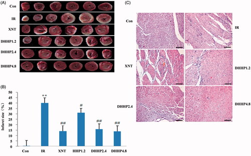
As shown in , after TTC staining, the infarct tissue was white, and the normal tissue was red (). The myocardial infarct size in the IR group was significantly higher than that in the control group, which was statistically significant (p < 0.01); The myocardial infarct size of DHHP various experimental groups and XNT groups (Positive groups) was significantly lower than that in the IR group, DHHP1.2 group (p < 0.05), DHHP2.4 group and DHHP 4.8 group (p < 0.01); however, there was no statistical significance detected between DHHP2.4 group and DHHP 4.8 group (). HE staining revealed that the myocardial fibres were neatly arranged, the nucleus was clear and was in the centre, the cell morphology and structure were complete, there were a few blood vessels in the myocardial interstitium and no vasodilation in the Con group. While in IR group, disordered arrangement of myocardial fibres, swelling and rupture of myocardial fibres, telangiectasia and hyperaemia, erythrocyte overflow, myocardial interstitial oedema, nuclear pyknosis and unclear nucleoli. DHHP various experimental groups pre-treatment obviously alleviated pathological damage in the MIRI model in vitro (). However, in the range of 2.4 − 4.8 g/kg, the protective effect of DHHP on MIRI did not increase with increasing dose.
Figure 2. Myocardial pathology and myocardial infarct size in every groups. (A) Photos of myocardium with TTC staining. (B) Myocardial infarct size (%). (C) Representative photos of myocardium stained with HE (200×). Data were expressed as the mean ± SD (n = 6). **p < 0.01 vs. Con; *p < 0.05 vs. Con; ##p < 0.01 vs. IR #p < 0.05 vs. IR.
Cell toxicity and cell viability assay
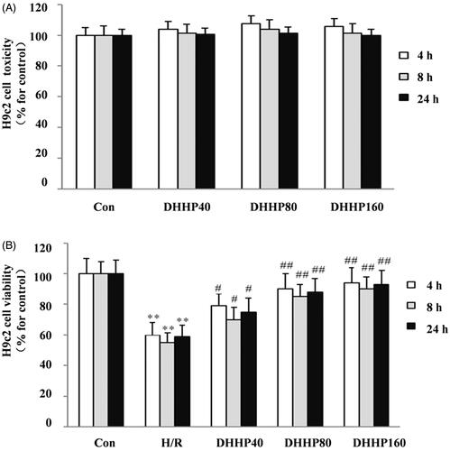
As shown in , in the concentration range of 40–160 μg/mL, DHHP was not cytotoxic regardless of in vitro culturing for 4, 8 and 24 h (). The cell viability of the H/R group was significantly lower than that of the Con group (p < 0.01); in the concentration range of 40–160 μg/mL, DHHP can increase cell viability as the concentration increases, DHHP 40 (p < 0.01), DHHP 80 (p < 0.01), DHHP 160 (p < 0.01); but at 80–160 μg/mL, there was no significant difference in cell viability ().
Figure 3. Effects of DHHP on Cell toxicity and Cell viability of H9C2. (A) The cell toxicity. (B) The cell viability. Data were expressed as the mean ± SD (n = 10). **p < 0.01 vs. Con; *p < 0.05 vs. Con; ##p < 0.01 vs. IR; #p < 0.05 vs. IR.
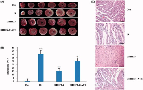
ATR reverses effects of DHHP on myocardial pathology and myocardial infarction
As shown in , the white tissue sections in the DHHP2.4 + ATR group were more than those in the DHHP2.4 group, indicating that the myocardial damage in the DHHP2.4 + ATR group was more serious than that in the DHHP2.4 group (). The area of myocardial infarction in the DHHP2.4 + ATR group was significantly larger than that in the DHHP2.4 group in (p < 0.05). HE staining revealed DHHP2.4 group pre-treatment obviously alleviated pathological damage in the MIRI model in vitro; however, ATR reverses the effects of DHHP on myocardial pathology ().
Figure 4. Myocardial pathology and infarct size. (A) Photos of myocardium after TTC staining. (B) Infarct size as a percentage of total size. (C) Photos of myocardium stained with HE (200×). Data were expressed as the mean ± SD (n = 6). ΔΔp < 0.01 vs. Con, **p < 0.01 vs. IR, #p < 0.05 vs. DHHP2.4.
ATR reverses effects of DHHP on apoptosis in the heart
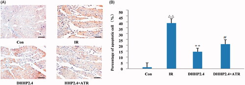
As shown in , cardiomyocyte apoptosis in the DHHP2.4 group was less than that in the IR group (p < 0.01), and cardiomyocyte apoptosis in the DHHP2.4 + ATR group was significantly increased than that in the DHHP2.4 group (p < 0.05).
Figure 5. The Cardiomyocyte apoptosis (400×). (A) TUNEL detected slices. (B) Quantification of TUNEL detection. Data were expressed as the mean ± SD (n = 6). ΔΔp < 0.01 vs. Con, **p < 0.01 vs. IR, #p < 0.05 vs. DHHP2.4.
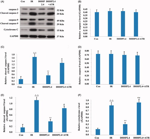
ATR reverses effects of DHHP on the cytochrome C, cleaved caspase-9 and cleaved caspase-3 protein levels
As shown in , the protein levels of cleaved caspase-3, cleaved caspase-9 and cytochrome C in the DHHP2.4 group were significantly lower than those significantly decreased in the IR group (p < 0.05). The protein levels of cytochrome C, cleaved caspase-9 and cleaved caspase-3 in the DHHP2.4 + ATR group were significantly higher than those in the DHHP2.4 group (p < 0.05).
Figure 6. ATR reverses effects of DHHP on the cytochrome c, cleaved caspase-9 and cleaved caspase-3 protein levels. (A) Western blot analysis of Cytochrome C, cleaved caspase-9 and cleaved caspase-3 protein expression. (B) The relative expression levels of Cytochrome C in each group. (C) The relative expression levels of cleaved Caspase-9 in each group. (D) The relative expression levels of cleaved Caspase-3 in each group. Data were expressed as the mean ± SD (n = 3), ΔΔp < 0.01 vs. Con, **p < 0.01 vs. IR, #p < 0.05 vs. DHHP2.4, ##p < 0.01 vs. DHHP2.4.
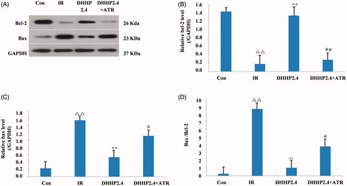
ATR reverses effects of DHHP on Bcl-2 and Bax protein expression
Western blot analysis, as shown in , the relative expression level of Bax protein and Bax/Bcl-2 in the DHHP2.4 group were significantly lower than those in the IR group (p < 0.05); the relative expression level of Bax protein and Bax/Bcl-2 in the DHHP2.4 + ATR group were significantly higher than those in the DHHP2.4 group (p < 0.05). As shown in , the relative expression level of Bcl-2 in the DHHP2.4 group was manifestly higher than those in the IR group (P < 0.05); the relative expression level of Bcl-2 in the DHHP2.4 + ATR group was manifestly lower than that in the DHHP2.4 group (p < 0.05).
Figure 7. Effects of DHHP on the Bcl-2 and Bax protein. (A) Western blot analysis of Bcl-2 and Bax protein expression. (B) The relative expression levels of Bcl-2 in each group. (C) The relative expression levels of Bax in each group. (D) The ratio of Bax and Bcl-2 in each group. Data were expressed as the mean ± SD (n = 3), Δ<0.05 vs. Con, ΔΔp < 0.01 vs. Con, *p < 0.05 vs. IR, **p < 0.01 vs. IR, #p < 0.05 vs. DHHP2.4, ##p < 0.01 vs. DHHP2.4.
ATR reverses effects of DHHP on myocardial mitochondria
As shown in , in the IR group, most mitochondria were swollen, and vacuoles and network structures disappeared. This mitochondrial damage was alleviated in DHHP2.4 group. However, this mitochondrial damage was not alleviated in the DHHP2.4 + ATR group.
Figure 8. The influence of DHHP on MPTP opening. (A) The representative electron images of myocardial mitochondria (5000×). (B) The content of NAD+ in ischaemic myocardium. (C) The sensitivity of MPTP to calcium. (D) The membrane potential of isolated mitochondria. Data were expressed as the mean ± SD (n = 6), Δp < 0.05 vs. Con, *p < 0.05 vs. IR, #p < 0.05 vs. DHHP2.4.
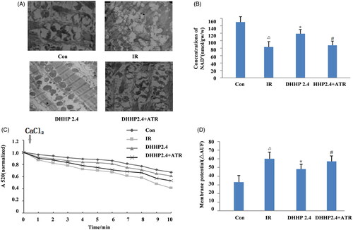
As shown in , ischaemic myocardium in the DHHP2.4 group retained higher NAD+ than that of the IR group (p < 0.05). NAD+ decreased in the DHHP2.4 + ATR group compared to the DHHP2.4 group (p < 0.05).
As shown in , isolated mitochondria in the DHHP2.4 group have higher resistance to Ca2+ stimulation than in the IR group, but the resistance to Ca2+ decreased in the DHHP2.4 + ATR group compared with DHHP2.4 group.
As shown in , the difference in the potential (measured as ΔAUF) was outstandingly higher in the DHHP2.4 group than that of the IR group (p < 0.05); the difference in the potential (measured as ΔAUF) was obviously lower in DHHP2.4 + ATR group than that of the DHHP2.4 group (p < 0.05).
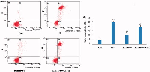
Detection of cell apoptosis with flow cytometry analysis
As shown in , flow cytometry results indicated that H/R significantly increased H9c2 cell apoptosis, compared with the Con group (p < 0.01). H9c2 cell apoptosis in the DHHP 80 group was obviously lower than that of the H/R group (p < 0.01). Compared with the DHHP 80 group, H/R-induced H9c2 cell apoptosis was significantly increased in DHHP80 + ATR group (p < 0.05).
Figure 9. Effects of DHHP on cell apoptosis. (A) Two-dimensional scatter plot of apoptosis. (B) Quantification of apoptosis. Data were expressed as the mean ± SD (n = 10). **p < 0.01 vs. Con; ##p < 0.01 vs. H/R; Δp < 0.05 vs. DHHP 80.
Discussion
The compatibility of traditional Chinese medicine aims to enhance the effects of the herbs, and/or eliminate or reduce the side effects of one of the herbs. The compatibility of danshen-honghua belongs to the former that danshen-honghua herb pairs could enhance the effects of danshen or honghua in CVD (Liu et al. Citation2011; Qu et al. Citation2017; Zhang and Ren Citation2017; Zhang et al. Citation2018; Wang et al. Citation2019). Literature studies show that mitochondria and cardiomyocyte apoptosis exerted an important role in MIRI (Wu et al. Citation2014; Li et al. Citation2019; Sun et al. Citation2019; Wang et al. Citation2019). Therefore, this study revealed the mechanisms of DHHP protecting MIRI based on mitochondria and apoptosis.
XNT are commonly clinically used in Chinese medicine for CVD and are used for the treatment of CVD such as angina pectoris, coronary heart disease and myocardial ischaemia reperfusion, so XNT is selected as positive drugs. By comparison between positive drugs and DHHP, the results showed that XNT and DHHP had similar protective effects on MIRI, suggesting that the positive control group was reasonable.
Under physiological conditions, MPTP remains closed or rarely opens, but the increase of Ca2+ in the mitochondrial matrix is the main factor inducing the opening of MPTP (He et al. Citation2020). Literature studies have shown that MIRI was related to the opening of MPTP (Morciano et al. Citation2017), and the sensitivity of MPTP to Ca2+ and the content of NAD+ in heart tissue are negatively associated with the opening degree of MPTP (Jia et al. Citation2018). However, the Bcl-2 protein family played a key role in the opening of MPTP (Tong et al. Citation2017). The content of NAD+ and the expression of Bcl-2 in DHHP group were higher, and more resistant to stimulation with Ca2+ and lower expression of Bax protein and Bax/Bcl-2, than that of the IR group. The result was consistent with literature studies, which indicate that DHHP inhibits MPTP openness by regulating the Bcl-2 protein family. In this study, we provided evidence that DHHP can inhibit the opening of MPTP in the course of MIRI.
Under physiological conditions, MPTP is in a closed state; under transient, mild ischaemia or chronic ischaemia, MPTP is in a reversible low-level open state; when long-term severe ischaemia and mitochondrial damage reaches a certain level, MPTP was at an irreversible high-level status. MPTP high level open can lead to mitochondrial proton-electrochemical gradient dissipation, ion homeostasis destruction, mitochondrial swelling, ATP hydrolysis, the permeability of mitochondrial membrane increasing which in turn causes the release of cytochrome C from mitochondria, cytochrome C binds to the apoptotic protease activator, and recruits caspase-9 to form apoptotic bodies, caspase-9 is activated and cleaved to cleaved-caspase-9 and then activated and cleaved the downstream apoptotic executive protein caspase-3 to cleaved-caspase-3, and induces apoptosis (Jia et al. Citation2018; Li et al. Citation2018). The study showed that DHHP reduced the expression of cytochrome C, cleaved-caspase-9, cleaved- caspase-3 protein, and decreased cell apoptosis which inhibits MIRI. Indirect evidence that MPTP opening was inhibited by DHHP treatment was provided in this study.
MPTP plays an important role in cell survival and apoptosis, which involves many fields such as ischaemia/reperfusion, tumour, ageing and neurodegeneration. ATR is an opening agent of MPTP. It is used to study the protective effects of DHHP on MIRI and potential mechanism based on apoptosis and mitochondria.
The isolated rat heart model and cardiomyocyte H9C2 used in this study are not regulated by nerves and body fluids, and cannot fully simulate the pathophysiological changes under the condition of MIRI in the clinic. Secondly, the upstream pathways of MPTP, such as GSK-3β, PI3K-Akt and MAPK/ERK1/2 pathways have not been confirmed. Therefore, the cardio-protection research of DHHP for MIRI should be carried out in vivo in the future, and the upstream pathway of MPTP should be studied.
Conclusions
The study found that the DHHP2.4 + ATR group had no significant difference in cardiomyocyte apoptosis and mitochondria of MIRI with IR group. However, the cardiomyocyte apoptosis and mitochondria were significantly different in the DHHP2.4 + ATR group compared with the DHHP2.4 group, suggesting that DHHP could alleviate MIRI by regulating Bax, Bcl-2, Cytochrome C, cleaved caspase-9, cleaved caspase-3 proteins and mitochondria. However, ATR can reverse the cardioprotective effect of DHHP on MIRI. Therefore, we considered that the protective effects of DHHP on MIRI were achieved by inhibiting the opening of MPTP.
Disclosure statement
No potential conflict of interest was reported by the author(s).
Additional information
Funding
References
- Benjamin EJ, Blaha MJ, Chiuve SE, Cushman M, Das SR, Deo R, de Ferranti SD, Floyd J, Fornage M, Gillespie C, et al. 2017. Heart disease and stroke statistics-2017 update: a report from the American Heart Association. Circulation. 135(10):e146–e603.
- Chen J, Wei J, Orgah J, Zhu Y, Ni J, Li L, Zhang H, Gao X, Fan G. 2019. Cardioprotective effect of danhong injection against myocardial infarction in rats is critically contributed by MicroRNAs. Evid Based Complement Alternat Med. 2019:4538985.
- Chen XJ, Ren SM, Dong JZ, Qiu CG, Chen YW, Tao HL. 2019. Ginkgo biloba extract-761 protects myocardium by regulating Akt/Nrf2 signal pathway. Drug Des Devel Ther. 13:647–655.
- Deng X, Xing X, Sun G, Xu X, Wu H, Li G, Sun X. 2017. Guanxin danshen formulation protects against myocardial ischemia reperfusion injury-induced left ventricular remodeling by upregulating estrogen receptor beta. Front Pharmacol. 8:e00777.
- Donato M, Evelson P, Gelpi RJ. 2017. Protecting the heart from ischemia/reperfusion injury: an update on remote ischemic preconditioning and postconditioning. Curr Opin Cardiol. 32(6):784–790.
- Guo J, Yong Y, Aa J, Cao B, Sun R, Yu X, Huang J, Yang N, Yan L, Li X, et al. 2016. Compound danshen dripping pills modulate the perturbed energy metabolism in a rat model of acute myocardial ischemia. Sci Rep. 6:37919.
- He F, Wu Q, Xu B, Wang X, Wu J, Huang L, Cheng J. 2017. Suppression of stim1 reduced intracellular calcium concentration and attenuated hypoxia/reoxygenation induced apoptosis in H9C2 cells. Biosci Rep. 37(6):e20171249.
- He Y, Fu Y, Xi M, Zheng H, Zhang Y, Liu Y, Zhao Y, Xi J, He Y. 2020. Zn2+ and mPTP mediate resveratrol-induced myocardial protection from endoplasmic reticulum stress. Metallomics. 12(2):290–300.
- Huang H, Lai S, Luo Y, Wan Q, Wu Q, Wan L, Qi W, Liu J. 2019. Nutritional preconditioning of apigenin alleviates myocardial ischemia/reperfusion injury via the mitochondrial pathway mediated by Notch1/Hes1. Oxid Med Cell Longev. 2019:e973098.
- Jia D. 2011. The protective effect of mitochondrial ATP-sensitive K + channel opener, nicorandil, combined with Na+/Ca2+ exchange blocker KB-R7943 on myocardial ischemia-reperfusion injury in rat. Cell Biochem Biophys. 60(3):219–224.
- Jia P, Liu C, Wu N, Jia D, Sun Y. 2018. Agomelatine protects against myocardial ischemia reperfusion injury by inhibiting mitochondrial permeability transition pore opening. Am J Transl Res. 10(5):1310–1323.
- Jiang S, Shi Z, Li C, Ma C, Bai X, Wang C. 2014. Hydroxysafflor yellow A attenuates ischemia/reperfusion-induced liver injury by suppressing macrophage activation. Int J Clin Exp Pathol. 7(5):2595–2608.
- Koo YE, Song J, Bae S. 2018. Use of plant and herb derived medicine for therapeutic usage in cardiology. Medicines. 5(2):38.
- Li D, Wang X, Huang Q, Li S, Zhou Y, Li Z. 2018. Cardioprotection of CAPE-oNO2 against myocardial ischemia/reperfusion induced ROS generation via regulating the SIRT1/eNOS/NF-κB pathway in vivo and in vitro. Redox Biol. 15:62–73.
- Li J, Zhao L, Zhao X, Wang P, Liu Y, Ruan J. 2018. Foxo1 attenuates NaF-induced apoptosis of LS8 cells through the JNK and mitochondrial pathways. Biol Trace Elem Res. 181(1):104–111.
- Li LM, Fu JH, Guo H, Han X, Li L, Xin GJ, Zhao YW, Zhang Q, Zheng QS, Liu JX. 2019. Protective effect of safflower yellow injection against rat MIRI by TLR-NF-kappa B inflammatory pathway. China J Chin Mater Med. 44(12):2566–2571.
- Li X, Jia P, Huang Z, Liu S, Miao J, Guo Y, Wu N, Jia D. 2019. Lycopene protects against myocardial ischemia-reperfusion injury by inhibiting mitochondrial permeability transition pore opening. Drug Des Devel Ther. 13:2331–2342.
- Liao Z, Liu D, Tang L, Yin D, Yin S, Lai S, Yao J, He M. 2015. Long-term oral resveratrol intake provides nutritional preconditioning against myocardial ischemia/reperfusion injury: involvement of VDAC1 downregulation. Mol Nutr Food Res. 59(3):454–464.
- Liu J, Zhang D, Li J, Feng J, Yang X, Shi D, Liang X. 2011. Effects of Salvia miltiorrhiza and Carthamus tinctorius aqueous extracts and compatibility on rat myocardial ischemic reperfusion injury. China J Chin Mater Med. 36(2):189–194.
- Liu S, Wu N, Miao J, Huang Z, Li X, Jia P, Guo Y, Jia D. 2018. Protective effect of morin on myocardial ischemia-reperfusion injury in rats. Int J Mol Med. 42(3):1379–1390.
- Miao J, Huang Z, Liu S, Li X, Jia P, Guo Y, Wu N, Jia D. 2019. Hydroxytyrosol protects against myocardial ischemia reperfusion injury by inhibiting mitochondrial permeability transition pore opening. Exp Ther Med. 17(1):671–678.
- Morciano G, Bonora M, Campo G, Aquila G, Rizzo P, Giorgi C, Wieckowski MR, Pinton P. 2017. Mechanistic role of mPTP in ischemia-reperfusion injury. Adv Exp Med Biol. 982:169–189.
- Pell VR, Spiroski AM, Mulvey J, Burger N, Costa A, Logan A, Gruszczyk AV, Rosa T, James AM, Frezza C, et al. 2018. Ischemic preconditioning protects against cardiac ischemia reperfusion injury without affecting succinate accumulation or oxidation. J Mol Cell Cardiol. 123:88–91.
- Qian J, Zhao X, Wang W, Zhang S, Hong Z, Chen X, Zhao Z, Hao C, Wang C, Lu S, et al. 2018. Transcriptomic study reveals recovery of impaired astrocytes contribute to neuroprotective effects of danhong injection against cerebral ischemia/reperfusion-induced injury. Front Pharmacol. 9:e00250.
- Qu C, Tang YP, Shi XQ, Zhou GS, Shang EX, Shang LL, Guo JM, Liu P, Zhao J, Zhao BC, et al. 2017. Comparative study on promoting blood effects of danshen-honghua herb pair with different preparations based on chemometrics and multi-attribute comprehensive index methods. China J Chin Mater Med. 42(15):3017–3025.
- Quan W, Ma S, Zhu Y, Shao Q, Hou J, Li X. 2020. Apigenin-7-O-β-d-(6″-p-coumaroyl)-glucopyranoside reduces myocardial ischaemia/reperfusion injury in an experimental model via regulating the inflammation response. Pharm Biol. 58(1):80–88.
- Shi X, Zhu H, Zhang Y, Zhou M, Tang D, Zhang H. 2017. Xuefuzhuyu decoction protected cardiomyocytes against hypoxia/reoxygenation injury by inhibiting autophagy. BMC Complement Altern Med. 17(1):325–334.
- Sun XH, Wang X, Zhang Y, Hui J. 2019. Exosomes of bone-marrow stromal cells inhibit cardiomyocyte apoptosis under ischemic and hypoxic conditions via miR-486-5p targeting the PTEN/PI3K/AKT signaling pathway. Thromb Res. 177:23–32.
- Tan ME, He CH, Jiang W, Zeng C, Yu N, Huang W, Gao ZG, Xing JG. 2017. Development of solid lipid nanoparticles containing total flavonoid extract from Dracocephalum moldavica L. and their therapeutic effect against myocardial ischemia-reperfusion injury in rats. Int J Nanomedicine. 12:3253–3265.
- Tong Z, Xie Y, He M, Ma W, Zhou Y, Lai S, Meng Y, Liao Z. 2017. VDAC1 deacetylation is involved in the protective effects of resveratrol against mitochondria-mediated apoptosis in cardiomyocytes subjected to anoxia/reoxygenation injury. Biomed Pharmacother. 95:77–83.
- Wang WD, Wang L, Cheng L, Yin XJ, Xu HY, Wang JL, Liang RX, Yang HJ. 2014. Protection effect of Yindan Xinnaotong Capsule and main compositions compatibility on myocardial ischemia/reperfusion injury. China J Chin Mater Med. 39(9):1690–1694.
- Wang XP, Wang PF, Bai JQ, Gao S, Wang YH, Quan LN, Wang F, Wang XT, Wang J, Xie YD. 2019. Investigating the effects and possible mechanisms of danshen- honghua herb pair on acute myocardial ischemia induced by isoproterenol in rats. Biomed Pharmacother. 118:109268.
- Wei Y, Xu M, Ren Y, Lu G, Xu Y, Song Y, Ji H. 2016. The cardioprotection of dihydrotanshinone I against myocardial ischemia-reperfusion injury via inhibition of arachidonic acid ω-hydroxylase. Can J Physiol Pharmacol. 94(12):1267–1275.
- Wu N, Zhang X, Guan Y, Shu W, Jia P, Jia D. 2014. Hypercholesterolemia abrogates the cardioprotection of ischemic postconditioning in isolated rat heart: roles of glycogen synthase kinase-3β and the mitochondrial permeability transition pore. Cell Biochem Biophys. 69(1):123–130.
- Xiao G, Lyu M, Wang Y, He S, Liu X, Ni J, Li L, Fan G, Han J, Gao X, et al. 2019. Ginkgo flavonol glycosides or ginkgolides tend to differentially protect myocardial or cerebral ischemia-reperfusion injury via regulation of TWEAK-Fn14 signaling in heart and brain. Front Pharmacol. 10:735.
- Xiao J, Ke ZP, Shi Y, Zeng Q, Cao Z. 2018. The cardioprotective effect of thymoquinone on ischemia-reperfusion injury in isolated rat heart via regulation of apoptosis and autophagy. J Cell Biochem. 119(9):7212–7217.
- Xie R, Li J, Zhao H. 2018. The underlying mechanisms involved in the protective effects of ischemic postconditioning. Cond Med. 1(2):73–79.
- Xu M, Li X, Song L. 2020. Baicalin regulates macrophages polarization and alleviates myocardial ischaemia/reperfusion injury via inhibiting JAK/STAT pathway. Pharm Biol. 58(1):655–663.
- Yan R, Li W, Yin L, Wang Y, Bo J. 2017. Cardiovascular diseases and risk-factor burden in urban and rural communities in high-, middle-, and low-income regions of China: a large community-based epidemiological study. J Am Heart Assoc. 6(2):e004445.
- Yang HX, Wang P, Wang NN, Li SD, Yang MH. 2021. Tongxinluo ameliorates myocardial ischemia-reperfusion injury mainly via activating parkin-mediated mitophagy and downregulating ubiquitin-proteasome system. Chin J Integr Med. 27(7):542–550.
- Yang P, Li JH, Li AL, Li J, Wang Y, Ren SY, Li XL. 2019. Garlicin post-conditioning suppresses adhesion molecules in a porcine model of myocardial ischemia-reperfusion injury. Chin J Integr Med. 25(1):31–36.
- Yuan X, Jing S, Wu L, Chen L, Fang J. 2014. Pharmacological postconditioning with tanshinone IIA attenuates myocardial ischemia-reperfusion injury in rats by activating the phosphatidylinositol 3-kinase pathway. Exp Ther Med. 8(3):973–977.
- Zhang CY, Ren WG. 2017. Pharmacokinetic research strategies of compatibilities and synergistic effects of classical danshen herb pairs based on pharmacokinetics of “danshen-bingpian” and “danshen-honghua”. China J Chin Mater Med. 42(12):2413–2419.
- Zhang Q, Tan CN, Wang YL, Liu WJ, Yang FQ, Chen H, Xia ZN. 2018. Adsorbed hollow fiber-based biological fingerprinting for the discovery of platelet aggregation inhibitors from danshen-honghua decoction. J Sep Sci. 41(12):2651–2660.
- Zhang X, Du Q, Yang Y, Wang J, Dou S, Liu C, Duan J. 2017. The protective effect of luteolin on myocardial ischemia/reperfusion (I/R) injury through TLR4/NF-κB/NLRP3 inflammasome pathway. Biomed Pharmacother. 91:1042–1052.