
 ?Mathematical formulae have been encoded as MathML and are displayed in this HTML version using MathJax in order to improve their display. Uncheck the box to turn MathJax off. This feature requires Javascript. Click on a formula to zoom.
Abstract
Context
Current medicine for Alzheimer’s disease (AD) cannot effectively reverse or block nerve injury. Traditional Chinese Medicine practice and research imply Aconiti lateralis Radix Praeparata (Fuzi) may meet this goal.
Objective
Analysing the anti-AD effect of Fuzi and its potential molecular mechanism.
Materials and methods
AD model cells were treated with Fuzi in 0-300 mg/mL for 24 h in 37 °C. The cell viability (CV) and length of cell projections (LCP) for each group were observed, analysed, and standardised using control as a baseline (CVs and LCPs). The Fuzi and AD relevant genes were identified basing on databases, and the molecular mechanism of Fuzi anti-AD was predicted by network analysis.
Results
Experiment results showed that Fuzi in 0.4 mg/mL boosted LCP (LCPs = 1.2533, p ≤ 0.05), and in 1.6–100 mg/mL increased CV (CVs from 1.1673 to 1.3321, p ≤ 0.05). Bioinformatics analysis found 17 Fuzi target genes (relevant scores ≥ 20), showing strong AD relevant signals (RMS_p ≤ 0.05, related scores ≥ 5), enriched in the pathways regulating axon growth, synaptic plasticity, cell survival, proliferation, apoptosis, and death (p ≤ 0.05). Especially, GRIN1 and MAPK1 interacted with APP protein and located in the key point of the “Alzheimer’s disease” pathway.
Discussion and conclusions
These results suggest that Fuzi may have therapeutic and prevention potential in AD, and GRIN1 and MAPK1 may be the core of the pathways of the Fuzi anti-AD process. Fuzi should be studied more extensively, especially for the prevention of AD.
Keywords:
Introduction
Alzheimer’s disease (AD), an age-related, neurodegenerative disease, causes progressive memory decline and cognitive dysfunction (Lane et al. Citation2018). The incidence rate of this disease is increasing each year, and it is estimated that more than 80 million patients worldwide will suffer from this disease by 2050 (Mangialasche et al. Citation2010; Prince et al. Citation2013; Van Cauwenberghe et al. Citation2016). However, the treatment of AD still plagues us, due to its complex pathogenesis and limited types of available drugs (Mangialasche et al. Citation2010; Briggs et al. Citation2016; De Strooper and Karran Citation2016). More seriously, these drugs can’t reverse or block the nervous system injury (Briggs et al. Citation2016; Lee and Kim Citation2017), and there is lack of high-quality evidence for their efficacy in mild cognitive impairment (Anderson Citation2019). So, other drugs are needed to reverse or block the nervous system damaged in AD, even in the mild cognitive impairment (MCI) stage.
Although the mechanism is not clear, Traditional Chinese Medicines (TCM) have been applied to AD treatment in practice in China to address both the symptoms and root causes with good effectiveness, one of which is Aconiti lateralis Radix Praeparata (Fuzi). Previous studies have confirmed that Fuzi-based prescriptions combining with existing AD drugs improve cognitive status effectively in AD patients (Yang et al. Citation2018) and inhibit nerve cell apoptosis (Sheng et al. Citation2004; Sun et al. Citation2018). Many ingredients of Fuzi, such as EIC, myristic acid, deltoin, etc., target to muscarinic acetylcholine receptor M1, cholinesterase and acetylcholinesterase (Ru et al. Citation2014), which play important roles in AD relevant processes: learning and memory processes, the metabolism of amyloid-β precursor (APP) protein, and neurotransmission (Ahmed et al. Citation2017; Lazarevic-Pasti et al. Citation2017; Felder et al. Citation2018; Scarpa et al. Citation2020). Moreover, asiatic acid, another component of Fuzi, plays an favorable role in the protection of brain nerves (Sirichoat et al. Citation2015; Chaisawang et al. Citation2017; Park et al. Citation2017; Ahmad Rather et al. Citation2018; Loganathan and Thayumanavan Citation2018). Fuzi protects the mitochondria (Kang et al. Citation2016; Lu et al. Citation2017; Huang et al. Citation2018) whose injury plays an important role in AD development. Fuzi has anti-aging effects by enhancing the antioxidant capacity and inhibiting cell apoptosis (Zhou et al. Citation2015), which is a prominent risk factor for AD (Trevisan et al. Citation2019).
The above information suggests that Fuzi, a multi-target Chinese herb, could be used as an anti-AD medicine. It may treat AD by anti-aging, and protecting mitochondria and nervous system. To study how Fuzi performs in anti-AD, we performed cellular experiments and bioinformatics analysis in this research. The results show that Fuzi is a promising medicine for AD treatment and protection.
Materials and methods
Culture of APP cells
SH-SY5Y cell line, over-expressing 695-amino-acid Swedish mutation amyloid-β precursor protein (APP cell) (Kong et al. Citation2015; Song et al. Citation2015), was kindly provided from Wang’s lab (Department of Laboratory Animal Science, Kunming Medical University, Kunming, China), which had been established and its effectiveness had been tested in 2017. The cells were cultured in DMEM medium (Hyclone, code: DMEIF-12 (AF29527017)) with 10% foetal bovine serum (Gibco, code: 2139378 cp), in 37 °C and 5% CO2 condition. The cells were resuscitated and identified before the experiment. The APP cells passing the check for contamination were used in the follow-up experiment.
CCK-8 method to detect cell viability
Fuzi granules extracts (Guangdong Pharmaceutical Co., LTD.) were dissolved in DMEM medium (Hyclone, code: DMEIF-12 (AF29527017)) with 1% foetal bovine serum (Gibco, code: 2139378 cp) to a final concentration of 400 mg/mL. APP cell was seeded in a 96-well plate with 5000 cells/well. The drug was administrated when cells grew to 70% confluence. The culture contained Fuzi with final concentration of 0, 0.4, 0.8, 1.6, 3.125, 6.25, 12.5, 25, 50, 100, 200, 250, and 300 mg/mL was used to replace the basal culture medium and the cell was cultured for 24 h. CCK-8 solution (10 μL) (Cell Counting Kit-8 of Wanleibo, code: WLP004) was added to each well and incubated for 1–4 h, then. The absorbance at 450 nm was determined by a microplate reader (BIOTEK, code: ELX-800). The cell viability that represents cell proliferation activity was calculated as below:
Adose: Absorbance of cells, CCK solution, and drug solution Ablank: Absorbance of the medium, CCK solution, but no cells A0 dose: Absorbance of cells, CCK solution, but no drug solution.
The cell viability (CV) of the cells treated without Fuzi (NC) was used as a baseline to standardise that of other groups, which were symbolised as CV (Supplementary Material).
Observation of cell morphology
When the APP cells were grown to about 70% confluence, culture containing 0, 0.4, 0.8, 1.6, 3.125, 6.25, 12.5, 25, 50, 100, 200, 250, 300 mg/mL of Fuzi replaced the basal culture medium. The cells were observed under inverted phase contrast microscope (Nikon, code: TE2000-U) after cultured for 24 h in 37 °C, 5% CO2 condition.
Measurement of the length of projections (LCP)
The total number and LCP in a visual field were measured by Viewpoint (BETA v1.0.0.0) software. The average projections length in the visual field was obtained by dividing the total LCP with the total cell number. Three visual fields were measured in each group, which were averaged to obtain the final LCP. LCP of different groups were standardised using NC as a baseline (Supplementary Material), which were symbolised as LCPs. The summary graphs were drawn with ImageJ and Adobe Illustrator.
Statistical analyzes
The data of CVs and LCPs were analysed by SPSS17.0. Multiple group comparisons were performed using one-way analysis of variance (ANOVA). After homogeneity of variances test, Fisher’s least significant difference (LSD) or Dunnett’s T3 test (uneven) were performed to determine significant differences between different groups. Data was expressed as means ± SD, and p ≤ 0.05 (*) and p ≤ 0.01 (**) was considered as statistically and extremely significant. The statistical analysis of enrichment analysis used the Fisher's test of the database.
Bioinformatics analyses
The related ingredients, diseases, and target genes were informed with keyword Fuzi in the Traditional Chinese Medicine systems pharmacology (TCMSP) database (http://tcmspw.com/tcmsp.php) (Ru et al. Citation2014) and summarised with the AD-related information. The ingredients of Fuzi and their target genes were searched with key words of Fuzi and Fupian (synonym for Fuzi) and filtered using score ≥ 10 and adjusted p-value ≤ 0.05 (p-value after Benjamini-Hochberg multiple testing correction) as the cut-off in the BATMAN-TCM database (http://bionet.ncpsb.org/batman-tcm/) (Liu et al. Citation2016). Then the genes with scores higher than five were selected to calculate root mean square (RMS) (Wang et al. Citation2014) of multiple correlation values for the same ingredients and same genes, in which genes with RMS score higher than 20 were defined as target genes of Fuzi. The ‘Alzheimer’s disease’ was searched as the keyword in the GeneCards database (http://www.genecards.org/) (Stelzer et al. Citation2016), and the relevant genes with a score higher than 10 were selected as AD correlated genes.
The UK Biobank and IGAP meta-analysis GWAS summary statistic of AD was download from the Psychiatric Genomics Consortium (http://web.pasteur-lille.fr/en/recherche/u744/igap/igap_download.php) (Lambert et al. Citation2013) and the SNPs with significant p values (p ≤ 0.05) were annotated by Annovar. The GEO data (GSE1297, GSE36980, GSE44772, GSE48350 and GSE5281) (Blalock et al. Citation2004; Liang et al. Citation2007; Berchtold et al. Citation2008; Zhang et al. Citation2013; Hokama et al. Citation2014) of Human AD expression profile was analysed by GEO2R for differential expression (Naoi et al. Citation2004), where different tissues were analysed independently. RMS of p values was calculated after genes were screened with the condition of p ≤ 0.05 in each independent analysis; candidate genes were selected with the condition of RMS_p ≤ 0.05 and the condition of p ≤ 0.05 in five independent analysis. Finally, the overlapping genes of the above analysis were obtained by the Perl script. String (https://string-db.org/) (Szklarczyk et al. Citation2019) and Reactome (http://reactome.org/) (Fabregat et al. Citation2018) were performed for protein interaction and pathway enrichment analysis, respectively. The summary graphs were drawn with R version 3.4.3 and Adobe Illustrator.
All candidate genes were located in the classical AD pathway by tools of the KEGG database (http://www.kegg.jp/) (Ghansah et al. Citation1993). The candidate genes relevant ingredients of Fuzi were figured out from the predicted list download from the BATMAN-TCM database (Liu et al. Citation2016) by Perl script. The Venn diagram was also drawn with R version 3.4.3 and Adobe Illustrator.
Results
The impact of Fuzi on cell changes with its different concentration
APP cell line as a classical cell model of AD (Kong et al. Citation2015; Song et al. Citation2015), were treated with the solution of Fuzi granules at a gradient concentration (0.4–300 mg/mL) for 24 h, then CV and LCP were analysed. The results showed that APP cells treated with a suitable concentration of Fuzi not only increased CV but also promoted LCP. However, when the cells were treated with an over-high concentration solution of Fuzi granules treatment, both the CV and LCP decreased significantly ().
Figure 1. The gene interaction and the change pattern of cells. (A) The modification pattern for the cell viability and the length of cell’s projections, when APP cells were treated by Fuzi solution of different concentration. Statistically (p ≤ 0.05) and extremely (p ≤ 0.01) significant difference were symbolled as “*” and “**”, and p ≤ 0.1 was symbolled as “#”. (B) The gene interaction pattern for the AD relevant Fuzi target genes.
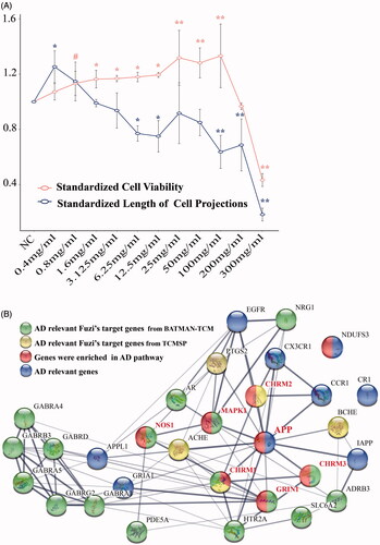
The CV of APP cells increased slowly when Fuzi concentration augmented from 0.4–12.5 mg/mL (CVs from 1.0719 to 1.1942), and from 1.6 mg/mL (CVs = 1.1673) the differences in the CV of APP cells treated with and without Fuzi (NC) reached a significant level (p ≤ 0.05). CV increased sharply in 25 mg/mL (CVs = 1.3182) and reached a peak at 100 mg/mL (CVs = 1.3321). In these concentrations, the CV of APP cells was extremely higher than that in NC (p ≤ 0.01). It plunged, then, when the concentration continued to increase, which decreased significantly comparing to NC when concentration was in more than 200 mg/mL (CVs = 0.9667, p ≤ 0.01, ).
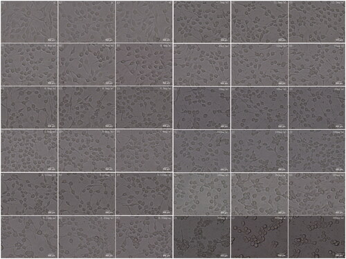
Comparatively, LCP were promoted in 0.4 mg/mL (LCPs = 1.2533) and 0.8 mg/mL (LCPs = 1.1467) Fuzi treated groups (), and the cells were in healthy spindle shape (). Especially in the 0.4 mg/mL group, LCP of APP cells was significantly higher than that of the NC group (p ≤ 0.05, ). Notably, it even show the opposite effect when the concentration was higher than 0.8 mg/mL. Although LCP increased a little at some Fuzi concentration, the processes of APP cells treated with Fuzi tended to be shorter than that of NC starting from 1.6 mg/mL (LCPs = 0.9900, ). The APP cells’ processes became extremely short when the concentration was more than 25 mg/mL (LCPs = 0.9167, p < 0.01, ), and the cell shape also turned from a spindle shape to nearly circle or triangle ().
Figure 2. The microscope images for cell morphology. NC symbol the APP cells were not treated by Fuzi. The other images symbol the APP cells were treated by Fuzi solution of different concentration, from 0.4 to 300 mg/ml.
The molecular mechanism of Fuzi’s anti-AD function is revealed in bioinformatics analysis
To reveal the molecular mechanism of Fuzi’s anti-AD function, the target genes of Fuzi were predicted by the tools of BATMAN-TCM database (Liu et al. Citation2016), and the AD relevant genes were obtained based on the analysis for the data from Psychiatric Genomics Consortium (Lambert et al. Citation2013), GEO (Naoi et al. Citation2004), and GeneCards (Stelzer et al. Citation2016) databases. As showed in materials and methods, we performed the analysis and found 17 target genes of Fuzi (relevant scores ≥ 20). The 17 target genes significantly differentially expressed between AD and normal samples (RMS_p ≤ 0.05), own high related scores (related scores ≥ 5) for AD in the GeneCards database (Stelzer et al. Citation2016) (), and 15 of them contained significant SNP sites in AD GWAS analysis. Moreover, the 17 genes are involved in the amyloid-β precursor protein (APP) interaction network with other AD relevant genes (), which were defined as methods showing. Importantly, five target genes of Fuzi (Cholinergic Receptor Muscarinic 1/3: CHRM1/3, Glutamate Ionotropic Receptor NMDA Type Subunit 1: GRIN1, Mitogen-Activated Protein Kinase 1: MAPK1, Nitric Oxide Synthase 1: NOS1) were enriched in the classical AD pathway of KEGG (, ). They were located in the important position of the pathway (), which are responsible for the regulation of mitochondrial dysfunction, apoptosis, neurofibrillary tangles, cell death, protein oxidation, DNA damage, inflammation and lipid peroxidation ().
Figure 3. The KEGG pathway of AD.
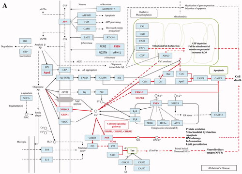
Table 1. The information for the 17 AD relevant target genes of Fuzi.
The functional annotation and enrichment analysis were performed by Reactome (Fabregat et al. Citation2018) for the 17 AD relevant Fuzi’s target genes. Then, the target genes were enriched in the pathway responsible for axon growth (Axon guidance (FDR = 4.70E-02), L1CAM interactions (FDR = 1.54E-02), cell apoptotic (Apoptotic factor-mediated response (FDR = 2.51E-02) and Cytochrome c-mediated apoptotic response (FDR = 2.10E-02)), the bi-directional regulation for cell death and proliferation (MAPK1/MAPK3 signalling (FDR = 8.15E-04)), cell survival (PIP3 activates AKT signalling (FDR = 1.54E-02), signalling by ERBB2 (FDR = 8.50E-03), signalling by NTRKs (FDR = 1.54E-02)) and the function of neuronal system (Neuronal System (FDR = 1.06E-05), transmission across Chemical Synapses (FDR = 1.87E-06), Negative regulation of NMDA receptor-mediated neuronal transmission (FDR = 3.74E-02) and MECP2 regulates neuronal receptors and channels (FDR = 4.42E-02) (). Interestingly, the screened genes MAPK1 and GRIN1 not only participate the pathways responsible for the function of neurons (Axon guidance, Neurotransmitter receptors, and postsynaptic signal transmission, and so on), but also work in the neuron survival relevant pathways (MAPK1/MAPK3 signalling, signalling by ERBB2, and so on). Moreover, the two participate in half of the pathways.
Table 2. The pathway enrichment results of 17 Fuzi’s target genes.
At the same time, the AD relevant target genes of Fuzi were also scanned in the TCMSP database (Ru et al. Citation2014). Five target genes (Prostaglandin G/H synthase 2: PTGS2, Cholinergic Receptor Muscarinic 1/2: CHRM1/2, Cholinesterase: BCHE, and Acetylcholinesterase: ACHE) () were found. These genes are also involved in the protein interaction network of AD relevant genes (). CHRM2 and CHRM1 were also detected by BATMAN-TCM database (Liu et al. Citation2016) analysis and were located in the classical AD pathway of KEGG (). Although PTGS2, BCHE, and ACHE do not participate in the AD pathway, they are responsible for the regulation of memory, learning, synaptic plasticity, acetylcholine catabolic, which play an important role in AD ().
Table 3. The structure and targets for AD relevant Fuzi’s compounds.
Compounds of Fuzi may play a key role in AD treatment
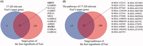
By TCMSP database (Ru et al. Citation2014) scanning, eight ingredients (arachic acid; 14-deoxy-11,12-didehroandrographolide; delphin_qt; deoxyandrographolide; myristic acid; mescaline; salsolinol; EIC; fuzitine) of Fuzi related to AD were figured out (). More importantly, four compounds of Fuzi ingredients targeting on MAPK1 and GRIN1, which have been implicated in the regulation of neuron survival and functional pathway, were selected from the predicted list download from the BATMAN-TCM database (Liu et al. Citation2016). Three of them (M-aminophenol, Ortho-aminophenol, and P-aminophenol) targeting on GRIN1, and one (Coryneine) on MAPK1 (related scores ≥ 20). Amazingly, except MAPK1 and GRIN1, they may also target on the other AD relevant Fuzi target genes: GABRB3, SLC6A2, GABRD, PDE5A, NRG1, GABRG2, GABRA5, ADRB3, GABRA1, and GABRA4 (). Enrichment analysis for all target genes (147) showed that the target genes are also responsible for axon growth, cell apoptosis, cell death, proliferation, and cell survival (Supplementary Material), and 45 pathways are shared by the four compounds and Fuzi ().
Figure 4. The relationship of 17 candidate genes and 4 compounds (m-aminophenol, o-aminophenol, p-aminophenol, and coryneine) related genes. (A) The crossover relationship of the genes. (B) The crossover relationship of enrichment pathways that the genes enriched in.
Discussion
In this study, the AD cell models were treated with Fuzi granules at a gradient concentration (0.4–300 mg/mL) for 24 h. The cellular improvement by Fuzi showed a clear preference in different concentrations. It mainly promoted the growth of cell projections at 0.4–0.8 mg/mL, it increases the cellular proliferation from 1.6 to 100 mg/mL, and it is responsible for the toxic aspect of Fuzi in high concentration (). In further bioinformatics analysis, 17 genes were closely related to AD by combined data from BATMAN-TCM (Liu et al. Citation2016), Psychiatric Genomics Consortium (Lambert et al. Citation2013), GEO (Naoi et al. Citation2004), and GeneCards (Stelzer et al. Citation2016) databases (). Five of them were located in the important place of the classical AD pathway (). In addition, the 17 genes all take part in pathways for the axon growth, the bi-directional regulation for cell death and proliferation, and the function of the neuronal system and five target genes of Fuzi from TCMSP database (Ru et al. Citation2014) were responsible for memory, learning, synaptic plasticity, acetylcholine catabolic regulating. These bioinformatics results suggest that these regulatory genes are vital for the process of Fuzi inhibit AD. Subsequently, the analysis found 8 AD relevant ingredients of Fuzi from the TCMSP database (Ru et al. Citation2014), and four ingredients from the BATMAN-TCM database (Liu et al. Citation2016), which targeted on GRIN1, MAPK1 and other AD relevant genes. It suggests these ingredients may be the basis for Fuzi to antagonise AD.
Fuzi in different concentration may be useful for the treatment of different phases of AD
In our research, Fuzi treatment with 0.4–0.8 mg/mL concentration increased the length of the cell’s projections and maintained cellular morphology. The length of SH-SY5Y cell projections was considered as the morphological marker for the SH-SY5Y cell line differentiated to own neuron function (Påhlman et al. Citation1981). Moreover, the bioinformatics analysis found that the target genes of Fuzi take part in the regulation of axon growth and synaptic plasticity. It implied that Fuzi might help neuron sprouting to form some new synapses to protect the function of nerve cells in the very low concentration. The current research showed that AD may originate from a cognitive decline caused by synaptic terminals loss (Ali et al. Citation2018), which means that the low concentration might be useful for preventing AD in the cognitive decline phase.
For the next stage of AD treatment, low to medium concentration Fuzi may be effective. Previous research pointed out that intracellular Aβ accumulation occurred before extracellular accumulation, damages not only the cell itself but also adjacent cells. With damage expanding and aggravating, large numbers of cells die, forming extracellular Aβ accumulation, which eventually leads to permanent dementia (Naoi et al. Citation2004). Therefore, in these stages, the neuron protection and injury inhibition are most important for AD treatment. The experiment and bioinformatics researches showed that the low to medium concentration Fuzi might target on some genes, which could regulate survival, proliferation, apoptotic, and death of cells, to improve the APP cells’ viability significantly. Particularly, Fuzi in 25 mg/mL significantly increases cell proliferation while ensuring that the cell length and cell morphology are in a good state. It means low to medium concentration Fuzi may play a positive role in these serious stages of AD. It may achieve unexpected results in the combination of existing AD drugs with Fuzi (25 mg/mL) or its active ingredients because the combination simultaneously achieves symptom relief and nerve protection.
Another notable point is that different AD relevant compounds of Fuzi have been found by TCMSP database (Ru et al. Citation2014) and BATMAN-TCM database (Liu et al. Citation2016), which plays a different role in AD regulation. Previous research on motor nerve terminal suggests that mescaline and coryneine could regulate ACh releases (Ghansah et al. Citation1993; Kimura et al. Citation1995; Nojima et al. Citation2000), which play a key role in AD by influencing synaptic loss and signal transform (Mangialasche et al. Citation2010; Ren et al. Citation2020; Wang et al. Citation2020; Zhu et al. Citation2020). Deoxyandrographolide activates PI3K/AKT pathways (Zhao et al. Citation2019), which are responsible for Alzheimer’s treatment by regulating the oxidative stress (Ali et al. Citation2018). While metabolites of salsolinol inducing apoptosis in dopamine neurons have been reported (Naoi et al. Citation2004). Combined the results that Fuzi boosts the cell projections in very low concentration, promotes the proliferation activity of cells in low to medium concentration, and causes injury of APP cells in high concentration (>100 mg/mL), we speculated that in different concentrations the core active ingredients of Fuzi might differ, and they regulate different pathways to anti-AD. Therefore, different Fuzi concentration may suit different treatment phases of AD.
GRIN1 and MAPK1 may be the key target genes for Fuzi anti-AD
The current study revealed that the 17 Fuzi target genes are closely related to AD and maybe the key for Fuzi anti-AD process, especially GRIN1 and MAPK1. The 17 genes are significantly differentially expressed between AD and normal samples (RMS_p ≤ 0.05). They posses high AD-related scores (related scores ≥ 5) in the GeneCards database (Stelzer et al. Citation2016). The 15 genes of them contain significant SNP sites (p ≤ 0.05) in AD GWAS analysis. Five genes of them directly interact with APP protein () and take part in the regulation of axon growth, apoptosis, death and proliferation, and the function of the neuronal system (), which are all playing an important role in AD (Zhao et al. Citation2019). These results provide compelling evidence for the potential function of the 17 Fuzi target genes for anti-AD. Intriguingly, the analysis further showed that GRIN1 and MAPK1 not only directly interact with APP protein in the key point of the AD regulation pathway, but also participate in half of the AD relevant pathways that the 17 genes are involved. In addition, in the m-aminophenol, o-aminophenol, and p-aminophenol target to GRIN1 showed very similar structures with carvacrol, which have therapeutic potential in preventing AD and protective effect on brain injury (Zhong et al. Citation2013; Ali-Shtayeh et al. Citation2020; Azizi et al. Citation2020; Shahrokhi Raeini et al. Citation2020).
For another, GRIN1 and MAPK1 have been closely related to neurodegeneration, synaptic plasticity, cell survival and AD in previous researches (Coyle et al. Citation2016; Preciados et al. Citation2016; Sun and Nan Citation2017; Lu and Malemud Citation2019). GRIN1 protein is a critical subunit of NMDA (Kaniakova et al. Citation2012), which is the target of memantine (Robinson and Keating Citation2006), and plays a key role in memory and learning by regulating the plasticity of synapses (Mori et al. Citation2011; Wang et al. Citation2011). GluN1 receptors and GRIN1 gene expression levels and location are significantly different in AD samples compared to controls (Leuba et al. Citation2014; Mohamed et al. Citation2015; Agca et al. Citation2020). The MAPK1 gene is believed as one age-dependent transcriptional changing gene that involves in the abnormal hyperphosphorylation of the tau-protein, causing aggregated neurofibrillary tangles (Kálmán et al. Citation2009). Moreover, galantamine could treat Alzheimer’s disease by attenuating the activation of MAPK1 (Noda et al. Citation2010). Taken these results together, we hypothesise that the complex regulation network with the core of GRIN1 and MAPK1 may play a key role in the process of Fuzi anti-AD.
Overall, our study suggests that different concentrations of Fuzi can be used in different treatment stages of AD by regulating the complex regulation network with the core of GRIN1 and MAPK1. Besides, the four compounds of Fuzi targeting GRIN1 and MAPK1 may be the key to anti-AD medicine. However, it still requires more neurobiological and animal experiments to verify the improvement of synaptic function by low-concentration of Fuzi and the effectiveness of different concentrations of Fuzi and compounds on the body function. At the same time, the molecular mechanisms of the Fuzi and the four ingredients’ anti-AD still need more molecular, neurobiological, and animal experiments. Furthermore, since Fuzi has long-term use as a food in some areas of Yunnan province, so an epidemiological survey for these areas may help to define the relationship between Fuzi and AD.
Author contributions
Y-T.W and H-X.Z performed experiment and analysis in the research and prepared figures and tables of this paper. J.W, M.Y and Q-Q.Z analysed experiment data and wrote the main manuscript and prepared figures and tables of this paper. S.Y, D-Y.Y, L-L.S, and L-H.Z gave some advice on data analysis and helped to revise the draft of the manuscript writing. L-M.W, H-X.W, and X.C designed the research, analysed the data and revise the draft of the manuscript writing.
Supplementary_Material.docx
Download ()Acknowledgement
We thank Prof. Jia Liu and Prof. Tinghua Wang for kindly providing the APP cells.
Disclosure statement
The authors report no declarations of interest. The authors alone are responsible for the content and writing of the paper.
Data availability statement
The authors confirm that the data supporting the findings of this study are available within the article [and/or] its Supplementary materials.
Additional information
Funding
References
- Agca C, Klakotskaia D, Stopa EG, Schachtman TR, Agca Y. 2020. Ovariectomy influences cognition and markers of Alzheimer’s disease. J Alzheimers Dis. 73(2):529–541.
- Ahmad Rather M, Justin Thenmozhi A, Manivasagam T, Dhivya Bharathi M, Essa MM, Guillemin GJ. 2018. Neuroprotective role of asiatic acid in aluminium chloride induced rat model of Alzheimer’s disease. Front Biosci (Schol Ed). 10:262–275.
- Ahmed T, Zahid S, Mahboob A, Farhat SM. 2017. Cholinergic system and post-translational modifications: an insight on the role in Alzheimer’s disease. Curr Neuropharmacol. 15(4):480–494.
- Ali-Shtayeh MS, Abu-Zaitoun SY, Dudai N, Jamous RM. 2020. Downy Lavender oil: a promising source of antimicrobial, antiobesity, and anti-Alzheimer’s disease agents. Evid Based Complement Alternat Med. 2020:5679408.
- Ali T, Kim T, Rehman SU, Khan MS, Amin FU, Khan M, Ikram M, Kim MO. 2018. Natural dietary supplementation of anthocyanins via PI3K/Akt/Nrf2/HO-1 pathways mitigate oxidative stress, neurodegeneration, and memory impairment in a mouse model of Alzheimer’s disease. Mol Neurobiol. 55(7):6076–6093.
- Anderson ND. 2019. State of the science on mild cognitive impairment (MCI). CNS Spectr. 24(1):78–87.
- Azizi Z, Salimi M, Amanzadeh A, Majelssi N, Naghdi N. 2020. Carvacrol and thymol attenuate cytotoxicity induced by amyloid β25-35 via activating protein kinase C and inhibiting oxidative stress in PC12 cells. Iran Biomed J. 24(4):243–250.
- Berchtold NC, Cribbs DH, Coleman PD, Rogers J, Head E, Kim R, Beach T, Miller C, Troncoso J, Trojanowski JQ, et al. 2008. Gene expression changes in the course of normal brain aging are sexually dimorphic. Proc Natl Acad Sci U S A. 105(40):15605–15610.
- Blalock EM, Geddes JW, Chen KC, Porter NM, Markesbery WR, Landfield PW. 2004. Incipient Alzheimer’s disease: microarray correlation analyses reveal major transcriptional and tumor suppressor responses. Proc Natl Acad Sci U S A. 101(7):2173–2178.
- Briggs R, Kennelly SP, O’Neill D. 2016. Drug treatments in Alzheimer’s disease. Clin Med (Lond). 16(3):247–253.
- Chaisawang P, Sirichoat A, Chaijaroonkhanarak W, Pannangrong W, Sripanidkulchai B, Wigmore P, Welbat JU. 2017. Asiatic acid protects against cognitive deficits and reductions in cell proliferation and survival in the rat hippocampus caused by 5-fluorouracil chemotherapy. PloS One. 12(7):e0180650.
- Coyle JT, Balu DT, Puhl MD, Konopaske GT. 2016. History of the concept of disconnectivity in Schizophrenia. Harv Rev Psychiatry. 24(2):80–86.
- De Strooper B, Karran E. 2016. The cellular phase of Alzheimer’s disease. Cell. 164(4):603–615.
- Fabregat A, Jupe S, Matthews L, Sidiropoulos K, Gillespie M, Garapati P, Haw R, Jassal B, Korninger F, May B, et al. 2018. The Reactome pathway knowledgebase. Nucleic Acids Res. 46(D1):D649–D655.
- Felder CC, Goldsmith PJ, Jackson K, Sanger HE, Evans DA, Mogg AJ, Broad LM. 2018. Current status of muscarinic M1 and M4 receptors as drug targets for neurodegenerative diseases. Neuropharmacology. 136(Pt C):449–458.
- Ghansah E, Kopsombut P, Malleque MA, Brossi A. 1993. Effects of mescaline and some of its analogs on cholinergic neuromuscular transmission. Neuropharmacology. 32(2):169–174.
- Hokama M, Oka S, Leon J, Ninomiya T, Honda H, Sasaki K, Iwaki T, Ohara T, Sasaki T, LaFerla FM, et al. 2014. Altered expression of diabetes-related genes in Alzheimer’s disease brains: the Hisayama study. Cereb Cortex. 24(9):2476–2488.
- Huang QQ, Zhao YL, Gao JF, Jing ZW, Zhang N, Ma t. 2018. [Effect of Dihuang Yinzi on mitochondrial structure and function in central nerve of Alzheimer’s disease rats]. Zhongguo Shi Yan Fang Ji Xue Za Zhi. 24:99–104. Chinese.
- Kálmán S, Pákáski M, Szucs S, Garab D, Domokos A, Zvara A, Puskás L, Bagdy G, Zelena D, Kálmán J. 2009. The transcription of the amyloid precursor protein and tryptophan 2,3-dioxygenase genes are increased by aging in the rat brain. Ideggyogy Sz. 62(9–10):326–332. Hungarian.
- Kang X, Liang ZK, Lu XG, Feng Q, Song JB, Wang Y, Liu J, Fan ZW, Song Y, Wang P. 2016. [Effect of Dahuang Fuzi Tang on mitochondrial structure and function of intestinal epithelial cells in rats with severe actue pancreatitis]. Zhong Hua Wei Sheng Ying Ji Dian Zi Za Zhi. 2:309–314. Chinese.
- Kaniakova M, Lichnerova K, Vyklicky L, Horak M. 2012. Single amino acid residue in the M4 domain of GluN1 subunit regulates the surface delivery of NMDA receptors. J Neurochem. 123(3):385–395.
- Kimura M, Kimura I, Muroi M, Nojima H, Diwan PV. 1995. Depolarizing neuromuscular blocking action of coryneine derived from aconite root in isolated mouse phrenic nerve-diaphragm muscles. Biol Pharm Bull. 18(5):691–695.
- Kong JJ, Zhang DD, Li P, Wei CY, Yu HJ, Zhang H, Zhang W, Wang YF, Cao YP. 2015. Nicorandil inhibits oxidative stress and amyloid-beta precursor protein processing in SH-SY5Y cells overexpressing APPsw. Int J Clin Exp Med. 8(2):1966–1975.
- Lambert J-C, Ibrahim-Verbaas CA, Harold D, Naj AC, Sims R, Bellenguez C, Jun G, DeStefano AL, Bis JC, Beecham GW, et al. 2013. Meta-analysis of 74,046 individuals identifies 11 new susceptibility loci for Alzheimer’s disease. Nat Genet. 45(12):1452–1458.
- Lane CA, Hardy J, Schott JM. 2018. Alzheimer’s disease. Eur J Neurol. 25(1):59–70.
- Lazarevic-Pasti T, Leskovac A, Momic T, Petrovic S, Vasic V. 2017. Modulators of acetylcholinesterase activity: from Alzheimer’s disease to anti-cancer drugs. Curr Med Chem. 24(30):3283–3309.
- Lee JK, Kim N-J. 2017. Recent advances in the inhibition of p38 MAPK as a potential strategy for the treatment of Alzheimer’s disease. Molecules. 22(8):1287–1309.
- Leuba G, Vernay A, Kraftsik R, Tardif E, Riederer BM, Savioz A. 2014. Pathological reorganization of NMDA receptors subunits and postsynaptic protein PSD-95 distribution in Alzheimer’s disease. Curr Alzheimer Res. 11(1):86–96.
- Liang WS, Dunckley T, Beach TG, Grover A, Mastroeni D, Walker DG, Caselli RJ, Kukull WA, McKeel D, Morris JC, et al. 2007. Gene expression profiles in anatomically and functionally distinct regions of the normal aged human brain. Physiol Genomics. 28(3):311–322.
- Liu Z, Guo F, Wang Y, Li C, Zhang X, Li H, Diao L, Gu J, Wang W, Li D, et al. 2016. BATMAN-TCM: a bioinformatics analysis tool for molecular mechanism of Traditional Chinese Medicine. Sci Rep. 6:21146.
- Loganathan C, Thayumanavan P. 2018. Asiatic acid prevents the quinolinic acid-induced oxidative stress and cognitive impairment. Metab Brain Dis. 33(1):151–159.
- Lu N, Malemud CJ. 2019. Extracellular signal-regulated kinase: a regulator of cell growth, inflammation, chondrocyte and bone cell receptor-mediated gene expression. IJMS. 20(15):3792.
- Lu X, Zhang L, Li P, Wang J, Li R, Huang Y, Wu M, Zhou H, Li Y, Wei S, et al. 2017. The protective effects of compatibility of Aconiti lateralis Radix Praeparata and zingiberis rhizoma on rats with heart failure by enhancing mitochondrial biogenesis via Sirt1/PGC-1α pathway. Biomed Pharmacother. 92:651–660.
- Mangialasche F, Solomon A, Winblad B, Mecocci P, Kivipelto M. 2010. Alzheimer’s disease: clinical trials and drug development. Lancet Neurol. 9(7):702–716.
- Mohamed NE, Lee JH, Francis PT, Esiri MM, Chen CP, Lai MK. 2015. Differential alterations of neocortical GluN receptor subunits in patients with mixed subcortical ischemic vascular dementia and Alzheimer’s disease. J Alzheimers Dis. 44(2):431–437.
- Mori F, Ribolsi M, Kusayanagi H, Siracusano A, Mantovani V, Marasco E, Bernardi G, Centonze D. 2011. Genetic variants of the NMDA receptor influence cortical excitability and plasticity in humans. J Neurophysiol. 106(4):1637–1643.
- Naoi M, Maruyama W, Nagy GM. 2004. Dopamine-derived salsolinol derivatives as endogenous monoamine oxidase inhibitors: occurrence, metabolism and function in human brains. Neurotoxicology. 25(1–2):193–204.
- Noda Y, Mouri A, Ando Y, Waki Y, Yamada SN, Yoshimi A, Yamada K, Ozaki N, Wang D, Nabeshima T. 2010. Galantamine ameliorates the impairment of recognition memory in mice repeatedly treated with methamphetamine: involvement of allosteric potentiation of nicotinic acetylcholine receptors and dopaminergic-ERK1/2 systems. Int J Neuropsychopharmacol. 13(10):1343–1354.
- Nojima H, Okazaki M, Kimura I. 2000. Counter effects of higenamine and coryneine, components of aconite root, on acetylcholine release from motor nerve terminal in mice. J Asian Nat Prod Res. 2(3):195–203.
- Påhlman S, Odelstad L, Larsson E, Grotte G, Nilsson K. 1981. Phenotypic changes of human neuroblastoma cells in culture induced by 12-O-tetradecanoyl-phorbol-13-acetate. Int J Cancer. 28(5):583–589.
- Park JH, Seo YH, Jang JH, Jeong CH, Lee S, Park B. 2017. Asiatic acid attenuates methamphetamine-induced neuroinflammation and neurotoxicity through blocking of NF-κB/STAT3/ERK and mitochondria-mediated apoptosis pathway. J Neuroinflammation. 14(1):240–254.
- Preciados M, Yoo C, Roy D. 2016. Estrogenic endocrine disrupting chemicals influencing NRF1 regulated gene networks in the development of complex human brain diseases. IJMS. 17(12):2086.
- Prince M, Bryce R, Albanese E, Wimo A, Ribeiro W, Ferri CP. 2013. The global prevalence of dementia: a systematic review and metaanalysis. Alzheimers Dement. 9(1):63–75.
- Ren JM, Zhang SL, Wang XL, Guan ZZ, Qi XL. 2020. Expression levels of the α7 nicotinic acetylcholine receptor in the brains of patients with Alzheimer’s disease and their effect on synaptic proteins in SH-SY5Y cells. Mol Med Rep. 22(3):2063–2075.
- Robinson DM, Keating GM. 2006. Memantine: a review of its use in Alzheimer’s disease. Drugs. 66(11):1515–1534.
- Ru J, Li P, Wang J, Zhou W, Li B, Huang C, Li P, Guo Z, Tao W, Yang Y, et, al. 2014. TCMSP: a database of systems pharmacology for drug discovery from herbal medicines. J Cheminform. 6:13–18.
- Scarpa M, Hesse S, Bradley SJ. 2020. M1 muscarinic acetylcholine receptors: a therapeutic strategy for symptomatic and disease-modifying effects in Alzheimer’s disease? Adv Pharmacol. 88:277–310.
- Shahrokhi Raeini A, Hafizibarjin Z, Rezvani ME, Safari F, Afkhami Aghda F, Zare Mehrjerdi F. 2020. Carvacrol suppresses learning and memory dysfunction and hippocampal damages caused by chronic cerebral hypoperfusion. Naunyn Schmiedebergs Arch Pharmacol. 393(4):581–589.
- Sheng YL, Jiang XD, Li HM, Pu JH, Wei XD, Ou Q, Zhang PX. 2004. [Experimental study to detect the effect of Baizhu Fuzi Rougui Heji on brain cell apoptosis in elderly mice]. Zhongguo Lao Nian Xue Za Zhi. 11:1055–1056. Chinese.
- Sirichoat A, Chaijaroonkhanarak W, Prachaney P, Pannangrong W, Leksomboon R, Chaichun A, Wigmore P, Welbat JU. 2015. Effects of asiatic acid on spatial working memory and cell proliferation in the adult rat hippocampus. Nutrients. 7(10):8413–8423.
- Song G, Li Y, Lin L, Cao Y. 2015. Anti-autophagic and anti-apoptotic effects of memantine in a SH-SY5Y cell model of Alzheimer’s disease via mammalian target of rapamycin-dependent and -independent pathways. Mol Med Rep. 12(5):7615–7622.
- Stelzer G, Rosen N, Plaschkes I, Zimmerman S, Twik M, Fishilevich S, Stein TI, Nudel R, Lieder I, Mazor Y, et al. 2016. The GeneCards Suite: from gene data mining to disease genome sequence analyses. Cur Protoc Bioinformatics. 54:1301–13033.
- Sun DY, Shan DH, Liu WJ, Wang SD. 2018. [Effects of Fuzi Lizhong pills on CREB, Sirt1, UCP2 expression, and ATP synthesis in small intestinal epithelial cells of rats with spleen Yang deficiency]. Shandong Yi Yao. 58:26–29. Chinese.
- Sun J, Nan G. 2017. The extracellular signal-regulated kinase 1/2 pathway in neurological diseases: a potential therapeutic target. Int J Mol Med. 39(6):1338–1346.
- Szklarczyk D, Gable AL, Lyon D, Junge A, Wyder S, Huerta-Cepas J, Simonovic M, Doncheva NT, Morris JH, Bork P, et al. 2019. STRING v11: protein-protein association networks with increased coverage, supporting functional discovery in genome-wide experimental datasets. Nucleic Acids Res. 47(D1):D607–D613.
- Trevisan K, Cristina-Pereira R, Silva-Amaral D, Aversi-Ferreira TA. 2019. Theories of aging and the prevalence of Alzheimer’s disease. Biomed Res Int. 2019:9171424.
- Van Cauwenberghe C, Van Broeckhoven C, Sleegers K. 2016. The genetic landscape of Alzheimer disease: clinical implications and perspectives. Genet Med. 18(5):421–430.
- Wang GD, Fan RX, Zhai W, Liu F, Wang L, Zhong L, Wu H, Yang HC, Wu SF, Zhu CL, et al. 2014. Genetic convergence in the adaptation of dogs and humans to the high-altitude environment of the Tibetan plateau. Genome Biol Evol. 6(8):2122–2128.
- Wang LP, Li F, Wang D, Xie K, Wang D, Shen X, Tsien JZ. 2011. NMDA receptors in dopaminergic neurons are crucial for habit learning. Neuron. 72(6):1055–1066.
- Wang X, Zhang D, Song W, Cai CF, Zhou Z, Fu Q, Yan X, Cao Y, Fang M. 2020. Neuroprotective effects of the aerial parts of Polygala tenuifolia Willd extract on scopolamine-induced learning and memory impairments in mice. Biomed Rep. 13(5):37.
- Yang X, Du SH, Zhang L, Yang JM, Luo QW. 2018. [Clinical study on 30 cases of Alzheimer’s disease treated by Ma Huang Fuzi Xixin decoction with donepezil]. Jiangsu Zhong Yi Yao. 50:33–35. Chinese.
- Zhang B, Gaiteri C, Bodea LG, Wang Z, McElwee J, Podtelezhnikov AA, Zhang C, Xie T, Tran L, Dobrin R, et al. 2013. Integrated systems approach identifies genetic nodes and networks in late-onset Alzheimer’s disease. Cell. 153(3):707–720.
- Zhao F, Siu JJ, Huang W, Askwith C, Cao L. 2019. Insulin modulates excitatory synaptic transmission and synaptic plasticity in the mouse hippocampus. Neuroscience. 411:237–254.
- Zhong Z, Wang B, Dai M, Sun Y, Sun Q, Yang G, Bian L. 2013. Carvacrol alleviates cerebral edema by modulating AQP4 expression after intracerebral hemorrhage in mice. Neurosci Lett. 555:24–29.
- Zhou G, Tang L, Zhou X, Wang T, Kou Z, Wang Z. 2015. A review on phytochemistry and pharmacological activities of the processed lateral root of Aconitum carmichaelii Debeaux. J Ethnopharmacol. 160:173–193.
- Zhu Z, Zhang L, Cui Y, Li M, Ren R, Li G, Sun X, Li Q. 2020. Functional compensation and mechanism of choline acetyltransferase in the treatment of cognitive deficits in aged dementia mice. Neuroscience. 442:41–53.