
Abstract
Context
Achyranthes bidentata Blume (Amaranthaceae) (ABR) and semen vaccariae (SV) are used commonly in the clinical treatment of erectile dysfunction in males with diabetes mellitus (DMED) to strengthen the kidney and promote blood circulation, and often achieve good curative effects.
Objective
Explore mechanistic details of ABR + SV treatment against DMED.
Materials and methods
Prediction of key targets by network pharmacology. A rat model of DM was established by streptozotocin injection (55 mg/kg). Apomorphine (100 μg/kg) was injected into rats to screen the DMED model. Group C (n = 6) and group M (n = 6) were gavaged with deionized water; group T (n = 6) was given Achyranthis bidentatae radix–semen vaccariae granule suspension (2.5 g/kg). It lasted 8 weeks. Real-time reverse transcription-quantitative polymerase chain reaction (RT-qPCR) and western blotting (WB) were used to measure the expression of tissue-related proteins and mRNA.
Results
The predicted key targets are albumin (ALB), caspase-3 (CASP3), vascular endothelial growth factor A (VEGFA), angiotensin-converting enzyme (ACE), and endothelial nitric oxide synthase (eNOS). Compared with the M group (0.52 ± 0.04; 0.50 ± 0.03; 0.49 ± 0.02; 0.23 ± 0.03), CASP3, VEGFA, and ACE protein expression reduced in the T group (0.39 ± 0.06; 0.34 ± 0.03; 0.39 ± 0.03), and eNOS protein expression increased (0.34 ± 0.03).
Conclusion
ABR + SV can improve erectile function in DMED rats. This study provides a potential mechanism for the treatment of DMED with ABR + SV and can benefit from more patients.
Introduction
Erectile dysfunction (ED) refers to the situation whereby the penis cannot continuously acquire or maintain sufficient erection to complete satisfactory sexual intercourse, and this state lasts for ≥3 months (Wang et al. Citation2013). ED is one of the main complications of diabetes mellitus (DM) (Shamloul and Ghanem Citation2013). The incidence of DM is increasing year by year worldwide and is closely related to age, vascular diseases, and metabolic syndrome (Banks et al. Citation2013). According to hyperglycaemia severity, the ED prevalence in DM patients can reach 71–75% (Lu et al. Citation2009), and the prevalence reaches a peak within 10 years after the diagnosis of DM (Penson and Wessells Citation2004). ED is common in men with DM, and its pathogenesis involves the vascular endothelium, nerve lesions, and muscle lesions (Thorve et al. Citation2011).
The most common treatments for erectile dysfunction in males with diabetes mellitus (DMED) are phosphodiesterase (PDE)-5 inhibitors, prosthesis implantation, and negative-pressure devices. However, these treatments have side effects and limitations. For instance, use of PDE-5 inhibitors can lead to facial redness, headaches, or gastrointestinal reactions (Vita et al. Citation2019; Yu et al. Citation2019). The indications for surgical treatment are limited and its efficacy is controversial (Yafi et al. Citation2016).
In recent years, traditional Chinese medicine (TCM) formulations have shown good effects against DMED (Qiu et al. Citation2021). Combined use of PDE-5 inhibitors with TCM formulations can promote the blood circulation and collateral blood vessels, and the clinical efficacy obtained is often better than that seen with use PDE-5 inhibitors alone (Li Citation2019; Luo et al. Citation2019). Modern research shows that the advantages of traditional Chinese medicine in the treatment of ED are reflected in the aspects of multisystem, multilevel, and multitarget. For example, shugan yiyang capsule can slow down the blood flow of penile veins, enhance sexual desire and ejaculation function, and improve the antihypoxia ability of penile endothelial cells (Yang et al. Citation2014). Compound Xuanju capsule can promote the secretion of luteinizing hormone (LH), and increase the synthesis of testosterone by acting on the Leydig cell (Wang et al. Citation2018).
A pathological mechanism of DMED has been postulated. This mechanism states that DM increases vascular permeability in the penis, damages the vascular endothelium and induces intracellular oxidative-stress reactions, which leads to production of oxygen free radicals and formation of a thrombosis, which affects blood flow in penile blood vessels (Hu et al. Citation2020). TCM theory suggests that DMED involves kidney deficiency and is marked by blood stasis (Li et al. Citation2020).
We have shown that use of leech-centipede medicines for activating blood circulation can improve ED by inhibiting activation of the protein kinase-C pathway in the treatment of DMED rats (Wang et al. Citation2021). Achyranthis bidentatae radix (ABR; Pinyin name Niu Xi; English name Achyranthis radix; Scientific name Achyranthes bidentata Blume [Amaranthaceae]) and semen vaccariae (SV; Pinyin name Wang Bu Liu Xing; English name cowherb seed; Scientific name Gypsophila vaccaria (L.) Sm [Caryophyllaceae]) are also used commonly in the clinical treatment of DMED to strengthen the kidney and promote blood circulation, and often achieve good curative effects (Li 2019; Zeng Citation2018). Hu (Citation2000) treated 56 patients with impotence using Vaccaria hispanica seeds, injection of Western medicine (prostaglandin E1), and an intravenous drip of Danshen: a curative effect was seen in 98% of patients. ABR can promote blood circulation and blood stasis, and strengthen the liver and kidney. SV can also promote blood circulation (Chen et al. Citation2016). ABR and SV in combination (ABR + SV) have a good clinical effect upon complex ED (Ma et al. Citation2017), but its mechanism of action is unclear, and relevant experimental studies have not been reported.
We employed network pharmacology to predict the key active components and targets of ABR + SV in DMED. Then, these targets and mechanisms were verified through animal experiments. In this way, we were able to predict the mechanism of action of ABR + SV in DMED treatment. But the gap of knowledge and hypothesis is that the experimental hypothesis is based on the theory of network pharmacology, which is still being supplemented and perfected and has certain timeliness.
Materials and methods
Biological networks
Full-spectrum analyses of metabolites
We identified the small- and medium-sized compounds in samples by high-performance liquid chromatography-tandem mass spectrometry (HPLC-MS) and then analysed them qualitatively and quantitatively (Chen et al. Citation2013). First, the samples were pre-treated (drying and grinding) and then weighed or diluted at constant volume. Particles were removed by filtration and centrifugation. Then, samples were tested on a LC-MS system (UltiMate™ 3000 LC, Q Exactive HF; Thermo Fisher, Waltham, MA, USA). A C18 column (Zorbax™ Eclipse; 1.8 µm × 2.1 × 100 mm; Sigma-Aldrich, St. Louis, MO, USA) was used for gas chromatography-MS. The conditions for chromatographic separation were set carefully: column temperature was 30 °C; flow rate was 0.3 mL/min; composition of the mobile phase was A (water + 0.1% formic acid) and B (pure acetonitrile); injection volume was 2 μL; automatic injector temperature was 4 °C. Compound Discoverer™ 3.1 (Thermo Fisher) was used for correction of retention times, peak identification, and peak extraction. According to secondary MS information, substances were identified by two online databases, mzCloud (www.mzCloud.org/) and mzVault (https://assets.thermofisher.com/), both of which are run by Thermo Fisher. Finally, we used Compound Discoverer 3.0 to extract some of the original data information (positive- and negative-ion modes) (Garcia and Barbas Citation2011).
Target collection
Through PubChem (https://pubchem.ncbi.nlm.nih.gov/) and Swiss Target Prediction (https://pubchem.ncbi.nlm.nih.gov/), the target of compounds from full-spectrum analyses of metabolites was obtained, and the inclusion criterion was probability >0 (Ma et al. Citation2020). We used the Human Gene Database (GeneCards; www.Genecards.org/) to obtain the target genes for DM and ED (Hao and Xiao Citation2014). The compound targets were intersected with DM and ED targets. The intersection targets were considered to be the potential target of ABR + SV in DMED treatment, which could be used for further construction and analyses of networks.
Protein–protein interactions (PPIs)
We used the Search Tool for the Retrieval of Interacting Genes/Proteins (STRING) database (https://STRING-db.org/) to identify the possible PPIs among the chosen targets. To improve data reliability, we used PPIs with a minimum interaction score ≥0.40 and filtered out the remaining PPIs (Tao et al. Citation2013).
Construction and analyses of networks
Cytoscape v3.7.1 (https://cytoscape.org/) was applied to build a herbal compound–target-disease network and PPI network. Afterwards, the PPI network was analysed further using “cytoHubba” (a Cytoscape plugin) to determine its indispensable targets.
Enrichment analyses using gene ontology (GO) and Kyoto encyclopedia of genes and genomes (KEGG) databases
The Database for Annotation, Visualization, and Integrated Discovery (DAVID) v6.8 (https://david.ncifcrf.gov/) provides a systematic and comprehensive biological and functional annotation of genes and proteins. We imported the related targets of ABR + SV treatment for DMED into the DAVID database by selecting the following parameters: “identifier” as the official gene symbol, “list type” as the gene list, and Homo sapiens as species. The results of pathway analyses using GO and KEGG databases were documented (Zhao and He Citation2018).
Experimental study
Drugs and reagents
The treatment drugs were ABR (catalog number, 19043162) and SV (19037911). The dosage form was granules. They were purchased from Beijing Kangrentang Pharmaceuticals (Beijing, China). The dose of ABR was 15 g and the dose of SV was 10 g, both of which were the adult daily dose. Granules of ABR (15 g) were mixed with those of SV (10 g) and ground into a fine powder. Then, 25 g was dissolved in 100 mL of deionized water and stored at 4 °C. The suspension was mixed thoroughly before administration to rats. According to the formula used to calculate the surface area of the human body and animals, the dose given to rats per kilogram of body weight was six times that of adults (Long et al. Citation2019).
Several reagents were purchased for our study. Streptozotocin (STZ; catalog number, s0130) and apomorphine (APO; a4393) were purchased from Sigma-Aldrich. A protein-extraction kit (GPP1815) and sodium dodecyl sulphate-polyacrylamide gel electrophoresis (SDS-PAGE) loading buffer (5×) (GPP1820) were obtained from Genepool (Beijing, China). Pentobarbital sodium solution was purchased from Guoyao Chemical Reagents (0037517; Shanghai, China). All primers were from Invitrogen (Carlsbad, CA, USA). We purchased a series of antibodies from BIOSS (Woburn, MA, USA) and Abcam (Cambridge, UK), including those for nitric oxide synthase (eNOS) (1:500 dilution; ab76198), vascular endothelial growth factor A (VEGFA; (1:1000; bs-20393r), albumin (1:500; bs-0945r), caspase-3 (1:500; 9662), angiotensin-converting enzyme (ACE; 1:500; bs-1004r) and actin (1:3000; ab6276). Fresco cryocentrifuge 17 (MA, USA) we use are produced by Thermo. Blood glucose metre and test paper are from Roche, Germany.
Laboratory animals
Forty male-specific pathogen-free Sprague-Dawley rats (12 weeks; mean body weight, 200 ± 20 g) were purchased from Beijing Huafukang Biotechnology (Animal Licence number: SCXK (Jing) 2014-0004). Rats were raised and tested in a clean animal laboratory of the Experimental Animal Centre of Dongzhimen Hospital within Beijing University of Traditional Chinese Medicine. They were allowed to consume deionized water and solid rat chow ad libitum. They were exposed to light for 10–12 h each day, indoor humidity was 55–80%, and the temperature was 18–25 °C. Before experimentation, the rats were shown to have a normal sexual function by mating with females (Itoga et al. Citation2020). The study protocol was approved (authorization number: 17–27) by the animal ethics committee of Beijing University of Traditional Chinese Medicine (Beijing, China).
Construction of a DM model
Rats fasted for 12 h before modelling. Six rats were selected randomly as the blank-control group. The other 34 rats were injected (i.p.) with STZ solution (55 mg/kg body weight) in the right lower abdomen. After administration, blood was collected from the caudal vein of pre-modelling rats 1 week later. The glucose concentration in blood was measured by a glucose metre (Roche, Basel, Switzerland). A DM model was considered to have been created if blood glucose > 16.7 mmol/L after 1 week, and urine volume increased (Yafi et al. Citation2016). Six rats died 72 h after modelling. One-week later, the blood glucose of five rats was <16.7 mmol/L. The other 23 DM rats showed significant weight loss, yellow hair, and increased urine volume.
Screening of a DM-induced ED model
Twenty three rats from the DM group and six rats from the blank-control group were placed in a dark observation box in a quiet room for 10 min. Then, APO was injected (100 μg/kg, s.c.) into the neck of rats. Penile erection of rats was monitored by the video camera (SONY, HDR-XR150E, Shanghai, China) within 30 min. This is the first assessment of erection. “Erection” is defined as protrusion, swelling, or lengthening of the penis (Park et al. Citation2018). DMED rats were considered to be DM rats without ED. Nineteen rats were identified as having DMED. After the establishment of animal models, 12 DMED rats were divided randomly into two groups of six: model (group M, n = 6) and treatment (group T, n = 6). Six rats without modelling were termed the normal (control) group (group C, n = 6). Rats in group C and group M underwent gavage with deionized water; group T was given a suspension of ABR + SV granules (2.5 g/kg; six-times human bodyweight) prepared in deionized water. All rats were fed routinely, and the corresponding drugs (1 mL/100 g body weight) were given at 10 am each day. The experiment lasted for 8 weeks. During experimentation, the body was weighed once a week and the dose adjusted. At the end of the study, all rats fasted for 10 h, then the tail vein blood glucose levels, body weights, and erectile function of all rats were measured. This is the second assessment of erection. After that, all rats were anaesthetized with 1% pentobarbital sodium (30 mg/kg body weight, i.p.), then the blood sample was collected from the abdominal aorta and centrifuged at 3000 rpm for 15 min to acquire the sera. And the rats were killed by abdominal aorta bloodletting, then the penile tissues were harvested stored at −80 °C until further analysis.
Histology
The central part of the newly dissected penis was fixed in 4% paraformaldehyde. Then, the samples were dehydrated and embedded in paraffin wax using a gradient of ethanol solutions. According to the standard protocol for histology examination, sections of thickness 4 µm were prepared for haematoxylin and eosin (H&E) staining. A light microscope equipped with a digital tube (Olympus BX51TF, Tokyo, Japan) was used to take photographs and analyse the morphology of the corpus cavernosum (CC). Simultaneously, CC tissue of length 1 mm was fixed in 1% osmium acid, and then dehydrated with a tissue processor (EM TP; Leica, Wetzlar, Germany) and placed in an embedding capsule. Using Lycra ultrathin slicer to slice the tissue: capsule cleaning: remove the capsule, add warm water, and stir on the mixer until the tissue is detached. Semithin section: first, repair the block, and remove part of the embedding agent around the tissue to expose the tissue. Then the tissue is clamped on the slicer with the trapezoid bottom at the bottom so that the knife is parallel to the plane of the tissue. After rough cutting, add warm water into the water tank, press the automatic slicing button, cut 2–3 pieces of 500 nm thickness, stick them on the glass, dry them, and then dye them with toluidine blue. After slicing, the penile cavernous tissue was stained with saturated uranium acetate and lead citrate. The ultrastructure and endothelial cells of penile cavernous tissue were observed under an electron microscope (HitachiH-7500, Tokyo, Japan).
Western blotting
We wished to measure protein expression of eNOS, VEGFA, ALB, CASP3, and ACE. The samples were taken from the middle and upper parts of the CC of each rat. According to the instructions of the protein-extraction kit, the protein concentration was adjusted with water and SDS-PAGE loading buffer (5×) and boiled for 10 min at 100 °C. Then, proteins were separated by SDS-PAGE using 10% alkyl sulphate polyacrylamide gels and transferred to polyvinylidene fluoride (PVDF) membranes. 50 μg protein was taken from each sample. We diluted the primary antibody, incubated it overnight at 4 °C, and washed it thrice with Tris buffer and Tween 20 buffer (TBST) in a decolourizing oscillator for 5 min each time at room temperature. Then, the secondary antibody was diluted (1:3000) with TBST buffer. After incubation for 30 min at room temperature, the PVDF membrane was washed three-times with TBST buffer on a decolourizing oscillator for 5 min each time at room temperature. Two types of electrochemiluminescence reagents (A and B) were mixed in a centrifugal tube in medium volume and full contact with the PVDF membrane. The Odyssey Dual-Colour Infra-red Laser Imaging System and Alpha software were used to scan and semi-quantitatively analyse the protein bands, respectively. β-Actin was used as an internal control to calculate the relative expression of CASP3, VEGFA, eNOS, ACE, and ALB protein.
Real-time reverse transcription-quantitative polymerase chain reaction (RT-qPCR)
A PCR instrument (slan-96p; Shanghai Hongshi Medical Technology, Shanghai, China) was used for RT-qPCR. Tissue (100 mg) was fully ground using a tissue homogenizer. The homogenate was then centrifuged, and 250 μL of trichloromethane was added to the supernatant. The sample was allowed to stand for 3 min and then was centrifuged again. The supernatant was collected and incubated at −20 °C for 15 min, followed by centrifugation at 4 °C for 10 min. The white precipitate at the bottom of the tube is deemed RNA. Next, 20 μL of water without RNA enzyme was added to the precipitate to dissolve the RNA. Reverse transcription was carried out according to the instructions of the cDNA reverse transcription kit. The PCR amplification conditions were as follows: predenaturation at 95 °C for 2 min, denaturation at 95 V for 15 s, extension at 55 °C–68 °C for 30 s, 35–45 cycles, and annealing at 45 °C for 20 s. The primer sequences are shown in .
Table 1. The primer sequences for RT-qPCR.
PCR was undertaken after the addition of the sample to a 96-well plate. The cycle number (CT value) of the fluorescence signal in each reaction tube was recorded when the set threshold was reached. The difference in gene expression was determined by the 2−△△Ct method (Hong et al. Citation2004).
Statistical analyses
All the data were expressed as the means ± standard deviation. Independent sample t-tests were employed for comparison between the two groups. All the data were analysed using SPSS 22.0 statistical software (SPSS Inc., Chicago, IL, USA). p < 0.05 was considered statistically significant.
Results
Biological networks
Full-spectrum analyses of metabolites
Seventy five compounds were obtained by full-spectrum analyses of metabolites.
Targets
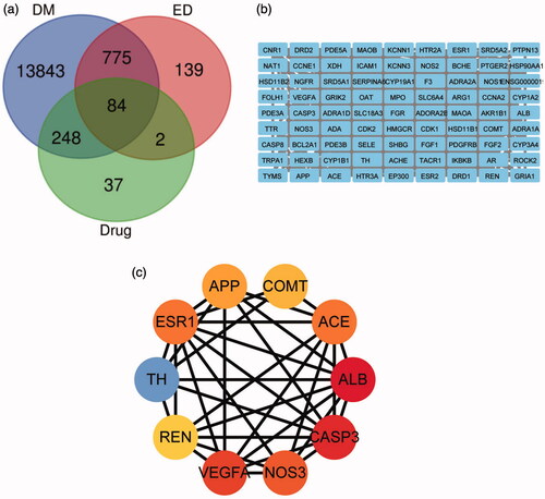
A total of 371 ABR + SV targets were obtained from predictions from PubChem and Swiss Target Prediction databases. A total of 14950 DM- and 1000 ED-related targets were obtained from the GeneCards database. Finally, 84 overlapping targets associated with ABR + SV and DMED were obtained (). Then, PPIs were analysed using the STRING database ().
Figure 1. (a) Intersection of ABR + SV targets, diabetes mellitus targets, and erectile dysfunction targets; (b) PPI network built by Cytoscape (3.7.1); (c) PPI network processed by Cytoscape (3.7.1) plug-in (cytohubba).
Network construction and topology analyses
The first ten targets of the “degree” parameter were considered to be the key targets. They are ALB, CASP3, VEGFA, eNOS, ACE, oestrogen receptor 1 (ESR1), tyrosine hydroxylase (TH), amyloid β precursor protein (APP), catechol-O-methyltransferase (COMT), and renin (REN) (, ). An herbal compound-target-disease network was constructed using Cytoscape ().
Figure 2. The network construction for herbal compound-target-disease.
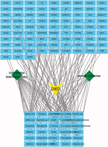
Table 2. Top 10 key targets.
Enrichment analyses using GO and KEGG databases
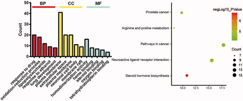
Enrichment analyses of 84 targets through DAVID v6.8 showed that 161 biological processes, 43 cellular components, 71 molecular functions, and 39 signalling pathways were involved. According to the p-value, the top-five biological processes and pathways were selected for display ().
Figure 3. Analyses of pathway enrichment using GO and KEGG databases.
Experimental study
Verification of erectile function
After APO induction, the number of erections in the M group was 0, which was significantly lower than that in the C group (p < 0.05). After 8 weeks of drug intervention, the number of erections in the T group was significantly higher than that in the M group (p < 0.05) ().
Table 3. Erectile times of rats in each group after drug intervention.
Body weight, blood glucose, insulin, and glucagon levels in each group
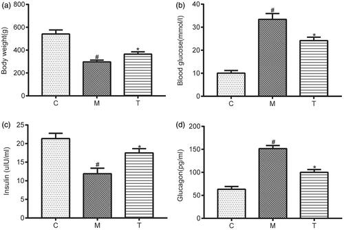
After drug intervention, the bodyweight of T rats was significantly higher than those of M rats (p < 0.05; ). The blood glucose levels in the T group were decreased significantly compared with those in the M group (p < 0.05; ). The blood insulin level was decreased significantly and the glucagon level was increased significantly in the M group compared with those in the C group (p < 0.05). After 8 weeks of drug intervention, the serum insulin level of rats was significantly higher and the level of glucagon was significantly lower in the T group than those in the M group (p < 0.05; ).
Figure 4. (a) The body weight in rats from the C, M, and T groups; (b) The blood glucose in rats from the C, M, and T groups; (c) The insulin in rats from the C, M, and T groups; (d. The glucagon in rats from the C, M, and T groups. Data are expressed as the mean ± SEM. Independent sample t-tests were employed for comparison between the two groups. Differences with p < 0.05 were considered statistically significant. #p< 0.05, the M group vs. the C group; *p < 0.05, the T group vs. the M group.
Structural analyses of the cavernous body of the penis
H&E staining showed that the trabeculae and blood sinuses were distributed evenly in group C, some red blood cells (RBCs) were in the sinus space, many smooth-muscle cells were in blood-containing sinus trabeculae, but the proliferation of interstitial tissue was not observed. Compared with group C, the number of blood-containing sinuses in the cavernous body of group M was reduced significantly and its distribution was disordered, the density of endothelial cells and smooth muscle cells was decreased, and the number of collagen fibres was increased. After 8 weeks of drug intervention, the distribution of blood-containing sinuses in group T was more regular than that in group M, the density of endothelial cells was increased, the number of collagen fibres was decreased, and RBCs were seen in some blood-containing sinuses ().
Figure 5 (a) H&E staining of the penile tissue of rats from the C, M, and T groups (×400). (i) In the C group: The trabeculae and blood sinuses were distributed evenly, and some red blood cells (RBCs) were in the sinus space, many smooth-muscle cells were in blood-containing sinus trabeculae. (Arrow ①②); (ii) In the M group: The number of blood-containing sinuses in the cavernous body was reduced significantly and its distribution was disordered, the density of endothelial cells and smooth muscle cells was decreased, and the number of collagen fibres was increased. (Arrow ③④); (iii) In the T group: the distribution of blood-containing sinuses was more regular than that in group M, the density of endothelial cells was increased, the number of collagen fibres was decreased, and RBCs were seen in some blood-containing sinuses. (Arrow ⑤⑥); (b) Ultrastructure of penile tissue of rats from the C, M, and T groups (×5000). (i) In the C group: the blood vessels of were normal, the morphology and structure of endothelial cells were basically normal, the nucleus was regular, the endothelial cells were closely connected, mitochondria and endoplasmic reticulum were observed in endothelial cells. (Arrow ①②); (ii) In the M group: Endothelial cells swelled, endothelial cell junction disappeared, endothelial cell mitochondria swelled and endoplasmic reticulum expanded. (Arrow ③④); (iii) In the T group: The blood vessels were normal, the morphology and structure of endothelial cells were basically normal, the endothelial cells were tightly connected, and mitochondria were seen in endothelial cells (Arrow ⑤⑥).
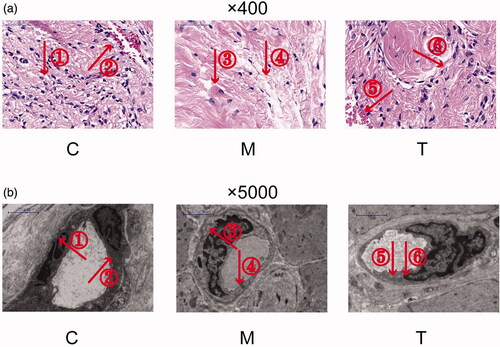
Under transmission electron microscope, the blood vessels of group C were normal, the morphology and structure of endothelial cells were basically normal, the nucleus was regular, the endothelial cells were closely connected, mitochondria and endoplasmic reticulum were observed in endothelial cells. In group M, endothelial cells swelled, endothelial cell junction disappeared, endothelial cell mitochondria swelled and endoplasmic reticulum expanded. After 8 weeks of drug intervention, the blood vessels were normal, the morphology and structure of endothelial cells were basically normal, the endothelial cells were tightly connected, and mitochondria were seen in endothelial cells ().
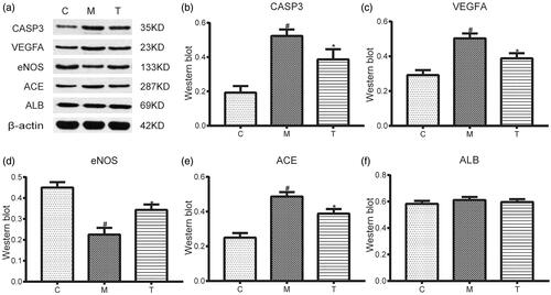
Western blotting
We measured protein expression of CASP3, VEGFA, eNOS, ACE, and ALB in penile cavernous tissue. Compared with group C (0.19 ± 0.04; 0.29 ± 0.03; 0.25 ± 0.03; 0.45 ± 0.03), protein expression of VEGFA, CASP3 and ACE in group M increased (0.52 ± 0.04; 0.50 ± 0.03; 0.49 ± 0.02) (p < 0.05), and protein expression of eNOS (0.23 ± 0.03) decreased (p < 0.05). Compared with group M (0.52 ± 0.04; 0.50 ± 0.03; 0.49 ± 0.02; 0.23 ± 0.03), protein expression of VEGFA, CASP3 and ACE in group T decreased (0.39 ± 0.06; 0.34 ± 0.03; 0.39 ± 0.03) (p < 0.05), and protein expression of eNOS increased (0.34 ± 0.03) (p < 0.05) (. There was no difference in protein expression of ALB among groups C, M, or T ().
Figure 6. (a) Electrophoretogram of five proteins (CASP3, VEGFA, eNOS, ACE, and ALB) in rats from the C, M, and T groups. (b–f) The expression levels of five proteins (CASP3, VEGFA, eNOS, ACE, and ALB) in rats from the C, M, and T groups were determined using western blotting. Data are expressed as the mean ± SEM. Independent sample t-tests were employed for comparison between the two groups. Differences with p < 0.05 were considered statistically significant. #p < 0.05, the M group vs. the C group; *p < 0.05, the T group vs. the M group.
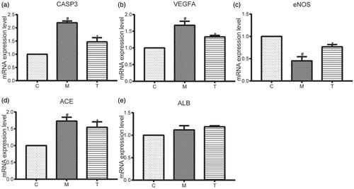
RT-qPCR
We measured mRNA expression of CASP3, VEGFA, eNOS, ACE, and ALB in penile cavernous tissue. Compared with group C (1.00 ± 1.00; 1.00 ± 1.00; 1.00 ± 1.00; 1.00 ± 1.00), mRNA expression of VEGFA, CASP3, and ACE in group M was increased (2.19 ± 0.07; 1.68 ± 0.12; 1.73 ± 0.11) (p < 0.05), and mRNA expression of eNOS was decreased (0.45 ± 0.09) (p < 0.05). Compared with group M (2.19 ± 0.07; 1.68 ± 0.12; 1.73 ± 0.11; 0.45 ± 0.09), mRNA expression of VEGFA, CASP3, and ACE in group T decreased (1.47 ± 0.16; 1.33 ± 0.05; 1.55 ± 0.16) (p < 0.05), and mRNA expression of eNOS was increased (0.77 ± 0.05) (p < 0.05) (. There was no difference in mRNA expression of ALB among groups C, M, or T ().
Figure 7. (a–e) The mRNA expression levels of five proteins (CASP3, VEGFA, eNOS, ACE, and ALB) in rats from the C, M, and T groups. determined using RT-qPCR. Data are expressed as the mean ± SEM. Independent sample t-tests were employed for comparison between the two groups. Differences with p < 0.05 were considered statistically significant. #p < 0.05, the M group vs. the C group; *p < 0.05, the T group vs. the M group.
Discussion
ABR and SV are commonly used TCM formulations employed for activating blood circulation and collateral blood vessels. ABR contains flavonoids, terpenoids, steroids, sugar, and other ingredients. ABR has several pharmacologic actions: inhibition of platelet aggregation and thrombosis; anti-atherosclerosis actions; inhibition of ischemia-reperfusion injury; anti-fibrosis actions; anti-inflammatory, analgesic, and antioxidant effects (Mo et al. Citation2020). Studies have shown that SV can inhibit the VEC injury induced by hyperglycosemia significantly (Zhu Citation2018).
DMED has become a research “hotspot” in recent years (Qiu et al. Citation2021). Penile erection is caused first by the body accepting conduction of a nerve impulse, which leads to relaxation of smooth muscle in the cavernous tissue of the penis, inflow of arterial blood, occlusion of venous vessels, increased blood volume in the penis, and then erection (Thorve et al. Citation2011). Any factors leading to decreased blood flow to the penis can lead to ED. Hyperglycaemia in DM patients can produce peripheral vascular lesions by affecting vascular walls and endothelial factors. These actions lead to the glycosylation of elastic fibres in vascular walls, which limits vasodilation in cavernous sinusoids, reduces blood flow in the cavernous body and, thus, affects penile erection (Richards and Vinik Citation2002; De Young et al. Citation2004). DMED pathogenesis may also be related to oxidative-stress injury caused by a long-term hyperglycaemic environment in DM patients. The products of oxidative stress (e.g., reactive oxygen species, malondialdehyde, 8-oxo-2′-deoxyguanosine) damage vascular endothelial cells (VECs), affect the release of nitric oxide (NO) and other factors released by endothelial cells, and can even cause VEC apoptosis (Cui et al. Citation2018).
Network pharmacology is an interactive network based on the concept of “disease-gene-target-drug”, which treats the intervention and effect of drugs on disease networks from a systematic and comprehensive perspective, so as to reveal the complex mechanism of drugs on the human body. Through the network pharmacology research, we found that ALB, CASP3, VEGFA, eNOS, and ACE are the key targets of ABR + SV in the treatment of DMED.
Caspase is a proteolytic enzyme present in cells in the form of inactive pro-caspase. If caspase is activated, it triggers a cascade which, ultimately, leads to apoptosis (Garrido et al. Citation2006). Caspase-3 is a major effector of apoptosis and is closely related to the morphologic changes observed in apoptosis (Mo et al. Citation2012). In DM rats, caspase-3 expression is increased (Li and Liu Citation2014; Yang et al. Citation2015), which induces VEC apoptosis in the cavernous body of the penis. Hence, caspase-3 may be an important target of DMED. VEGFA is one of the major pro-angiogenic factors involved in angiogenesis (Melincovici et al. Citation2018). Our network-pharmacology analyses predicted that VEGFA may be an important target of DMED. Scaldaferrif and colleagues (Citation2009) found that VEGFA overexpression can promote the generation of mucosal blood vessels and the adhesion of white blood cells, thereby leading to the increased risk of vascular thrombosis. Therefore, excessive expression of VEGFA in diabetic hyperglycaemia (which in turn leads to blood clots in penile vessels and ED) may be one of the mechanisms of DMED. ACE is a vasoconstrictor synthesized after VEC injury. However, non-enzymatic glycosylation occurs in DM patients due to long-term hyperglycaemia, abnormal fluctuations of blood glucose levels, or increases in blood viscosity. These actions in turn cause damage to VECs, which synthesize a large amount of ACE (Xie et al. Citation2020), which can lead to ED. Studies have shown that there are three subtypes of NOS: eNOS, neuronal NOS (nNOS) and inducible NOS (iNOS). Of these, eNOS and nNOS have major roles in penile erection (Lau and Adaikan Citation2014; Yafi et al. Citation2016). DM can lead to the accumulation of advanced glycation end products (AGEs) in the CC of the penis. AGEs bind to eNOS in cells and inhibit their function, thereby reducing NO synthesis. Such binding also leads to inactivation of free NO, which results in loss of the main neurotransmitter during penile erection, and leads to ED (Neves Citation2013).
Our western blotting and RT-qPCR results showed that protein expression of caspase-3, VEGFA, and ACE in group C was the least, and eNOS protein was the most. Also, protein expression of caspase-3, VEGFA and ACE in group M was the most, and eNOS protein was the least. However, after ABR + SV treatment, protein expression of caspase-3, VEGFA, and ACE in group T decreased significantly, whereas protein expression of eNOS increased significantly. Hence, DMED rats may have suffered ED due to increased expression of caspase-3, VEGFA, and ACE, and reduced protein expression of eNOS. ABR + SV administration may have downregulated protein expression of caspase-3, VEGFA, and ACE in penile CC, and increased eNOS expression to improve ED in DMED rats.
It is interesting to note that INS protein is the key target of ABR + SV therapy for DMED predicted through network pharmacology. But a few studies have focussed on albumin-related ED. In our rat experiments, there was no significant difference in ALB expression in rats of each group. These data suggested that the predicted results of network pharmacology may have limitations, and may not be fully consistent with experimental results. Nevertheless, network pharmacology and experimental research were combined innovatively in our study to preliminarily explain the mechanism of DMED and the possible targets of ABR + SV treatment of DMED. Our data could provide innovations for the clinical treatment of DMED.
Our study still has many limitations; for example, we did not measure the concentration of CA + in penile tissues and the cells with functional gain and loss. IC50, ED50, LD50, MIC, etc., were not measured. And we did not consider sex hormone-related indicators in the rat experiments. Further experiments will be carried out in the future.
Disclosure statement
No potential conflict of interest was reported by the author(s).
Additional information
Funding
References
- Banks E, Joshy G, Abhayaratna WP, Kritharides L, Macdonald PS, Korda RJ, Chalmers JP. 2013. Erectile dysfunction severity as a risk marker for cardiovascular disease hospitalisation and all-cause mortality: a prospective cohort study. PLOS Med. 10(1):e1001372.
- Chen GH, Sun DL, Jin BF, Zhang XD, Chen B. 2016. Analysis of Jin Baofang's experience in treating erectile dysfunction. Liaoning J Tradit Chin Med. 43(01):141–143.
- Chen W, Gong L, Guo Z, Wang W, Zhang H, Liu X, Yu S, Xiong L, Luo J. 2013. A novel integrated method for large-scale detection, identification, and quantification of widely targeted metabolites: application in the study of rice metabolomics. Mole Plant. 6:1769–1780.
- Cui K, Tang Z, Li CC, Wang T, Rao K, Wang SG, Liu JH, Chen Z. 2018. Lipoxin A4 improves erectile dysfunction in rats with type I diabetes by inhibiting oxidative stress and corporal fibrosis. Asian J Androl. 20(2):166–172.
- De Young L, Yu D, Bateman RM, Brock GB. 2004. Oxidative stress and antioxidant therapy: their bimpact in diabetes-associated erectile dysfunction. J Androl. 25(5):830–836.
- Garcia A, Barbas C. 2011. Gas chromatography-mass spectrometry (GC-MS)-based metabolomics. Methods Mol Biol. 708:191–204.
- Garrido C, Galluzzi L, Brunet M, Puig PE, Didelot C, Kroemer G. 2006. Mechanisms of cytochrome C release from mitochondria. Cell Death Differ. 13:1423–1433.
- Hao da C, Xiao PG. 2014. Network pharmacology: a Rosetta stone for traditional Chinese medicine. Drug Dev Res. 75:299–312.
- Hong KM, Najjar H, Hawley M, Press RD. 2004. Quantitative real-time PCR with automated sample preparation for diagnosis and monitoring of cytomegalovirus infection in bone marrow transplant patients. Clin Chem. 50:846–856.
- Hu R, Zhang W, Ou NJ, Liang Z, Yang YJ, Liu L, Liu XQ. 2020. The effect of insulin therapy on the expression of miR-126, VEGF and eNOS in the penile tissue of diabetic rats. J Tianjin Med Univ. 26:422–428.
- Hu ZC. 2000. 56 Cases of impotence treated by integrated traditional Chinese and western medicine. Shaanxi J Trad Chin Med. (11):503.
- Itoga A, Zha X, Nagase K, Aoki Y, Ito H, Yokoyama O. 2020. Correcting imbalance of sex hormones by a phosphodiesterase 5 inhibitor improves copulatory dysfunction in male rats with type 2 diabetes. BMJ Open Diab Res Care. 8(1):e001111.
- Lau LC, Adaikan PG. 2014. Possibility of inhibition of calcium-activated chloride channel rescuing erectile failures in diabetes. Int J Impot Res. 26(4):151–155.
- Li X. 2019. Clinical and experimental study of huoxue tongluo qiwei decoction on diabetic erectile dysfunction [dissertation]. China (CN): Beijing Univ Chin Med.
- Li XR, Zhang YS, Wang JS, Ding J, Sheng W, Shang JW. 2020. Research on the mechanism and treatment of diabetic erectile dysfunction in Chinese and Western medicine. Chin J Sex. 29(01):111–115.
- Li Y, Liu F. 2014. The expression of Cyt C and Caspase 3 in the lens epithelial cells of STZ-induced diabetic rats. Recent Advan Ophthalmol. 34:228–231.
- Long P, Yan W, Liu J, Li M, Chen T, Zhang Z, An J. 2019. Therapeutic effect of traditional Chinese medicine on a rat model of branch retinal vein occlusion. J Ophthalmology. 2019:1–13.
- Lu CC, Jiann BP, Sun CC, Lam HC, Chu CH, Lee JK. 2009. Association of glycemic control with risk of erectile dysfunction in men with type 2 diabetes. J Sex Med. 6(6):1719–1728.
- Luo ES, Yang QF, Xu WL. 2019. The efficacy of low-dose tadalafil combined with Xuefu Zhuyu tablets in the treatment of diabetic erectile dysfunction. China Prac Med. 14(24):111–112.
- Ma HF, Liu Y, Wang B, Dang J, Ma JX, Zhu YT, Li HS. 2017. Clinical randomized controlled study of tongluo xifeng qiwei decoction in the treatment of erectile dysfunction. Chin J Sex Sci. 26 (02):78–81.
- Ma TT, Zhang GL, Dai CF, Zhang BR, Cao KX, Wang CG, Yang GW, Wang XM. 2020. Scutellaria barbata and Hedyotis diffusa herb pair for breast cancer treatment: potential mechanism based on network pharmacology. J Ethnopharmacol. 259:112929.
- Melincovici CS, Boşca AB, Şuşman S, Mărginean M, Mihu C, Istrate M, Moldovan IM, Roman AL, Mihu CM. 2018. Vascular endothelial growth factor (VEGF) – key factor in normal and pathological angiogenesis. Rom J Morphol Embryol. 59:455–467.
- Mo J, Cui QL, Yao J, Zhang FT, Tan XH. 2012. The effect of recombinant human interleukin-10 on the apoptosis of human microvascular endothelial cells under hypoxia. J Prac Pediatr. 27:1107–1110.
- Mo Q, Hao EW, Qin WH, Hou XT, Deng JG, Wu Y. 2020. Research progress on the qualitative basis and pharmacological effects of drugs in smoothing blood circulation and removing blood stasis. Chin J Exp Formulas. 26(01):205.
- Neves D. 2013. Advanced glycation end-products: a common pathway in diabetes and age-related erectile dysfunction. Free Radic Res. 47 (sup1):49–69.
- Park J, Chai JS, Kim SW, Paick JS, Cho MC. 2018. Inhibition of Jun N-terminal kinase improves erectile function by alleviation of cavernosal apoptosis in a rat model of cavernous nerve injury. Urology. 113:253.e9–253.e16.
- Penson DF, Wessells H. 2004. Erectile dysfunction in diabetic patients. Diabetes Spectr. 17(4):225–230.
- Qiu WC, Guo XM, Zhu MLM, Chen KC, Zhu Y. 2021. Research progress in treatment of diabetic nephropathy with traditional Chinese medicine. J Liaoning Univ Trad Chin Med. 23(04):157–162.
- Richards D, Vinik A. 2002. Etiology and treatment of erectile failure in diabetes mellitus. Curr Diab Rep. 2:501–509.
- Scaldaferri F, Vetrano S, Sans M, Arena V, Straface G, Stigliano E, Repici A, Sturm A, Malesci A, Panes J, et al. 2009. VEGF-A links angiogenesis and inflammation in inflammatory bowel disease pathogenesis. Gastroenterology. 136:585–595.
- Shamloul R, Ghanem H. 2013. Erectile dysfunction. Lancet. 381(9861):153–165.
- Tao W, Xu X, Wang X, Li B, Wang Y, Li Y, Yang L. 2013. Network pharmacology-based prediction of the active ingredients and potential targets of Chinese herbal radix curcumae formula for application to cardiovascular disease. J Ethnopharmacol. 145(1):1–10.
- Thorve VS, Kshirsagar AD, Vyawahare NS, Joshi VS, Ingale KG, Mohite RJ. 2011. Diabetes-induced erectile dysfunction: epidemiology, pathophysiology and management. J Diabetes Complications. 25:129–136.
- Vita R, Benvenga S, Giammusso B, La Vignera S. 2019. Determinants of early response to low-intensity extracorporeal shockwaves for the treatment of vasculogenic erectile dysfunction: an open-label, prospective study. JCM. 8(7):1017.
- Wang JG, Tan Y, Wang WR, Xie ZP, Li T. 2018. Compound xuanju capsule combined with tadalafil in the treatment of erectile dysfunction with abdominal obesity. J Hubei Univ Med. 37:338–340.
- Wang XF, Zhu JC, Deng CH. 2013. Guidelines for the diagnosis and treatment of andrological diseases in China. 2013 ed. China, CN: People Health Publishing House; p. 71–78.
- Wang JS, Li X, Chen ZL, Feng JL, Bao BH, Deng S, Dai HH, Meng FC, Wang B, Li HS. 2021. Effect of leech-centipede medicine on improving erectile function in DIED rats via PKC signalling pathway-related molecules. J Ethnopharmacol. 267:113463.
- Xie J, Niu MZ, Zhang R. 2020. The changes of NAGL, sfrp5, ACE levels in T2DM patients with hypertension and the relationship with left ventricular remodeling. J Integrated Tradi Chin West Med Cardio-Cerebrovascular Dis. 18(08):1270.
- Yafi FA, Jenkins L, Albersen M, Corona G, Isidori AM, Goldfarb S, Maggi M, Nelson CJ, Parish S, Salonia A, et al. 2016. Erectile dysfunction. Nat Rev Dis Primers. 2:16003.
- Yang KB, Huang XJ, Zhang XG, Chen G, Bo J. 2014. Efficacy of low-dose tadalafil combined with shuganyishen capsule in the treatment of mild to moderate erectile dysfunction. Nat J Androl China. 20:267–272.
- Yang ZH, Song Y, Chu GP, Qiu P, Peng WP, Meng XF. 2015. Salidroside inhibits high glucose-damaged neuronal activity caspase 3 and promotes BDNF expression. Chin J Hist Cyto. 24:439–442.
- Yu XD, Wang JS, Zuo G. 2019. Traditional Chinese medicine on treating diabetic mellitus erectile dysfunction: protocol for a systematic review and meta-analysis. Medicine. 98(13):e14928.
- Zeng J. 2018. Clinical observation on the treatment of diabetic erectile dysfunction with the method of invigorating the kidney and promoting blood circulation combined with sildenafil. Beijing: Beijing University of Chinese Medicine.
- Zhao RL, He YM. 2018. Network pharmacology analysis of the anti-cancer pharmacological mechanisms of Ganoderma lucidum extract with experimental support using Hepa1-6-bearing C57 BL/6 mice. J Ethnopharmacol. 210:287–295.
- Zhu XX. 2018. Study on the protective effect and mechanism of flavonoid glycosides from wang buliu xing on vascular endothelial cell injury induced by high glucose [dissertation]. China, CN: Jiangnan University.