
Abstract
Context
The effect of kaempferol, a regulator of oestrogen receptors, on atherosclerosis (AS) and the underlying mechanism is elusive.
Objective
To explore the effect and mechanism of kaempferol on AS.
Methods and materials
In vivo, C57BL/6 and apolipoprotein E (APOE)–/– mice were randomly categorized into six groups (C57BL/6: control, ovariectomy (OVX), high-fat diet (HFD); APOE–/–: OVX-HFD, OVX-HFD + kaempferol (50 mg/kg) and OVX-HFD + kaempferol (100 mg/kg) and administered with kaempferol for 16 weeks, intragastrically. Oil-Red and haematoxylin–eosin (HE) staining were employed to examine the effect of kaempferol. In vitro, human aortic endothelial cells (HAECs) were pre-treated with or without kaempferol (5, 10 or 20 μM), followed by administration with kaempferol and oxidized low-density lipoprotein (ox-LDL) (200 μg/mL). The effect of kaempferol was evaluated using flow cytometry, and TdT-mediated dUTP Nick-End Labelling (TUNEL).
Results
In vivo, kaempferol (50 and 100 mg/kg) normalized the morphology of blood vessels and lipid levels and suppressed inflammation and apoptosis. It also activated the G protein-coupled oestrogen receptor (GPER) and PI3K/AKT/nuclear factor-erythroid 2-related factor 2 (Nrf2) pathways. In vitro, ox-LDL (200 μg/mL) reduced the cell viability to 50% (IC50). Kaempferol (5, 10 or 20 μM) induced-GPER activation increased cell viability to nearly 10%, 19.8%, 30%, and the decreased cellular reactive oxygen species (ROS) generation (16.7%, 25.6%, 31.1%), respectively, consequently attenuating postmenopausal AS. However, the protective effects of kaempferol were blocked through co-treatment with si-GPER.
Conclusions
The beneficial effects of kaempferol against postmenopausal AS are associated with the PI3K/AKT/Nrf2 pathways, mediated by the activation of GPER.
Introduction
Globally, most deaths from coronary heart disease are associated with atherosclerosis (AS) (Wang C et al. Citation2018). Although AS occurs in both men and women, reports from epidemiological studies show that postmenopausal women exhibit a higher prevalence of atherosclerotic disease than premenopausal women, suggesting the potential role of oestrogen in the progression of atherosclerotic disease (Aryan et al. Citation2020). Oestrogen is essential in the regulation of lipid metabolism (Morselli et al. Citation2018), inflammation (Mu et al. Citation2016) and vascular homeostasis (Filardo Citation2018). Natural oestrogen or selective oestrogen receptor (ER) modulators are crucial in cardiovascular diseases as they regulate the lipid level in blood vessels.
In clinics, AS patients are managed with statins (Okuyama et al. Citation2015), aspirin (Petri et al. Citation2017), nitroglycerin (Cho et al. Citation2011) and other drugs (Li et al. Citation2019). The lack of oestrogen is attributed to the loss of its protective effects in postmenopausal women, for example, anti-inflammation, antioxidation, low blood lipids, cholesterol, etc., subsequently contributing to the high incidence of AS. Therefore, oestrogen replacement therapy is administered as the basic clinical treatment of postmenopausal AS. Although this management option achieves a particular short-term effect, some long-term side effects occur, including fatigue, uterine leiomyoma and canceration (Malekmohammad et al. Citation2019). Accordingly, researchers have increasingly shifted their focus to explore safer and alternative strategies for managing AS. In China, traditional Chinese medicines (TCM) have been extensively adopted to manage various diseases for thousands of years (Hu et al. Citation2019; Jian et al. Citation2019). TCM exerts excellent effects in clinical AS therapy (McClelland et al. Citation2015; Zhu et al. Citation2020) and has attracted considerable attention from scholars. TCM contains phytoestrogen, with numerous pharmacological effects, and has been applied for the therapeutic management of human diseases (Kiyama Citation2017). Recent exploration of anti-AS mechanisms of phytoestrogen has uncovered more active ingredients which can be used to synthesize better therapeutic drugs (Gencel et al. Citation2012).
Oestrogens, produced in endocrine organs and reproductive systems, play a vital role in humans and animals. Phytoestrogens, as a family of exogenous oestrogens, widely exist in plants and food and are considered a promising therapeutic target for postmenopausal atherosclerotic (Morselli et al. Citation2017). Kaempferol, another natural flavonoid, is widely distributed in plants and possesses various pharmacological activities, including anti-inflammatory, antiapoptotic and anticancer effects (Zhong et al. Citation2018; Sharma et al. Citation2019). Notably, it plays a vital role in cancer therapy (Kim et al. Citation2018; Wu et al. Citation2018) and is a therapeutic agent for various diseases, including diabetes and obesity (Alkhalidy et al. Citation2018; Torres-Villarreal et al. Citation2019). Recent evidence indicates that kaempferol alleviates oxidized low-density lipoprotein (ox-LDL)-induced endothelial cell injury, associated with the inflammation-dependent pathway (Che et al. Citation2017). Another study in rats found that kaempferol exerted therapeutic effects on AS (Zhang et al. Citation2016). However, additional studies are warranted to certify the anti-postmenopausal atherosclerotic effect of kaempferol.
Compelling evidence confirmed that membrane G protein-coupled oestrogen receptor (GPER), a newly discovered ER activated by classic oestrogen and phytoestrogens, is associated with numerous diseases (Barton and Prossnitz Citation2015; Grande et al. Citation2020). In particular, the expression of GPER modulated by the regulator changed the concentration of oestrogen and further affected the deterioration of AS in postmenopausal women. In addition, GPER regulators stimulate the PI3K signal pathway to fight various diseases (Fan et al. Citation2018); this pathway is associated with the expression level of oestrogen (Pollard and Daniel Citation2019). Notably, PI3K/AKT/Nrf2 pathway is the most widely studied. However, whether kaempferol, as an activator of GPER, attenuates AS via modulation of the PI3K/AKT/Nrf2 signalling pathway is elusive.
In this article, we revealed the effects of kaempferol on postmenopausal AS in ovariectomy (OVX)+high fat diet-induced apolipoprotein E (APOE)–/– mice and in ox-LDL induced human aortic endothelial cells (HAECs) and explored the underlying molecular mechanisms. Also, we have elucidated the role of GPER in postmenopausal AS.
Materials and methods
Reagents
HAECs were purchased from Shanghai Bioleaf Biotech Co., Ltd. (Shanghai, China). The HAECs medium, Dulbecco’s modified Eagle’s medium (DMEM) and foetal bovine serum (FBS) were acquired from the Gibco-BRP Company (Gaithersburg, MD). Kaempferol (purity >98%) was purchased from Meilun Biotechnology Co. Ltd. (Dalian, China). Fresh plasma was acquired from the blood bank. For the cell viability test, we employed the Cell Count Kit-8 (CCK-8) assay, provided by Proteintech Group (Wuhan, China). TUNEL Staining Kit, purchased from Beyotime Biotechnology (Haimen, China), was employed to assess the level of cell apoptosis. Intracellular Ros was estimated using the ROS Assay Kit (Beyotime Biotechnology, Haimen, China). Antibodies specific for Bax, B-cell lymphoma-2 (Bcl-2), caspase 3, AKT, P-AKT, heme oxygenase-1 (HO-1), Nrf2, PI3K, intercellular cell adhesion molecule-1 (ICAM-1) and vascular cell adhesion molecule-1 (VCAM-1) were obtained from the Proteintech Group (Wuhan, China), whereas antibody against GPER was purchased from BOSTER Biological Technology (Pleasanton, CA). The BCA Protein Assay Kit was purchased from the Beyotime Institute of Biotechnology (Shanghai, China). Histopathology test was conducted using the Oil Red staining kit, Nanjing Jiancheng Bioengineering Institute (Nanjing, China).
Animal experiments
Eight-week-old female C57BL/6 mice were obtained from the Laboratory Animal Center of Dalian Medical University (Dalian, China). APOE–/– mice were purchased from Vital River Laboratory Animal Technology Co., Ltd. (Beijing, China). All animal studies adhered to the guidelines for the Care and Use of Laboratory Animals of the National Institutes of Health. Approval was issued by the ethics committee of Dalian Medical University. The living space had controlled temperature and a 12 h light/dark cycle. Mice accessed free water and were fed on 6 g normal food or high-fat diet (HFD, purchased from Jiangsu Medicine Ltd. (Lianyungang, China), containing 1.25% cholesterol and 0.5% sodium cholate. After one-week of adaptive feeding, C57BL/6 mice were randomly categorized into three groups, n = 8 for per group: A (control), B (OVX), C (HFD); APOE–/– mice were randomly divided into three groups, n = 8 for per group: D (OVX-HFD), E (OVX-HFD + kaempferol 50 mg/kg) and F (OVX-HFD + kaempferol 100 mg/kg), and then subjected to bilateral OVX or sham OVX (sham OVX). The mice in groups A, B, C and D were intragastrically administered with 0.5% CMC-Na per day, whereas mice in groups E and F were given kaempferol (dissolved with 0.5% CMC-Na) via the same route. After 16 weeks, all mice were anaesthetized using 4% chloral hydrate (Sigma, St. Louis, MO). Then, blood samples were drawn from the abdominal aorta. The aorta tissues were collected for further analysis.
Method of OVX: the mice were fixed with prone position, dorsal skin depilated and iodophor disinfected. The skin and muscle layer were cut and the fat tissue was clipped with tweezers and pull it out the wound. Further, the blood vessels were ligated at the connection between the fallopian tube and uterine horn was cut and the ovary moved out. Next, ovaries and surrounding fat tissue were removed, the incision was closed by suturing the muscles and stapling the skin. Finally, mice were given intramuscular injection of penicillin.
Oil red O staining
For tissue preparation, a longitudinal cut was made in the aortic arch, thoracic and abdominal aortae, and pinned flat. After rinsing in 60% isopropanol and PBS, tissues were stained with oil red O. We calculated the ratio of oil red O positive staining to the total surface area to estimate the face lesion size.
Haematoxylin–eosin (HE) staining
Arterial tissues for atherosclerotic lesions morphometric analysis were immersed in 10% formalin for 30 min, dehydrated in 75% ethanol overnight. The tissues were then embedded in paraffin and sliced into 4 μm sections. Haematoxylin and eosin-stained sections were subjected to histopathological examination.
Biochemical analysis
Plasma samples were centrifuged at 3000 rpm at 4 °C for 10 min and preserved at −80 °C, awaiting subsequent analysis. The levels of serum total cholesterol (TC), triglyceride (TG), high-density lipoprotein cholesterol (HDLC), low-density lipoprotein cholesterol (LDL-C), superoxide dismutase (SOD), glutathione (GSH) and malondialdehyde (MDA) were assessed using commercial kits purchased from the Nanjing Jiancheng Bioengineering Institute (Nanjing, China).
Enzyme-linked immunosorbent assay (ELISA)
HAECs were seeded and cultured in the six-well plates and then treated with indicated drugs (kaempferol and/or ox-LDL). Cells were collected in a lysate buffer. Plasma was centrifuged (2000 rpm, 4 °C, 5 min). The levels of tumour necrosis factor-α (TNF-α) and interleukin-6 (IL-6) were measured using TNF-α and IL-6 ELISA kits (Huamei, Wuhan, China) in serum and cells. The values of cell TNF-α and IL-6 were normalized based on cell protein concentrations assessed using the BCA Protein Assay Kit.
Western blot
Protein samples were extracted from aorta tissues or HAECs lysates according to the protein extraction kit (Nanjing Jiancheng, Nanjing, China). Extracts were subjected to sodium dodecyl sulphate-polyacrylamide mini gels (SDS-PAGE) and transferred to polyvinylidene fluoride (PVDF) membranes (Millipore, Bedford, MA). The membranes were blocked using 5% milk for 2 h and then incubated with specific primary antibodies at 4 °C overnight. This was followed by three times wash and continuous incubation with corresponding secondary antibodies at room temperature for 2 h. Eventually, a Bio-Rad imaging system with a Chemi HR camera 410 was used to capture the emitted light from the membranes exposed to enhanced chemiluminescence plus reagents (Beyotime, Haimen, China). The density of bands was analysed using Gel-Pro Analyser Version 4.0 (Media Cybernetics, Rockville, MD).
Cell culture and experiment design
HAECs were cultured in DMEM containing 10% FBS (Gibco, Gaithersburg, MD) in the incubator under the following conditions: humidity, 95%; temperature, 37 °C; CO2, 5%. All examinations of HAECs were conducted at 24 h after replenishing the common medium with the basal serum-free medium containing kaempferol (5, 10 or 20 μM) or ox-LDL (200 μg/mL, final).
Preparation of ox-LDL
To isolate the plasma, the blood sample was centrifuged for 5 min at 7000 rpm and 4 °C. The plasma was further subjected to discontinuous density-gradient hypervelocity centrifugation with Beckman Coulter Optima l-100 XP super centrifuge. It was then dialysed in phosphate-buffered saline (PBS) at 4 °C for 24 h to prepare the original low-density lipoprotein (LDL, density: 1.019–1.063 g/mL). Original LDL was oxidized in the presence of 50 μM CuSO4 at 37 °C for 24 h and terminated with 0.01% EDTA in PBS and sterilized via filtration (0.22 μm). Ox-LDL was preserved in the dark and prepared every two weeks.
CCK-8 assay
HAECs seeded into the 96-well plate to a density of 1 × 10⁵ cells/mL were then treated with specific drugs and grown to approximately 80% confluence. The complete medium was replenished with serum-free medium containing ox-LDL (0–300 μg/mL), kaempferol (0–200 μM) or combining ox-LDL (200 μg/mL) with kaempferol (0–20 μM). After 24 h incubation, 10 μL CCK-8 solution was added and further incubated for 1.5 h at 37 °C. A microplate reader (Tecan, Grödig, Austria) was used to measure the absorbance at 450 nm. The Prism GraphPad was employed to calculate the IC50 values (La Jolla, CA).
Cell apoptosis assay
HAECs cells were seeded and cultured in six-well plates. After treatment with indicated drugs, cells were collected to evaluate the apoptotic level using TUNEL Staining Kit. Briefly, the apoptotic rate was calculated as apoptotic cells/total cells, which emitted green and blue fluorescence, respectively, under an inverted fluorescence microscope.
Intracellular ROS assay
We applied 2′,7′-dichlorodihydrofluorescein diacetates (DCFH-DA) as the probe to estimate intracellular reactive oxygen species (ROS) of cells incubated with ox-LDL (200 μg/mL) or ox-LDL combined with kaempferol (5, 10 and 20 μM). Briefly, after incubation with indicated drugs, HAECs were further incubated with a serum-free medium containing DCFH-DA (10 μM) at 37 °C for 30 min. The BD FACS Calibur system (Becton, Dickinson and Company, Franklin Lakes, NJ) was employed to evaluate the fluorescence intensity with excitation and emission wavelengths of 488 nm and 525 nm, respectively.
siRNA transfection
The plasmid DNA and Lipofectamine 2000 were premixed in DMEM for 20 min. HAECs were left to grow to 50% confluence, and then GPER siRNA (20 nM) with Lipofectamine 2000 was added to complete the transfection. The medium was replenished with a serum-free medium at 6 h post transfection.si-RNA sequence: GCTGTACATTGAGCAGAAA.
Statistical analysis
All results were presented as the mean ± SD from at least three independent experiments. The data were analysed using the Prism program (Version 6.0, GraphPad, San Diego, CA). Statistical analysis was performed using one-way ANOVA followed by Tukey’s post hoc test or unpaired t-test. p Values <0.05 denoted statistically significant differences.
Results
Effects of kaempferol on the vasculature and lipid homeostasis in HFD-OVX induced APOE–/– mice
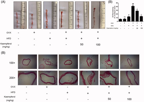
Oil red O staining revealed a larger lesion area of mice in the HFD-OVX group compared to mice in OVX, HFD and normal groups. Besides, kaempferol treatment significantly reduced the lesion area and thickness (). HE staining results demonstrated that the intimal lesion area contained larger necrotic cores in the aorta at the HFD-OVX group, whereas kaempferol administration potentially reduced the lesion areas (). These observations affirmed the inhibitory effects of kaempferol on the progression of atherosclerotic lesions.
Figure 1. Kaempferol reduced atherosclerotic lesion size and lesion lipid deposition in mice. Atherosclerotic lesion formation detected by red oil O staining (A); the quantitative data of Oil Red staining (B); Atherosclerotic lesion formation detected by HE staining (C) (n = 5).
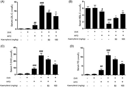
Based on the assay of levels of serum TC, LDLc, HDLc and TG, we examined the effect of kaempferol on lipid levels. Higher serum levels of TCH-O, TG, LDL-C and lower serum levels of HDL-C were found in the HFD-OVX group compared to OVX, HFD and normal groups (). In the groups treated with kaempferol, these changes were remarkedly reversed compared to the HFD-OVX group.
Figure 2. Kaempferol regulated lipid accumulation in mice. (A–D) Changes in the levels of serum LDL-C, HDL-C, T-CHO and TG in different groups. The results were analysed with one-way ANOVA and unpaired t-test and showed as the mean ± SD. #p < 0.05, ##p < 0.01, and ###p < 0.001 vs. the control group; *p < 0.05, and **p < 0.01 vs. the OVX-HFD group (n = 5).
Effects of kaempferol on the biochemical indices and inflammatory factors in HFD-OVX induced APOE–/– mice
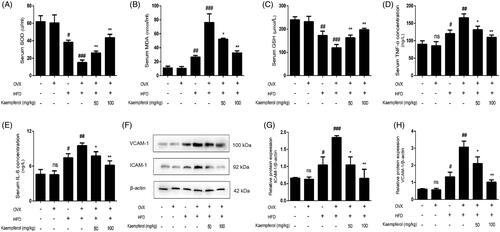
Oxidative stress is recognized as one of the pivotal factors of the AS pathological process. With kaempferol treatment, higher levels of serum SOD and GSH and lower serum level of MDA were reported compared to the HFD-OVX group (); also, it substantially lowered serum TNF-α and IL-6 levels compared to the HFD-OVX group. Western blot results demonstrated that the protein expression of ICAM and VCAM was significantly down-regulated by kaempferol (). Overall, these findings provide insights that kaempferol significantly lowered inflammation and inhibit oxidative stress.
Figure 3. Kaempferol mitigated the oxidative stress and inflammation in vivo. (A–E) Changes in the levels of serum SOD, MDA, GSH, TNF-α and IL-6 in different groups. (F–H) Changes in the arterial tissue protein expression of ICAM-1 and VCAM-A in different groups. The results were analysed with one-way ANOVA and unpaired t-test and showed as the mean ± SD. #p < 0.05, ##p < 0.01, and ###p < 0.001 vs. the control group; *p < 0.05, and **p < 0.01 vs. the OVX-HFD group; ns: not significant (p > 0.05 vs. the control group) (n = 5).
Effects of kaempferol treatment on apoptosis in HFD-OVX induced APOE–/– mice
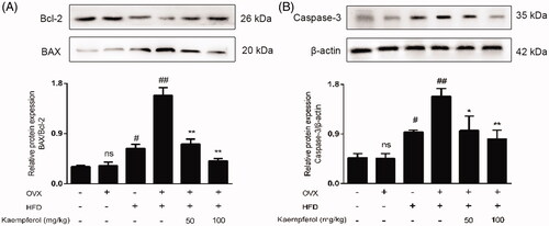
It has been widely confirmed that apoptosis of HAECs promoted the initiation and progression of AS by forming necrotic cores (Yang et al. Citation2019). Thus, the expression of apoptosis-related protein Bax, Bcl-2 and caspase 3 was assessed. In the HFD-OVX group, the ratio of Bax/Bcl-2 and expression level of caspase 3 were higher compared to OVX, HFD and normal groups (), whereas kaempferol treatment markedly reversed this tendency. Kaempferol (100 mg/kg) induced a 2.65-fold and 2.1-fold reduction in the expression of Bax/Bcl-2 and caspase 3, respectively, compared to the OVX-HFD group. Thus, we concluded that kaempferol-mediated reduction in apoptosis potentially improves the condition of AS.
Figure 4. Kaempferol inhibited the apoptosis in vivo. Changes of Bax/Bcl-2 ratio (A) and caspase 3 expression (B) in arterial tissue. Data were analysed with one-way ANOVA and unpaired t-test and presented as the mean ± SD. #p < 0.05, and ##p < 0.01 vs. the control group; *p < 0.05, and **p < 0.01 vs. the OVX-HFD group; ns: not significant (p > 0.05 vs. the control group) (n = 5).
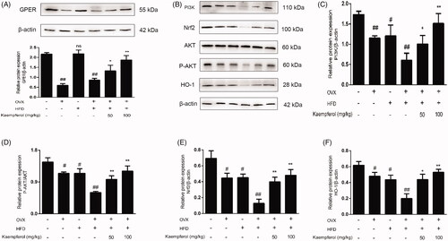
Effects of kaempferol treatment on GPER expression and PI3K/AKT/Nrf2 pathway in HFD-OVX induced APOE–/– mice
Recent findings suggested that GPER was involved in the progression of AS (Aryan et al. Citation2020). To explore the relationship between the beneficial effect of kaempferol on AS and GPER expression as well as PI3K/AKT/Nrf2 pathway. The protein expressions of GPER, PI3K, Nrf-2 and HO-1 were measured by western blotting in vivo. The level of GPER expression was markedly decreased in OVX and OVX-HFD groups compared to the control group; however, no significant change was reported in the HFD group. Compared to the OVX-HFD group, kaempferol administration was associated with a significantly higher expression of GPER (). Similarly, down-regulatory effects of expression of PI3K, NRF-2, HO-1 and P-AKT/AKT were observed in OVX, HFD and OVX-HFD groups. Notably, kaempferol (50 and 100 mg/kg) induced remarkedly high expression of the above proteins (). Collectively, these findings provide evidence on the potential benefits of kaempferol on AS through upregulation of GPER expression and activation of the PI3K/AKT/Nrf2 pathway.
Figure 5. Kaempferol upregulated GPER and then activated PI3K/AKT/Nrf2 pathway. Changes in expression level of GPER (A), PI3K (C), Nrf-2 (E), HO-1 (F) and P-AKT/AKT ratio (D) in arterial tissue. All date was analysed with one-way ANOVA and unpaired t-test and showed as the mean ± SD. #p < 0.05, and ##p < 0.01 vs. the control group; *p < 0.05, and **p < 0.01 vs. the OVX-HFD group; ns: not significant (p > 0.05 vs. the control group) (n = 5).
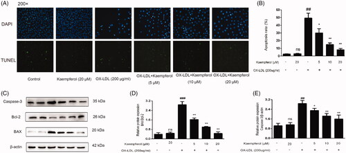
Effects of kaempferol on cell viability and apoptosis in HAECs treated with ox-LDL
We performed a CCK-8 kit assay to detect the effect of kaempferol on cell viability in HAECs. Notably, the cell viability decreased with elevated concentrations of ox-LDL (); the IC50 of ox-LDL to the cell was approximated at 200 μg/mL. Moreover, kaempferol (concentration range, 0–20 μM) did not exert any toxicity effect on HAECs cells but exhibited a beneficial effect on cell viability in HAECs (). Thus, we applied 200 μg/mL ox-LDL and 5, 10 and 20 μM kaempferol for the follow-up experiments. Evaluation of the effect of kaempferol on cell apoptosis via TUNEL staining revealed few TUNEL-positive cells in the control group and kaempferol alone group. However, ox-LDL induced the generation of numerous apoptosis cells (). Notably, compared to the ox-LDL group, co-administration with kaempferol (5, 10 and 20 μM) significantly lowered apoptosis. We further evaluated the expression of apoptotic-related proteins via Western blotting to assess the antiapoptotic effects of kaempferol on HAECs. Of note, ox-LDL administration induced higher expression of apoptotic-related proteins caspase 3 and Bax, lower expression of antiapoptotic-related protein Bcl-2 compared to control groups. Also, co-treatment with kaempferol lowered the expression of caspase 3 and Bax/Bcl-2 compared to the HFD-OVX group ().
Figure 6. Cell viability after the treatment of various of concentrations of ox-LDL and kaempferol. The effect of ox-LDL (A) kaempferol (B) and combination ox-LDL with kaempferol (C) on cell viability in HASCs cells. All results were analysed with one-way ANOVA and unpaired t-test and showed as the mean ± SD. ##p < 0.01 and ###p < 0.01 vs. the control group; *p < 0.05, **p < 0.01 and ***p < 0.001 vs. the ox-LDL group; ns: not significant (p > 0.05 vs. the control group) (n = 5).
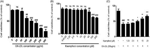
Figure 7. Kaempferol inhibited the ox-LDL induced apoptosis in HAEC cells. TUNEL staining results in different groups (A, B). The changes in the expression of caspase 3 and Bax/Bcl-2 ratio in HAEC cells (C–E). Data were analysed by one-way ANOVA and unpaired t-test and presented as the mean ± SD. ##p < 0.01 and ###p < 0.001 vs. the control group; *p < 0.05, and **p < 0.01 vs. the ox-LDL group; ns: not significant (p > 0.05 vs. the control group) (n = 5).
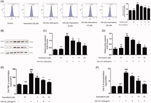
Effects of kaempferol on the ROS accumulation and the level of inflammatory factors in HAECs treated with ox-LDL
Following the assessment of the effects of kaempferol on oxidative stress and inflammatory factors in vitro, we found that ox-LDL-induced intracellular ROS accumulation was significantly suppressed after kaempferol co-treatment (). Similarly, ox-LDL elevated the intracellular concentrations of IL-6 and TNF-α, whereas kaempferol administration obviously decreased the level of inflammatory factors (). Further evaluation of the expression of intercellular and vascular adhesion molecules (ICAM and VCAM) through western blotting indicated that ox-LDL-induced expression of ICAM and VCAM was higher, more than twofold, compared to the control group. On the contrary, kaempferol, at concentrations of 5, 10 and 20 μM, lowered the expression of ICAM and VCAM. Notably, following treatment with 20 μM kaempferol, the expression of ICAM and VCAM recovered to an approximately normal level compared to the control group (). Therefore, it was deduced that kaempferol attenuated oxidative stress and blocked the release of inflammatory factors in HAECs.
Figure 8. Kaempferol inhibited the ROS accumulation and reduced the level of inflammation factors. HAECs cells were treated with kaempferol or free-foetal medium. Then, the cells were treated with indicated drugs or complete medium. The changes in ROS accumulation (A), the expression of ICAM-1 (C) and VCAM-1 (D) and level of TNF-α (E) and IL-6 (F) in HAEC cells were shown. Data are analysed by one-way ANOVA and unpaired t-test and presented as the mean ± SD. ##p < 0.01 and ###p < 0.001 vs. the control group. *p < 0.05 and **p < 0.01 and ***p < 0.01 vs. the ox-LDL group. ns: not significant (p > 0.05 vs. the control group) (n = 5).
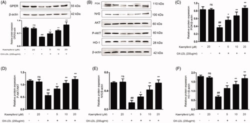
Effects of kaempferol treatment on GPER expression and PI3K/AKT/Nrf2 pathway in HAECs treated with ox-LDL
After treatment, HAECs cells were collected to detect the protein expression of GPER, PI3K, P-AKT/AKT, Nrf2 and HO-1. Compared to the control group, GPER protein expression was significantly lower after ox-LDL administration, whereas kaempferol increased GPER expression (). Besides, ox-LDL lowered the expression of PI3K, HO-1, Nrf2 and the ratio of p-AKT/AKT. Similarly, kaempferol treatment elevated the expression of PI3K, HO-1, Nrf2 and the ratio of p-AKT/AKT (). These results implied that kaempferol induced the activation of GPER and upregulation of the PI3K/AKT/Nrf2 pathway.
Figure 9. Kaempferol activated the GPER and regulated the PI3K/AKT/Nrf2 Pathway. HAECs cells were treated with kaempferol or free-foetal medium. Then, the cells were treated with indicated drugs or complete medium. The expression of GPER (A), PI3K (C), P-AKT/AKT (D), Nrf2 (E) and HO-1 (F) was detected by Western blot assay. Data are analysed by one-way ANOVA and unpaired t-test and presented as the mean ± SD. ##p < 0.01 vs. the control group. *p < 0.05 and **p < 0.01 vs. the ox-LDL group. ns: not significant (p > 0.05 vs. the control group) (n = 5).
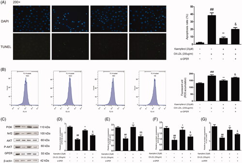
GPER activation is associated with the protective effect of kaempferol against AS
In this experiment, we evaluated the potential role of GPER in the protective effect of kaempferol against AS via a transient transfection experiment which demonstrated rare TUNEL-positive cells in the control group but more TUNEL-positive cells in the ox-LDL group. Thus, it was evident that ox-LDL induced apoptosis in HAECs (). However, kaempferol treatment notably inhibited ox-LDL-induced apoptosis, which was largely reversed after transfection of si-GPER. This contributed to the release of more apoptotic cells. In parallel to the apoptotic assay, the inhibition of kaempferol in the ROS generation diminished after si-GPER transfection (). Also, we explored the effect of si-GPER on the protein expression of PI3K, Nrf-2 and P-AKT. GPER, PI3K, Nrf-2 and the ratio of p-AKT/AKT expression were markedly decreased in HAECs exposed to ox-LDL, whereas kaempferol (20 μM) increased the protein expression of GPER, PI3K, Nrf-2 and ratio of p-AKT/AKT. However, the modulation of kaempferol on protein expression of GPER, PI3K, Nrf-2 and the ratio of p-AKT/AKT was significantly abolished after si-GPER transfection (). These findings suggested that kaempferol-induced GPER activation potentially regulates the PI3K/AKT/Nrf2 pathway, consequently alleviating AS.
Figure 10. GPER is involved in the effect of kaempferol on apoptosis, ROS and PI3K/AKT/Nrf2 pathway. HAECs cells were treated with kaempferol or kaempferol and si-GPER or free-foetal medium. Then, the cells were treated with indicated drugs or complete medium. The changes in apoptosis (A), ROS accumulation (B), GPER and PI3K/AKT/Nrf2 pathway (C–G) are shown. Data are analysed by one-way ANOVA and unpaired t-test and presented as the mean ± SD. ##p < 0.01 vs. the control group. *p < 0.05 and **p < 0.01 vs. the ox-LDL group. &p < 0.05 and &&p < 0.01 vs. the kaempferol + ox-LDL group (n = 5).
Discussion
Atherosclerosis gradually became the main factor attributed to the high incidence and mortality in developed and developing countries. For a long time, many research groups and scholars have dedicated their efforts towards curbing this disease (Fredman and Tabas Citation2017). In most cases, animal models are employed to explore the aetiology, pathological changes in AS, and the beneficial effects of anti-atherosclerotic medicine (Lee et al. Citation2017). Recent studies have demonstrated that postmenopausal women are associated with a higher risk of AS than premenopausal women, implying the potential association of oestrogen with AS (Huang et al. Citation2020). For instance, a study found that female mice who underwent OVX were more susceptible to AS than normal mice (Jing et al. Citation2017). In the present study, by evaluating the potential cardiovascular effects of kaempferol in HFD-OVX induced APOE–/– mice, we found more severe vascular lesions in the OVX-HFD group compared to normal mice. This indicated that a successful model of postmenopausal AS was achieved in mice. Additionally, kaempferol treatment alleviated pathological aortic changes in OVX-HFD induced APOE–/– mice.
Compelling evidence has confirmed that oestrogen plays a core role in the cardiovascular system by regulating the vascular physiological and pathological conditions, implicating oestrogen as a potential cardioprotective agent (Dworatzek and Mahmoodzadeh Citation2017). However, many side effects have been reported in patients treated with oestrogen replacement therapy (Schierbeck Citation2015). Contrarily, plant-derived oestrogen is associated with minimal side effects, therefore, has a broad application prospect. The effect of oestrogen on cardiovascular function is mediated by nuclear and membrane ERs, including oestrogen receptor α (ERα), oestrogen receptor β (ERβ) and G protein-coupled ER (GPR30 or GPER) (Han et al. Citation2013). Recent evidence found that GPER knockout aggravated the progression of AS in ovariectomized mice, whereas GPER agonist-treatment alleviated postmenopausal AS in ovariectomized mice (Yang et al. Citation2017). Herein, we found low expression of GPER and severe AS was reported in the OVX group. Besides, kaempferol treatment elevated GPER expression and alleviated vascular lesions. Recent study suggested that oestrogen loss was associated with lipid levels, inflammation and apoptosis in AS (Yang et al. Citation2019). In the present study, OVX-HFD was associated with high LDL-C, TG, T-CHO levels and low HDL-C level; however, these changes were reversed following kaempferol treatment. Moreover, OVX-HFD induced elevated oxidative stress level, release of inflammatory factors and apoptosis. Similarly, kaempferol reduced the over-generation of ROS, level of inflammation and apoptosis. Based on these observations, we concluded that kaempferol-induced GPER activation potentially inhibit inflammation, oxidative stress and apoptosis to protect against AS.
PI3K/AKT/Nrf2 signalling pathway is associated with inflammation, oxidative stress, free radical release and apoptosis (Chen et al. Citation2019; Xiong et al. Citation2020). Several studies have reported that activation of this pathway alleviates the injury in numerous organs, including the retina, lung, liver, kidney, etc. (Reddy et al. Citation2015; Zhang G et al. Citation2016; Xu et al. Citation2017; Thangapandiyan et al. Citation2019). Also, there is a close relationship between the PI3K/AKT/Nrf2 signalling pathway and the level of oestrogen; this may impact the progression of AS (Hwang and Jeong Citation2010). Similarly, Gu et al. found that the upregulation of PI3K could attenuate the damage of endothelial cells and inhibit AS progression in APOE mice (Gu et al. Citation2019). Therefore, the PI3K/AKT/Nrf2 signalling pathway potentially exerts beneficial effects in AS. We found that kaempferol is associated with high expression of PI3K, AKT, Nrf2 and low level of inflammatory factors, which consequently alleviate AS-induced injury. However, the correlation between GPER expression and PI3K/AKT/Nrf2 signalling pathway remains elusive and warrants further exploration.
Numerous reports revealed that loss of oestrogen induces the activation of oxidative stress and increases inflammation and apoptosis. ox-LDL is an essential factor in AS. It acts on endothelial cells, macrophages, platelets, fibroblasts and smooth muscle cells, aggravating the formation and vulnerability of atherosclerotic plaques and eventually contributes to inflammation and apoptosis (Kattoor et al. Citation2019). Chang et al. administrated ox-LDL, an agent for establishing the model, to endothelial cells and macrophages to investigate the mechanism of AS in vitro (Chang et al. Citation2019). Our present results showed that the IC50 of ox-LDL to HAECs cells was approximately 200 μg/mL (), whereas kaempferol exerted no toxicity effects to HAECs cells below 40 μM (). On this basis, ox-LDL 200 μg/mL and kaempferol were co-treated in HAECs to explore the protective effects of kaempferol on AS. The results demonstrated that kaempferol increased cell viability and alleviated ox-LDL-induced apoptosis and inflammation. Therefore, we validated the in vitro protective effect of kaempferol on AS. Moreover, the GPER gene knockout experiment was carried out to further explore the mechanism of kaempferol protecting against AS and the relationship between GPER and PI3K/AKT/Nrf2 signal pathway. After the knockdown of GPER, the inhibiting effects of kaempferol on oxidative stress, inflammation and apoptosis were significantly weakened, and the PI3K/AKT/Nrf2 signal pathway was down-regulated (). These results suggest that kaempferol-induced GPER upregulation attenuated AS by modulating the PI3K/AKT/Nrf2 pathway to inhibit oxidative stress, inflammation and apoptosis.
For a long time, plant-derived oestrogen has been adopted as a therapeutic agent for human diseases, and currently, it plays an important role in health care practice. Recent findings indicate that the administration of phytoestrogens may be beneficial for the management of cancer, osteoporosis and neurodegenerative diseases (Lagari and Levis Citation2013; Langasco et al. Citation2019; Peiffer et al. Citation2020). With the advancement in science and more in-depth research, researchers extracted and separated effective phytoestrogens from Chinese herbal medicine and transformed the traditional medicine treatment to elucidate the underlying molecular mechanism (Wang X et al. Citation2006). Attractively, numerous studies found that phytoestrogens present satisfactory results as potential therapeutic options for AS (Kirichenko et al. Citation2017). In our current study, kaempferol alleviated OVX-HFD-derived AS in vivo and ox-LDL-induced injury in vitro. However, there are many obstacles to overcome a successful translation from basic experiments to clinical application. Future experiments should be conducted to explore the mechanism of kaempferol in alleviating AS to bring prosperity for its prevention and treatment, especially postmenopausal AS.
Conclusions
The present work elucidated the protective effects of kaempferol on postmenopausal AS. It uncovers a new foresight into the further molecular mechanisms of kaempferol against postmenopausal AS. Of note, in ovariectomized mice treated with HFD, kaempferol pre-administration improved the morphology of blood vessels, normalized the lipid levels, and suppressed the level of ROS over-generation, inflammation and apoptosis significantly. Kaempferol was associated with high GPER expression and upregulation of the PI3K/AKT/Nrf2 signal pathway. Besides, the assay of GPER specific siRNA transfection indicated that the protective effect of kaempferol on postmenopausal AS is involved in activating the PI3K/AKT/Nrf2 pathway via the activation of GPER.
Author contributions
Huijun Sun conceived and designed this study. Zhuo Feng conducted the experiments, collected the data and drafted the manuscript. Yue, Jin, Changyuan, Wang, Qiang Meng and Jingjing Wu provided technological support and contributed to other works. All the authors read and approved this manuscript.
Disclosure statement
All authors declare that there are no conflicts of interest.
Data availability statement
The data used to support the findings of this study are available from the corresponding author upon request.
Additional information
Funding
References
- Alkhalidy H, Moore W, Wang Y, Luo J, McMillan RP, Zhen W, Zhou K, Liu D. 2018. The flavonoid kaempferol ameliorates streptozotocin-induced diabetes by suppressing hepatic glucose production. Molecules. 23:2338.
- Aryan L, Younessi D, Zargari M, Banerjee S, Agopian J, Rahman S, Borna R, Ruffenach G, Umar S, Eghbali M. 2020. The role of estrogen receptors in cardiovascular disease. Int J Mol Sci. 21:4314.
- Barton M, Prossnitz ER. 2015. Emerging roles of GPER in diabetes and atherosclerosis. Trends Endocrinol Metab. 26(4):185–192.
- Chang W, Zhu F, Zheng H, Zhou Z, Miao P, Zhao L, Mao Z. 2019. Glucagon-like peptide-1 receptor agonist dulaglutide prevents ox-LDL-induced adhesion of monocytes to human endothelial cells: an implication in the treatment of atherosclerosis. Mol Immunol. 116:73–79.
- Che J, Liang B, Zhang Y, Wang Y, Tang J, Shi G. 2017. Kaempferol alleviates ox-LDL-induced apoptosis by up-regulation of autophagy via inhibiting PI3K/Akt/mTOR pathway in human endothelial cells. Cardiovasc Pathol. 31:57–62.
- Chen S, Li X, Wang Y, Mu P, Chen C, Huang P, Liu D. 2019. Ginsenoside Rb1 attenuates intestinal ischemia/reperfusion-induced inflammation and oxidative stress via activation of the PI3K/Akt/Nrf2 signaling pathway. Mol Med Rep. 19:3633–3641.
- Cho SH, Jeong MH, Sim DS, Hong YJ, Park HW, Kim JH, Ahn Y, Cho JG, Park JC, Kang JC. 2011. Diagnostic value of nitroglycerin-induced headache as a negative predictor of coronary atherosclerosis. Chonnam Med J. 47(1):14–19.
- Dworatzek E, Mahmoodzadeh S. 2017. Targeted basic research to highlight the role of estrogen and estrogen receptors in the cardiovascular system. Pharmacol Res. 119:27–35.
- Fan DX, Yang XH, Li YN, Guo L. 2018. 17β-Estradiol on the expression of G-protein coupled estrogen receptor (GPER/GPR30) mitophagy, and the PI3K/Akt signaling pathway in ATDC5 chondrocytes in vitro. Med Sci Monit. 24:1936–1947.
- Filardo EJ. 2018. A role for G-protein coupled estrogen receptor (GPER) in estrogen-induced carcinogenesis: dysregulated glandular homeostasis, survival and metastasis. J Steroid Biochem Mol Biol. 176:38–48.
- Fredman G, Tabas I. 2017. Boosting inflammation resolution in atherosclerosis: the next frontier for therapy. Am J Pathol. 187(6):1211–1221.
- Gencel VB, Benjamin MM, Bahou SN, Khalil RA. 2012. Vascular effects of phytoestrogens and alternative menopausal hormone therapy in cardiovascular disease. Mini Rev Med Chem. 12(2):149–174.
- Grande F, Occhiuzzi MA, Lappano R, Cirillo F, Guzzi R, Garofalo A, Jacquot Y, Maggiolini M, Rizzuti B. 2020. Computational approaches for the discovery of GPER targeting compounds. Front Endocrinol. 11:517.
- Gu L, Ye P, Li H, Wang Y, Xu Y, Tian Q, Lei G, Zhao C, Gao Z, Zhao W, et al. 2019. Lunasin attenuates oxidant-induced endothelial injury and inhibits atherosclerotic plaque progression in ApoE–/– mice by up-regulating heme oxygenase-1 via PI3K/Akt/Nrf2/ARE pathway. FASEB J. 33(4):4836–4850.
- Han G, Li F, Yu X, White RE. 2013. GPER: a novel target for non-genomic estrogen action in the cardiovascular system. Pharmacol Res. 71:53–60.
- Hu H, Wang C, Jin Y, Meng Q, Liu Q, Liu Z, Liu K, Liu X, Sun H. 2019. Catalpol inhibits homocysteine-induced oxidation and inflammation via inhibiting Nox4/NF-κB and GRP78/PERK pathways in human aorta endothelial cells. Inflammation. 42(1):64–80.
- Huang D, Jia Z, Chang L, Zhou H, Wu X, Wei C. 2020. The role of collateral disease theory in the prevention and treatment of atherosclerosis in post-menopausal women: a narrative review. Ann Palliat Med. 9(4):2314–2322.
- Hwang YP, Jeong HG. 2010. Ginsenoside Rb1 protects against 6-hydroxydopamine-induced oxidative stress by increasing heme oxygenase-1 expression through an estrogen receptor-related PI3K/Akt/Nrf2-dependent pathway in human dopaminergic cells. Toxicol Appl Pharmacol. 242(1):18–28.
- Jian X, Liu Y, Zhao Z, Zhao L, Wang D, Liu Q. 2019. The role of traditional Chinese medicine in the treatment of atherosclerosis through the regulation of macrophage activity. Biomed Pharmacother. 118:109375.
- Jing Y, Cai D, Chen Q, Xiong Q, Hu T, Yao Y, Lin C, Sun X, Lu Y, Kong X, et al. 2017. Liuwei Dihuang soft capsules attenuates endothelial cell apoptosis to prevent atherosclerosis through GPR30-mediated regulation in ovariectomized ApoE-deficient mice. J Ethnopharmacol. 208:185–198.
- Kattoor AJ, Kanuri SH, Mehta JL. 2019. Role of Ox-LDL and LOX-1 in atherogenesis. Curr Med Chem. 26(9):1693–1700.
- Kim TW, Lee SY, Kim M, Cheon C, Ko SG. 2018. Kaempferol induces autophagic cell death via IRE1-JNK-CHOP pathway and inhibition of G9a in gastric cancer cells. Cell Death Dis. 9(9):875.
- Kirichenko TV, Myasoedova VA, Orekhova VA, Ravani AL, Nikitina NA, Grechko AV, Sobenin IA, Orekhov AN. 2017. Phytoestrogen-rich natural preparation for treatment of climacteric syndrome and atherosclerosis prevention in perimenopausal women. Phytother Res. 31(8):1209–1214.
- Kiyama R. 2017. Estrogenic potentials of traditional Chinese medicine. Am J Chin Med. 45(7):1365–1399.
- Lagari VS, Levis S. 2013. Phytoestrogens in the prevention of postmenopausal bone loss. J Clin Densitom. 16(4):445–449.
- Langasco R, Fancello S, Rassu G, Cossu M, Cavalli R, Galleri G, Giunchedi P, Migheli R, Gavini E. 2019. Increasing protective activity of genistein by loading into transfersomes: a new potential adjuvant in the oxidative stress-related neurodegenerative diseases? Phytomedicine. 52:23–31.
- Lee YT, Lin HY, Chan YW, Li KH, To OT, Yan BP, Liu T, Li G, Wong WT, Keung W, et al. 2017. Mouse models of atherosclerosis: a historical perspective and recent advances. Lipids Health Dis. 16(1):12.
- Li TT, Wang ZB, Li Y, Cao F, Yang BY, Kuang HX. 2019. The mechanisms of traditional Chinese medicine underlying the prevention and treatment of atherosclerosis. Chin J Nat Med. 17(6):401–412.
- Malekmohammad K, Sewell RDE, Rafieian-Kopaei M. 2019. Antioxidants and atherosclerosis: mechanistic aspects. Biomolecules. 9:301.
- McClelland RL, Jorgensen NW, Budoff M, Blaha MJ, Post WS, Kronmal RA, Bild DE, Shea S, Liu K, Watson KE, et al. 2015. 10-Year coronary heart disease risk prediction using coronary artery calcium and traditional risk factors: derivation in the MESA (multi-ethnic study of atherosclerosis) with validation in the HNR (Heinz Nixdorf Recall) study and the DHS (Dallas Heart Study). J Am Coll Cardiol. 66(15):1643–1653.
- Morselli E, Santos RS, Criollo A, Nelson MD, Palmer BF, Clegg DJ. 2017. The effects of oestrogens and their receptors on cardiometabolic health. Nat Rev Endocrinol. 13(6):352–364.
- Morselli E, Santos RS, Gao S, Ávalos Y, Criollo A, Palmer BF, Clegg DJ. 2018. Impact of estrogens and estrogen receptor-α in brain lipid metabolism. Am J Physiol Endocrinol Metab. 315(1):E7–E14.
- Mu PW, Jiang P, Wang MM, Chen YM, Zheng SH, Tan Z, Jiang W, Zeng LY, Wang TH. 2016. Oestrogen exerts anti-inflammation via p38 MAPK/NF-κB cascade in adipocytes. Obes Res Clin Pract. 10(6):633–641.
- Okuyama H, Langsjoen PH, Hamazaki T, Ogushi Y, Hama R, Kobayashi T, Uchino H. 2015. Statins stimulate atherosclerosis and heart failure: pharmacological mechanisms. Expert Rev Clin Pharmacol. 8(2):189–199.
- Peiffer DS, Ma E, Wyatt D, Albain KS, Osipo C. 2020. DAXX-inducing phytoestrogens inhibit ER + tumor initiating cells and delay tumor development. NPJ Breast Cancer. 6:37.
- Petri MH, Laguna-Fernandez A, Arnardottir H, Wheelock CE, Perretti M, Hansson GK, Bäck M. 2017. Aspirin-triggered lipoxin A4 inhibits atherosclerosis progression in apolipoprotein E–/– mice. Br J Pharmacol. 174(22):4043–4054.
- Pollard KJ, Daniel JM. 2019. Nuclear estrogen receptor activation by insulin-like growth factor-1 in neuro-2A neuroblastoma cells requires endogenous estrogen synthesis and is mediated by mutually repressive MAPK and PI3K cascades. Mol Cell Endocrinol. 490:68–79.
- Reddy NM, Potteti HR, Vegiraju S, Chen HJ, Tamatam CM, Reddy SP. 2015. PI3K-AKT signaling via Nrf2 protects against hyperoxia-induced acute lung injury, but promotes inflammation post-injury independent of Nrf2 in mice. PLOS One. 10(6):e0129676.
- Schierbeck L. 2015. Primary prevention of cardiovascular disease with hormone replacement therapy. Climacteric. 18(4):492–497.
- Sharma D, Gondaliya P, Tiwari V, Kalia K. 2019. Kaempferol attenuates diabetic nephropathy by inhibiting RhoA/Rho-kinase mediated inflammatory signalling. Biomed Pharmacother. 109:1610–1619.
- Thangapandiyan S, Ramesh M, Hema T, Miltonprabu S, Uddin MS, Nandhini V, Bavithra Jothi G. 2019. Sulforaphane potentially ameliorates arsenic induced hepatotoxicity in albino wistar rats: implication of PI3K/Akt/Nrf2 signaling pathway. Cell Physiol Biochem. 52:1203–1222.
- Torres-Villarreal D, Camacho A, Castro H, Ortiz-Lopez R, de la Garza AL. 2019. Anti-obesity effects of kaempferol by inhibiting adipogenesis and increasing lipolysis in 3T3-L1 cells. J Physiol Biochem. 75(1):83–88.
- Wang C, Niimi M, Watanabe T, Wang Y, Liang J, Fan J. 2018. Treatment of atherosclerosis by traditional Chinese medicine: questions and quandaries. Atherosclerosis. 277:136–144.
- Wang X, Jia W, Zhao A, Wang X. 2006. Anti-influenza agents from plants and traditional Chinese medicine. Phytother Res. 20(5):335–341.
- Wu P, Meng X, Zheng H, Zeng Q, Chen T, Wang W, Zhang X, Su J. 2018. Kaempferol attenuates ROS-induced hemolysis and the molecular mechanism of its induction of apoptosis on bladder cancer. Molecules. 23:2592.
- Xiong Y, Zhao B, Zhang W, Jia L, Zhang Y, Xu X. 2020. Curcumin promotes osteogenic differentiation of periodontal ligament stem cells through the PI3K/AKT/Nrf2 signaling pathway. Iran J Basic Med Sci. 23(7):954–960.
- Xu YP, Han F, Tan J. 2017. Edaravone protects the retina against ischemia/reperfusion-induced oxidative injury through the PI3K/Akt/Nrf2 pathway. Mol Med Rep. 16(6):9210–9216.
- Yang K, Zhang H, Luo Y, Zhang J, Wang M, Liao P, Cao L, Guo P, Sun G, Sun X. 2017. Gypenoside XVII prevents atherosclerosis by attenuating endothelial apoptosis and oxidative stress: insight into the ERα-mediated PI3K/Akt pathway. Int J Mol Sci. 18:77.
- Yang Q, Wang C, Jin Y, Ma X, Xie T, Wang J, Liu K, Sun H. 2019. Disocin prevents postmenopausal atherosclerosis in ovariectomized LDLR–/– mice through a PGC-1α/ERα pathway leading to promotion of autophagy and inhibition of oxidative stress, inflammation and apoptosis. Pharmacol Res. 148:104414.
- Zhang G, Wang Q, Zhou Q, Wang R, Xu M, Wang H, Wang L, Wilcox CS, Liu R, Lai EY. 2016. Protective effect of tempol on acute kidney injury through PI3K/Akt/Nrf2 signaling pathway. Kidney Blood Press Res. 41(2):129–138.
- Zhang K, Zhang Y, Zhang M, Gu L, Liu Z, Jia J, Chen X. 2016. Effects of phospholipid complexes of total flavonoids from persimmon (Diospyros kaki L.) leaves on experimental atherosclerosis rats. J Ethnopharmacol. 191:245–253.
- Zhong X, Zhang L, Li Y, Li P, Li J, Cheng G. 2018. Kaempferol alleviates ox-LDL-induced apoptosis by up-regulation of miR-26a-5p via inhibiting TLR4/NF-κB pathway in human endothelial cells. Biomed Pharmacother. 108:1783–1789.
- Zhu Q, Kang J, Xu G, Li J, Zhou H, Liu Y. 2020. Traditional Chinese medicine Shenqi compound to improve lower extremity atherosclerosis of patients with type 2 diabetes by affecting blood glucose fluctuation: study protocol for a randomized controlled multicenter trial. Medicine. 99(11):e19501.