
 ?Mathematical formulae have been encoded as MathML and are displayed in this HTML version using MathJax in order to improve their display. Uncheck the box to turn MathJax off. This feature requires Javascript. Click on a formula to zoom.
Abstract
Context
Myristicin is a natural active compound that has inflammatory, antimicrobial and anti-proliferative properties. Yet, its effect on hepatic carcinoma has not been investigated.
Objective
To explore the role and related molecular mechanism of myristicin in hepatic carcinoma in vitro.
Materials and methods
Human hepatic carcinoma cell lines (Huh-7 and HCCLM3 cells) were treated with different concentrations of myristicin (0.5, 1 and 5 mM) for 24, 48 and 72 h. Then, (3-(4,5-dimethylthiazol-2-yl)-2,5-diphenyltetrazolium bromide) tetrazolium assay (MTT), flow cytometer (FCM) analysis and transwell assay were performed to determine cell proliferation, apoptosis and migration/invasion, respectively. Protein levels of B-cell lymphoma-2 (Bcl-2), Bcl-2 associated X (Bax), E-cadherin, N-cadherin and phosphatidylinositol 3-kinase (PI3K)/protein kinase B (AKT)/mammalian target of rapamycin (mTOR) signalling pathway-related proteins were detected using Western blot assay. Gene expression was determined using quantitative real time-polymerase chain reaction (qRT-PCR).
Results
Myristicin inhibited cell proliferation and induced apoptosis in Huh-7 and HCCLM3 cells; suppressed cell migration and invasion ability, and increased E-cadherin expression and decreased N-cadherin expression, thereby inhibiting epithelial–mesenchymal transition (EMT). Finally, the findings indicated that myristicin decreased phosphorylated (p)-mTOR and p-AKT expression at the protein level.
Discussion and conclusions
Myristicin exerts an efficient therapeutic effect on hepatic carcinoma by suppressing PI3K/Akt/mTOR signalling pathway; thus, it may be used as a new potential drug for hepatic carcinoma treatment.
Introduction
Hepatic carcinoma is one of the most common malignant tumours in China and the third most common cause of cancer death worldwide (Chedid et al. Citation2017; Hartke et al. Citation2017). It originates in liver cells or intrahepatic gallbladder epithelial cells, and there are obvious regional differences in its distribution. The main features of advanced hepatic carcinoma include liver pain, keratitis, jaundice, ascites and some other symptoms (El Jabbour et al. Citation2019). It is an aggressive type of tumour with a high rate of metastasis and recurrence that presents with no early symptoms; thus, its early diagnosis may be challenging, and its prognosis is poor (Yang et al. Citation2017). More than one million new cases are detected each year, among which 700,000 do not survive (Bray et al. Citation2018; Siegel et al. Citation2019). The increasing incidence has been found among 45–65 years old; yet, with the passing of time and the development of society, the incidence of hepatic carcinoma has been increasing among younger generations (FaRazi and Depinho Citation2006). The main treatments include tumour resection, chemotherapy and radiotherapy. However, the five-year survival rate of hepatic carcinoma patients remains low, especially due to drug resistance.
Myristicin (1-allyl-5-methoxy-3,4-methylenedioxybenzene) is a natural alkenylbenzene compound found in nutmeg (Lee et al. Citation2005; Martins et al. Citation2011), and in many favoured foods and dietary supplements, such as fennel, cinnamon, cloves, fennel, coriander, star anise and dried celery (Hallström and Thuvander Citation1997), and in some medicinal plants, such as Todaroa aurea (Sol.) Parl. (Apiaceae), Daucus glochidiatus (Labill.) Fisch. & C.A.Mey. (Apiaceae) and Pseudorlaya pumila (L.) Grande (Apiaceae). Previous research showed that myristicin possesses anti-inflammatory, antimicrobial and anti-proliferative properties (Lee and Park Citation2011; Stefano et al. Citation2011). In traditional medicine, myristicin has been used to treat cholera, stomach cramps, nausea, diarrhoea and anxiety (Martins et al. Citation2011). The inhibitory effects of myristicin on tumorigenesis have also been reported (Zheng et al. Citation1992; Lee et al. Citation2005). In this study, we explored the anticancer effect of myristicin on hepatic carcinoma and analysed the underlying regulatory mechanism.
Materials and methods
Cells
Human hepatic carcinoma cell (HCC) line Huh7 and HCCLM3 were obtained from American Tissue Culture Collection (ATCC). Cells were cultured in Dulbecco's modified Eagle medium (DMEM; Gibco, Grand Island, NY) medium supplemented with 10% foetal bovine serum (FBS; Gibco, Grand Island, NY) and 1% penicillin/streptomycin (Gibco, Grand Island, NY) in a humidified atmosphere containing 5% CO2/95% air at 37 °C.
MTT assay
Cells were cultured in a 96-well plate at a density of 1.5 × 10⁴ cells per well and treated with different concentrations of myristicin (0.5, 1 and 5 mM) (Lee et al. Citation2005) for 24, 48 and 72 h. Cells without myristicin treatment were considered as the control. At each time point, 20 μL of sterile MTT dye (5 mg/mL, Sigma, St. Louis, MO) was added to each well and incubated for another 4 h at 37 °C. The absorbance was measured at 570 nm by a microplate reader (Bio-Rad, Hercules, CA). The data were analysed as the means ± standard deviation (S.D.) of three separate experiments.
FCM assay
Cell apoptosis was performed by using the annexin-V/propidium iodide (PI) Apoptosis Detection Kit (BD Biosciences, Franklin Lakes, NJ). Briefly, after cells were treated with different concentrations of myristicin (0.5, 1 and 5 mM) for 48 h. Cells without myristicin treatment were considered as the control. Cells were collected, centrifuged at low temperature at high speed, and resuspended in 100 μL of FITC-binding buffer. Subsequently, samples were mixed with 5 μL ready-to-use annexin V-FITC and 5 μL PI into the buffer for 30 min at room temperature in dark. Annexin V-FITC and PI fluorescence were assessed by BD FACSCalibur flow cytometer (BD Technologies, Franklin Lakes, NJ).
Transwell assay
We used a transwell experiment to detect the cell migration and invasion ability. The difference between migration and invasion experiments was whether Matrigel (BD Biosciences, Franklin Lakes, NJ) was presented. Huh-7 or HCCLM3 cells were plated in a 24-wells plate and treated with myristicin for 48 h. Cells without myristicin treatment were considered as the control. Cells were then digested and resuspended in a serum-free DMEM medium. Next, they were placed in an upper chamber, while a DMEM medium with 20% FBS was placed in a lower chamber. After 24 h, the cells in the lower chamber were washed, fixed and then stained with 0.1% crystal violet. Finally, the invasive and migratory cells were observed with a microscope.
Caspase-3 activity detection
We detect caspase-3 activity by using a caspase-3 activity detection kit (Beyotime Biotechnology, Shanghai, China). Briefly, cells were collected into an EP tube, centrifuged at 600×g for 5 min at 4 °C, and lysed for 15 min in an ice bath. After centrifugation for 10–15 min, the supernatant was collected and placed in an ice-bath pre-cooled centrifuge tube, after which the caspase-3 enzyme activity was measured at 405 nm.
qRT-PCR assay
Total RNA was acquired using TRIzol (TaKara, Kusatsu, Japan) following the manufacturer’s instructions. The RNA was transformed into cDNA by reverse transcription using cDNA (Vazyme, Nanjing, China). Subsequently, cDNA was used for amplification. We performed qPCR with SYBR Green PCR kit (Vazyme, Nanjing, China) followed by reference manual. GAPDH was used as endogenous control. Primer sequences for PCR were listed as following:
GAPDH, forward: 5′-CTTTGGTATCGTGGAAGGACTC-3′;
reverse: 5′-GTAGAGGCAGGGATGATGTTCT-3′;
Bcl-2, forward: 5′-GATCCTCGAGATGGCGCACGCTGGGAGAAC-3′;
reverse: 5′-GATCGGATCCTCATGGCTGAGCGCAG-3′;
Bax, forward: 5′-GGACGAACTGGACAGTAACATGG-3′;
reverse: 5′-GCAAAGTAGAA-AAGGGCGACAAC-3′;
E-cadherin, forward, 5′-CGAGAGCTACACGTTCACGG-3′;
reverse: 5′-GGGTGTCGAGGGAAAAATAGG-3′;
N-cadherin, forward, 5′-TTTGATGGAGGTCTCCTAACACC-3′;
reverse: 5′-ACGTTTAACACGTTGGAAATGTG-3′.
The
method (Livak and Schmittgen Citation2001) was used to quantify relative gene expression.
Western blot assay
The cells were lysed, and total protein was obtained by using RIPA buffer (Solarbio, Beijing, China). A bicinchoninic acid assay kit (Pierce, Appleton, WI) was used to quantify the total protein. An equal number of proteins were separated by 12% sodium dodecyl sulphate polyacrylamide gel electrophoresis (SDS-PAGE) for 40 min and then transferred to polyvinylidene fluoride (PVDF) membranes. The membranes were blocked for 1.5 h with 5% non-fat milk to prevent non-specific binding and then incubated with primary antibodies including anti-B-cell lymphoma-2 (Bcl-2) (cat. no. ab32124; dilution: 1:1000; Abcam, Cambridge, UK), anti-E-cadherin (cat. no. ab227639; dilution: 1:1000; Abcam, Cambridge, UK), anti-N-cadherin (cat. no. ab76011; dilution: 1:1000; Abcam, Cambridge, UK), anti-mTOR (cat. no. ab134903; dilution: 1:1000; Abcam, Cambridge, UK), anti-p-mTOR (cat. no. ab137133; dilution: 1:1000; Abcam, Cambridge, UK), anti-AKT (cat. no. 9272; dilution: 1:1000; Cell Signaling Technology, Inc., Boston, MA), anti-p-AKT (cat. no. 4060; dilution: 1:1000; Cell Signaling Technology, Inc., Boston, MA) and anti-Bcl-2 associated X (Bax) (cat. no. ab32503; 1:1000, Abcam, Cambridge, UK) at 4 °C overnight, and then with horseradish peroxidase-conjugated anti-rabbit immunoglobulin G secondary antibody (cat no. 7074; 1:2,000; Cell Signaling Technology, Inc., Boston, MA) at room temperature for 2 h. The protein bands were visualized by the enhanced chemiluminescence method (GE Healthcare Life Sciences, Piscataway, NJ). GAPDH (1:1000, Abcam, Cambridge, UK) served as a loading control for normalization. Protein expression was quantified using ImageJ software (version 1.46; National Institutes of Health, Bethesda, MD).
Statistical analysis
Results were analysed by GraphPad Prism 6.0 software (La Jolla, CA). Statistical significance between groups was determined by Student’s t-test or one-way analysis of variance (ANOVA) followed by Tukey’s post hoc tests. Data were from three independent experiments. A p < 0.05 was considered to be statistically significant.
Results
The effect of myristicin on HCC proliferation
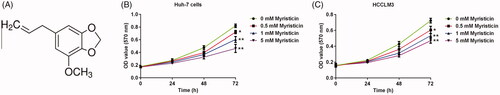
Myristicin is a natural alkenylbenzene compound. Its chemical formula is shown in . MTT assay was used to explore the effect of myristicin on HCC viability. Our results showed that compared to the control group, myristicin significantly suppresses Huh-7 () and HCCLM3 () cell proliferation.
Figure 1. Myristicin suppresses HCC proliferation in a dose-dependent manner. (A) The chemical formula of myristicin. (B, C) MTT analysis revealing cell viability when Huh-7 cells and HCCLM3 cells were treated with 0.5, 1 and 5 mM myristicin for 24 h, 48 h and 72 h. *,**p < 0.05, 0.01 vs. 0 mM myristicin treatment group.
The effect of myristicin on HCC apoptosis
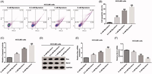
The effect of myristicin on early and late apoptosis was examined using a flow cytometer (FCM) assay to detect cell apoptosis. FCM assay showed that compared to the control group, myristicin significantly induced apoptosis and improved caspase-3 activity in Huh-7 cells () and HCCLM3 cells (). Meanwhile, myristicin decreased Bcl-2 expression and increased Bax expression at protein and mRNA levels in Huh-7 cells () and HCCLM3 cells (). Taken together, myristicin promoted cell apoptosis.
Figure 2. Myristicin induces cell apoptosis in Huh-7 cells in a dose-dependent manner. (A) FCM assay. (B) Cell apoptosis ratio analysed by a GraphPad Prism 6.0 software (La Jolla, CA). (C) Caspase3 activity was measured by the caspase3 detection kit. (D) Bcl-2 and Bax protein expression analysis by Western blot. (E, F) Bcl-2 and Bax mRNA expression analysis by qRT-PCR. *,**p < 0.05, 0.01 vs. 0 mM myristicin treatment group.
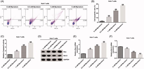
Figure 3. Myristicin induces cell apoptosis in HCCLM3 cells. (A) FCM assay. (B) Cell apoptosis ratio analysed by a GraphPad Prism 6.0 software (La Jolla, CA). (C) Caspase-3 activity was measured by the caspase-3 detection kit. (D) Bcl-2 and Bax protein expression analysis by Western blot. (E, F) Bcl-2 and Bax mRNA expression analysis by qRT-PCR. *,**p < 0.05, 0.01 vs. 0 mM myristicin treatment group.
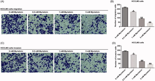
The effect of myristicin on the migration and invasion of HCC
Our results demonstrated that with the increase of myristicin concentration, migration () and invasion () ability of Huh-7 cells was significantly inhibited compared with the control group. The same results were observed in HCCLM3 cells ().
Figure 4. Myristicin suppressed migration and invasion of Huh-7 cells. (A) Transwell analysis of cell migration without matrigel in the lower chamber. (B) The number of migratory cells expressed as mean ± S.D. (C) Transwell analysis of cell invasion with matrigel in the lower chamber. (D) The number of invasive cells expressed as mean ± S.D. *,**p < 0.05, 0.01 vs. 0 mM myristicin treatment group.
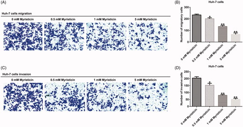
Figure 5. Myristicin suppressed migration and invasion of HCCLM3 cells. (A) Transwell analysis of HCCLM3 cell migration. (B) The number of migratory cells. (C) Transwell analysis of HCCLM3 cell invasion. (D) The number of invasive cells. *,**p < 0.05, 0.01 vs. 0 mM myristicin treatment group.
The effect of myristicin on EMT of HCC
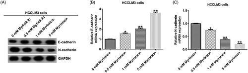
Epithelial–mesenchymal transition (EMT) is an important sign of cancer metastasis. We detected EMT-related protein expression. Western blot assay indicated that compared to the control group, myristicin increased E-cadherin expression and decreased N-cadherin expression in HCC cells ( and ). In addition, quantitative real time-polymerase chain reaction (qRT-PCR) assay showed that compared to the control group, E-cadherin was significantly up-regulated ( and ) and N-cadherin was down-regulated ( and ) in HCC cells.
Figure 6. Myristicin inhibits EMT in Huh-7 cells. Huh-7 cells were treated with 0.5, 1 and 5 mM myristicin for 48 h. (A) Western blot analysis of E-cadherin and N-cadherin expression. (B) qRT-PCR analysis of E-cadherin expression. (C) qRT-PCR analysis of N-cadherin expression. *,**p < 0.05, 0.01 vs. 0 mM myristicin treatment group.
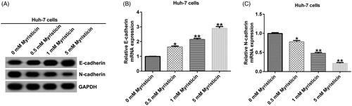
Figure 7. Myristicin inhibits EMT in HCCLM3 cells. (A) Western blot analysis of E-cadherin and N-cadherin expression. (B) qRT-PCR analysis of E-cadherin expression. (C) qRT-PCR analysis of N-cadherin expression. *,**p < 0.05, 0.01 vs. 0 mM myristicin treatment group.
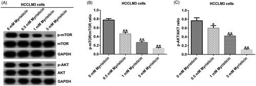
Myristicin suppressed PI3K/Akt/mTOR signalling pathway in HCC
To explore the mechanism of myristicin in HCC, we detected some protein expression in phosphatidylinositol 3-kinase (PI3K)/Akt/mTOR signalling pathway. Western blot assay showed that in Huh-7 and HCCLM3 cells, myristicin decreased p-mTOR and p-AKT expression ( and ) and significantly decreased the ratio of p-mTOR/mTOR ( and ) and p-AKT/AKT ( and ) in HCC cells.
Figure 8. Myristicin suppresses the PI3K/Akt/mTOR signalling pathway in Huh-7 cells. (A) Western blot analysis of p-mTOR and p-AKT expression in Huh-7 cells. (B) The ratio of p-mTOR/mTOR in Huh-7 cells. (C) The ratio of p-AKT/AKT in Huh-7 cells. *,**p < 0.05, 0.01 vs. 0 mM myristicin treatment group.
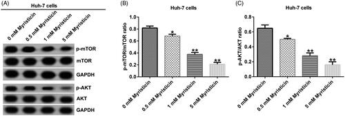
Figure 9. Myristicin suppresses the PI3K/Akt/mTOR signalling pathway in HCCLM3 cells. (A) Western blot analysis of p-mTOR and p-AKT expression in HCCLM3 cells. (B) The ratio of p-mTOR/mTOR in HCCLM3 cells. (C) The ratio of p-AKT/AKT in HCCLM3 cells. *,**p < 0.05, 0.01 vs. 0 mM myristicin treatment group.
Discussion
The development of hepatic carcinoma has seriously affected people’s living standards. In recent years, although the medical level has been continuously improved, hepatic carcinoma has a high mortality rate due to the difficulty of early diagnosis of hepatic carcinoma patients, fewer therapeutic targets and poor postoperative recovery. After surgical resection of hepatic carcinoma patients, the number of patients who survived one year after surgery was more than 80%, and the number of patients who survived 5 years was more than 50% (Margarit et al. Citation2005). Therefore, it is very important to continuously explore new treatment strategies to treat hepatic carcinoma patients. In the present study, we provided some evidence that myristicin is a natural compound that can be used for HCC treatment to a certain extent.
Myristicin is the main active ingredient of nutmeg (Jana and Shekhawat Citation2010; Muchtaridi et al. Citation2010) and coriander leaf oil (Zheng et al. Citation1992; Wei and Shibamoto Citation2007), which is mainly used as a flavouring agent, but also as a spice in the cosmetics industry. Previous research showed that myristicin could produce excitement and hallucinogenic effects. It may also be potentially toxic, as overdose can cause hallucinations and, in some cases, even coma (Stein et al. Citation2001; Sivathanu et al. Citation2014). Lee and Park (Citation2011) demonstrated that myristicin has anti-inflammatory effects by inhibiting inflammatory factor expression. Lee et al. (Citation2005) further found that myristicin induces early apoptotic events in human neuroblastoma, including cytochrome c release, caspase-3 activation and PARP lysis, leading to the death of SK-N-SH cells. In our study, we investigated the effect of myristicin on hepatic carcinoma. Our results showed that myristicin suppressed cell proliferation and induced apoptosis in Huh-7 and HCCLM3 cells.
Hepatic carcinoma is an aggressive type of tumour with a high rate of metastasis. As the continuous metastasis of tumours leads to high incidence and mortality, it is necessary to effectively treat hepatic carcinoma from the perspective of inhibiting cancer metastasis (Wu et al. Citation2015). Tumour metastasis depends on the migration and invasion capabilities of cancer cells (Hua et al. Citation2018). In this study, myristicin inhibited cell migration and invasion.
EMT is a process that refers to the transformation of epithelium to mesenchymal cells, which gives cells the ability to transfer and invade (Lamouille et al. Citation2014). EMT is currently considered an important reminder of cancer metastasis (Ye et al. Citation2017). In addition, during the early stage of cancer metastasis, the adhesion of cancer cells can be separated by reducing EMT. The main markers of EMT include epithelial cell marker (E-cadherin) and mesenchymal cell markers (N-cadherin). In this study, we found that myristicin suppressed EMT by increasing E-cadherin expression and decreasing N-cadherin expression.
PI3K/Akt/mTOR signalling pathway is involved in development of many cancers (Costa et al. Citation2018; Alzahrani Citation2019; Ediriweera et al. Citation2019). Activation of this signalling pathway can promote cell growth, proliferation and participate in angiogenesis (Alzahrani Citation2019). It can also exert a crucial role in mediating cell movement, invasion and metastasis (Jeong et al. Citation2014; Ramadan et al. Citation2020). PI3K/Akt/mTOR signalling pathway inhibition has been shown to be used to inhibit HCC (Kudo Citation2011; Ye et al. Citation2019). Hua et al. (Citation2018) indicated that ruscogenin inhibits HCC migration and invasion by regulating PI3K/Akt/mTOR signalling pathway. Furthermore, Song et al. (Citation2020) indicated that miR-29-3p overexpression suppresses HCC growth by inhibition of Robo1 and inactivation of PI3K/Akt/mTOR signalling pathway. Also, Sun et al. (Citation2019) showed that miR-1914 exerted the inhibiting effect through the PI3K/Akt/mTOR signalling pathway in HCC. Consistent with the previous research results, our data suggested that myristicin suppresses the activation of the PI3K/Akt/mTOR signalling pathway in hepatic carcinoma.
However, this study is a preliminary in vitro study of the effect of myristicin on hepatocellular carcinoma. In order to make myristicin's effect on hepatocellular carcinoma more convincing, more in-depth research is needed. For example, the role of myristicin in other types of hepatic carcinoma cell lines should be studied. Besides, the effect of more doses of myristicin (a minimum of five doses is required) on HCC cells should be studied. Moreover, additional in vivo studies are required to confirm these findings. Therefore, in our next study, we plan to examine the effect of more dose of myristicin in other types of hepatic carcinoma cell lines and in a mouse model bearing an HCC tumour.
Conclusions
Myristicin prevents the malignant biological behaviour of hepatic carcinoma cells by inhibiting the PI3K/Akt/mTOR signalling pathway. Thus, myristicin is a potential therapeutic agent for hepatic carcinoma.
Disclosure statement
The authors declare no conflict of interest.
Additional information
Funding
References
- Alzahrani AS. 2019. PI3K/Akt/mTOR inhibitors in cancer: at the bench and bedside. Semin Cancer Biol. 59:125–132.
- Bray F, Ferlay J, Soerjomataram I, Siegel RL, Torre LA, Jemal A. 2018. Global cancer statistics 2018: GLOBOCAN estimates of incidence and mortality worldwide for 36 cancers in 185 countries. CA Cancer J Clin. 68(6):394–424.
- Chedid MF, Kruel CRP, Pinto MA, Grezzana-Filho TJM, Leipnitz I, Kruel CDP, Scaffaro LA, Chedid AD. 2017. Hepatocellular carcinoma: diagnosis and operative management. Arq Bras Cir Dig. 30(4):272–278.
- Costa RLB, Han HS, Gradishar WJ. 2018. Targeting the PI3K/AKT/mTOR pathway in triple-negative breast cancer: a review. Breast Cancer Res Treat. 169(3):397–406.
- Ediriweera MK, Tennekoon KH, Samarakoon SR. 2019. Role of the PI3K/AKT/mTOR signaling pathway in ovarian cancer: biological and therapeutic significance. Semin Cancer Biol. 59:147–160.
- El Jabbour T, Lagana SM, Lee H. 2019. Update on hepatocellular carcinoma: pathologists' review. World J Gastroenterol. 25(14):1653–1665.
- Farazi PA, DePinho RA. 2006. Hepatocellular carcinoma pathogenesis: from genes to environment. Nat Rev Cancer. 6(9):674–687.
- Hallström H, Thuvander A. 1997. Toxicological evaluation of myristicin. Nat Toxins. 5(5):186–192.
- Hartke J, Johnson M, Ghabril M. 2017. The diagnosis and treatment of hepatocellular carcinoma. Semin Diagn Pathol. 34(2):153–159.
- Hua H, Zhu Y, Song YH. 2018. Ruscogenin suppressed the hepatocellular carcinoma metastasis via PI3K/Akt/mTOR signaling pathway. Biomed Pharmacother. 101:115–122.
- Jana S, Shekhawat GS. 2010. Anethum graveolens: an Indian traditional medicinal herb and spice. Pharmacogn Rev. 4(8):179–184.
- Jeong YJ, Choi Y, Shin JM, Cho HJ, Kang JH, Park KK, Choe JY, Bae YS, Han SM, Kim CH, et al. 2014. Melittin suppresses EGF-induced cell motility and invasion by inhibiting PI3K/Akt/mTOR signaling pathway in breast cancer cells. Food Chem Toxicol. 68:218–225.
- Kudo M. 2011. mTOR inhibitor for the treatment of hepatocellular carcinoma. Dig Dis. 29(3):310–315.
- Lamouille S, Xu J, Derynck R. 2014. Molecular mechanisms of epithelial–mesenchymal transition. Nat Rev Mol Cell Biol. 15(3):178–196.
- Lee BK, Kim JH, Jung JW, Choi JW, Han ES, Lee SH, Ko KH, Ryu JH. 2005. Myristicin-induced neurotoxicity in human neuroblastoma SK-N-SH cells. Toxicol Lett. 157(1):49–56.
- Lee JY, Park W. 2011. Anti-inflammatory effect of myristicin on RAW 264.7 macrophages stimulated with polyinosinic–polycytidylic acid. Molecules. 16(8):7132–7142.
- Livak KJ, Schmittgen TD. 2001. Analysis of relative gene expression data using real-time quantitative PCR and the 2−ΔΔCT method. Methods. 25(4):402–408.
- Margarit C, Escartín A, Castells L, Vargas V, Allende E, Bilbao I. 2005. Resection for hepatocellular carcinoma is a good option in Child-Turcotte-Pugh class A patients with cirrhosis who are eligible for liver transplantation. Liver Transpl. 11(10):1242–1251.
- Martins C, Doran C, Laires A, Rueff J, Rodrigues AS. 2011. Genotoxic and apoptotic activities of the food flavourings myristicin and eugenol in AA8 and XRCC1 deficient EM9 cells. Food Chem Toxicol. 49(2):385–392.
- Muchtaridi A, Subarnas A, Apriyantono R. 2010. Identification of compounds in the essential oil of nutmeg seeds (Myristica fragrans Houtt.) that inhibit locomotor activity in mice. Int J Mol Sci. 11:4771–4781.
- Ramadan F, Fahs A, Ghayad SE, Saab R. 2020. Signaling pathways in rhabdomyosarcoma invasion and metastasis. Cancer Metastasis Rev. 39(1):287–301.
- Siegel RL, Miller KD, Jemal A. 2019. Cancer statistics, 2019. CA Cancer J Clin. 69(1):7–34.
- Sivathanu S, Sampath S, David HS, Rajavelu KK. 2014. Myristicin and phenytoin toxicity in an infant. BMJ Case Rep. 2014:bcr2013203000.
- Song Q, Zhang H, He J, Kong H, Tao R, Huang Y, Yu H, Zhang Z, Huang Z, Wei L, et al. 2020. Long non-coding RNA LINC00473 acts as a microRNA-29a-3p sponge to promote hepatocellular carcinoma development by activating Robo1-dependent PI3K/AKT/mTOR signaling pathway. Ther Adv Med Oncol. 12:1758835920937890.
- Stefano VD, Pitonzo R, Schillaci D. 2011. Antimicrobial and antiproliferative activity of Athamanta sicula L. (Apiaceae). Pharmacogn Mag. 7(25):31–34.
- Stein U, Greyer H, Hentschel H. 2001. Nutmeg (myristicin) poisoning-report on a fatal case and a series of cases recorded by a poison information centre. Forensic Sci Int. 118(1):87–90.
- Sun L, Wang L, Chen T, Yao B, Wang Y, Li Q, Yang W, Liu Z. 2019. microRNA-1914, which is regulated by lncRNA DUXAP10, inhibits cell proliferation by targeting the GPR39-mediated PI3K/AKT/mTOR pathway in HCC. J Cell Mol Med. 23(12):8292–8304.
- Wei A, Shibamoto T. 2007. Antioxidant activities and volatile constituents of various essential oils. J Agric Food Chem. 55(5):1737–1742.
- Wu YJ, Neoh CA, Tsao CY, Su JH, Li HH. 2015. Sinulariolide suppresses human hepatocellular carcinoma cell migration and invasion by inhibiting matrix metalloproteinase-2/-9 through MAPKs and PI3K/Akt signaling pathways. Int J Mol Sci. 16(7):16469–16482.
- Yang Y, Chen L, Gu J, Zhang H, Yuan J, Lian Q, Lv G, Wang S, Wu Y, Yang YT, et al. 2017. Recurrently deregulated lncRNAs in hepatocellular carcinoma. Nat Commun. 8:14421.
- Ye L, Mayerle J, Ziesch A, Reiter FP, Gerbes AL, De Toni EN. 2019. The PI3K inhibitor copanlisib synergizes with sorafenib to induce cell death in hepatocellular carcinoma. Cell Death Discov. 5:86–97.
- Ye X, Brabletz T, Kang Y, Longmore GD, Nieto MA, Stanger BZ, Yang J, Weinberg RA. 2017. Upholding a role for EMT in breast cancer metastasis. Nature. 547(7661):E1–E3.
- Zheng GQ, Kenney PM, Zhang J, Lam LKT. 1992. Inhibition of benzo[a]pyrene-induced tumorigenesis by myristicin, a volatile aroma constituent of parsley leaf oil. Carcinogenesis. 13(10):1921–1923.