
Abstract
As stated in the comprehensive diagnostic criteria for IgG4-related disease (IgG4-RD), IgG4-RD is characterized by elevated serum IgG4 level and pathological findings, characterized by infiltration of IgG4-positive plasma cells. In addition to fibrotic changes, dysregulated activation of lymphocytes is considered as one of major pathogenic events in IgG4-RD. Among lymphocytes, the importance of plasmablast, T follicular helper (Tfh) cells, T type 2 helper (Th2) cells, T regulatory (Treg) cells, and CD4 positive T cells with cytotoxic activity has been reported. Conversely, comprehensive immunophenotyping in patients with IgG4-RD revealed that there are two different axes consisting plasmablast-Tfh cells and Treg cells. There is need for research to seek out molecules associated with these immunocompetent cell interactions. It is believed that this will contribute to the future application to disease-specific treatment for IgG4-RD.
Introduction
IgG4-related disease (IgG4-RD) is a relatively new concept with a Japanese origin. This is triggered by the report of high serum IgG4 in sclerosing pancreatitis [Citation1]. IgG4-RD is characterized by hyperimmunoglobulinaemia G4, marked infiltration of IgG4-positive plasma cells into swollen or hypertrophic organs, and fibrosis [Citation2]. The comprehensive diagnostic criteria for IgG4-RD [Citation3] are commonly used for the diagnosis of IgG4-RD. This is based on elevated serum IgG4 levels and pathological findings. The histopathological findings include infiltration of lymphocytes and IgG4-positive plasmablasts, along with characteristic fibrosis and sclerosis of tissues [Citation4]. As is clear from these findings, lymphocyte seems to have a central role in the pathogenesis of IgG4-RD.
This article outlines the pathological processes of IgG4-RD, with emphasis on the immunophenotypes.
Lymphocytes involved in the pathological processes of IgG4-RD
The pathology of IgG4-RD is not fully understood. One of the major barriers for elucidating pathological processes of IgG4-RD is that the characteristic absence of IgG4 in mice. In humans, there are four subclasses namely; IgG1, IgG2, IgG3, and IgG4. However, only three classes (IgG1, IgG2a, IgG2b, and IgG3) exists in the mice. Therefore, there is no suitable animal (mouse) model for this disorder. The role of IgG4 in the pathophysiology of IgG4-RD is not fully understood, making it difficult to elucidate the pathology. The involvement of human leucocyte antigens such as HLA DRB 1*04:05-DQB1*04:01 has been suggested especially in autoimmune pancreatitis [Citation5]. However, in contrast to other autoimmune diseases such as systemic lupus erythematosus, familial occurrence has not been reported for this disease. Also, elucidation of pathological conditions using genetic techniques is limited. Currently, comprehensive single nucleotide polymorphism analysis is performed in Japan, and the results are expected to generate new findings.
In addition to basic research for IgG4-RD, there are hypothesis that cannot be explained theoretically in the clinical setting. In laboratory examinations, elevated serum IgG4, elevated IgE, hypocomplementemia, and low inflammatory response are generally seen in patients with IgG4-RD. However, the pathological significance of IgG4 in patients with IgG4-RD is not known, as mentioned earlier. IgG1 and IgG3 cause inflammation through complement activation and opsonization, while IgG4 lacks these functions. Furthermore, high level of IgG4 is sometimes seen even in patients who are in clinical remission. It is unknown whether IgG4 in IgG4-RD acts as an exacerbating factor. Meanwhile, since hypocomplementemia is seen as a result of the formation of strong immune complex, serious clinical conditions manifests in patients with hypocomplementemia. For example, activity of lupus nephritis is known to be strongly associated with hypocomplementemia. In contrast, there are no evidences of autoantibody production and involvement of immune complex in patients with IgG4-RD, although hypocomplementemia is seen. In order to resolve these clinical questions, there is need for the elucidation of the pathological processes that can offer a theoretical explanation of these phenomenon.
On the other hand, the involvement of lymphocyte is conclusive from the viewpoint that immunosuppressive therapy is effective in patients with IgG4-RD. Although sensitive to glucocorticoid treatment, the disease has been reported to relapse in nearly half the cases [Citation6]. In order to prevent relapse, rituximab which is a B cell depletion therapy have been used off-label in Europe and United States and seems to be effective [Citation7,Citation8]. In addition, there is a report showing the effectiveness of abatacept from Japan [Citation9]. Since these lymphocyte-targeted therapies have positive effects, lymphocyte involvement in IgG4-RD is strongly inferred. However, there are no drugs (including rituximab) that demonstrated high efficacy in a double blind, placebo-controlled trial. In clinical settings, other medications besides are used in IgG4-RD patients. These medications include azathioprine, methotrexate, and mycophenolate mofetil [Citation10]. In other words, because the pathogenesis of IgG4-RD is unclear, the treatments are selected empirically. In order to create a more effective treatment, clarification of pathology and disease-specific treatment is needed.
The subsets of T and B lymphocytes
Differentiation of CD4+ T helper cells into functionally distinct helper T subsets is critical not only for host defense but immune-related diseases [Citation11]. T helper1 (Th1) cells, which express T-bet and selectively produce interferon (IFN)-γ and protect the host against intracellular infections. Th2 cells, which express GATA3 and produce interleukin (IL)-4, IL-5 and IL-13 and mediate host defense against helminths. Th17 cells selectively produce IL-17 and express transcription factor RORγt. T regulatory (Treg) cells are another CD4+ lineage with essential immunosuppressive functions and expression of the master regulator transcription factor Foxp3.
The newest T helper subset, T follicular helper (Tfh) cells, have emerged as a critical regulator of autoimmunity. Tfh cells provide B cell help by promoting class switching of B cells and are defined by the expression of master regulator Bcl6 and effector cytokine IL-21, along with key surface molecules, such as PD-1, CXCR5, CD40L, and ICOS [Citation12]. Thus, the interaction between B cell and Tfh cell is necessary for antibody production and play an important role in antibody-driven diseases, such as IgG4-RD.
On the other hand, B cells produce antibodies and control the immune system through cytokine production and antigen presentation. CD19+ B cells are classified into three subsets based on CD27 and IgD expression: naive B cells (CD27-IgD+), unswitched memory B cells (CD27+IgD+), and class-switched memory B cells (CD27+IgD-). In addition, B cell subsets exhibit effector functions and differentiate into plasmablasts that secrete more antibodies than B cells.
Immune cells involved in IgG4-RD
Until now, the importance of several immune cells has been reported in the basic research of IgG4-RD (). From this aspect, plasmablast is viewed as a representative immunocompetent cell. In patients with IgG4-RD, it has been reported that the number of plasmablast, which expresses CD19+CD20-CD38+CD27+, is increased in comparison to healthy individuals [Citation13]. In addition, these plasmablasts express IgG4 and proliferates oligoclonally. Meanwhile, another report has shown the correlation between plasmablast and organ disorders [Citation14]. The coefficient for these correlations was higher than that between serum IgG4 levels and organ disorders, suggesting involvement in the pathology of plasmablast. It is known that the use of rituximab [Citation14] and glucocorticoids [Citation15] result in the reduction of these increased plasmablasts.
Table 1. Immune cells in IgG4-related disease.
On the other hand, there have been assumed that plasmablast contributes to excessive production of gamma globulin in patients with IgG4-RD, resulting in hypergammaglobulinemia. On the other hand, there have been several reports on T cells as well. In particular, an increase in Tfh cells has been reported [Citation16]. Tfh cells play an essential role in the formation of germinal centers, and hyperplasia of germinal center while infiltration of Tfh cells are observed in the target organ in patients with IgG4-RD. In another report, when Tfh cells from IgG4-RD patients were cultured with naive B cells, the differentiation into plasmablasts expressing IgG4 was found [Citation17]. Increase in peripheral Tfh cells can imply that Tfh play a role during the pathological processes in IgG4-RD. It is generally accepted that Tfh cells play an important role in the differentiation of B cells by producing IL-21. However, the interaction between Tfh cells and B cells in IgG4-RD is still unknown. Further investigation on both direct cell–cell contact and indirect paracrine signaling in the interaction among these cells will be needed.
Meanwhile, it is known that many patients with IgG4-RD are predisposed to allergies, and the history of atopic dermatitis and asthma are often seen. Eosinophilia and increased level of IgE in the peripheral blood also indicate allergic diathesis. In this respect, Th2 cells have also been considered important in the pathological processes of IgG4-RD. From previous papers, involvement of Th2 has been shown in patients with IgG4-RD concomitant with atopic dermatitis [Citation18,Citation19]. However, it has been reported that the involvement of GATA3-positive Th2 cells were only seen in cases concomitant with atopic dermatitis [Citation18], and the contribution of Th2 cell itself in the pathogenesis of IgG4-RD may be lower than that of Tfh cells and plasmablast. Recently, increased expression of IL-33 produced by macrophages in the salivary glands in patients with IgG4-RD has been shown [Citation20], and the involvement of Th2 cells, which highly express IL-33 receptor, has been noted. In addition, we have shown that Tfh cells possess plasticity [Citation21] and multifunctionality [Citation22]. Subclass of Tfh cells could produce Th2 cytokines [Citation23], suggesting that plasticity of Tfh cells play a role in pathogenesis.
It is believed that Treg cells play a key role in the onset or pathological processes in patients with IgG4-RD [Citation24]. In fact, it has been reported that expressions of IL-4, IL-10, and IL-13 in target organ were enhanced [Citation25,Citation26]. Histologically, the increased number of Treg cells in autoimmune pancreatitis [Citation27] and IgG4-related sclerosing cholangitis [Citation28], the increased number of CD4+CD25high T cells in autoimmune pancreatitis [Citation29], and the increased number of CD4+Foxp3+ T cells in the salivary glands of Mikulicz disease [Citation30] have been reported. Thus, Treg cells can be thought to infiltrate in almost all target organs of IgG4-RD. In general, Treg cells strongly express Foxp3 and negatively regulate immune responses via CTLA-4 expression and IL-10 production. In this theory, Treg cells may contribute to prevention of disease onset and disease progression. By contrast, in addition to natural Treg cells which mature in the thymus [Citation31], inducible Treg cells in periphery by TGF-β and small amount of IL-2 are known. The plasticity of inducible Treg cells to Th17 cells has been shown [Citation32]. From these aspects, the interpretation of the role of Treg cells in the pathogenesis is controversial.
Moreover, the importance of CD4+ T cells with cytotoxic activity (CD4+CTL) has been reported [Citation33,Citation34]. This report showed that CD4+ T cells, which were clonally proliferated in the target organ, expressed cytotoxic proteins such as granzyme B and perforin. These cells also produced important cytokines for fibrosis such as TGF-β and expressed SLAM7 which regulates B cell proliferation, suggesting an indirect association with the pathogenesis of IgG4-RD.
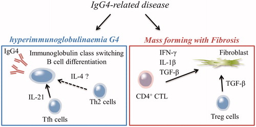
All of these reports are important for exploring the therapeutic target of IgG4-RD and have contributed to clarifying the pathology of IgG4-RD (). However, previous reports focused on a single or several immunocompetent cells. Therefore, the significance of immunocompetent cells in the immune network and their relationships among themselves are unknown.
Figure 1 Scheme of two important clinical findings in patients with IgG4-RD.
Immunophenotype in patients with IgG4-RD
In order to overcome this issue, we designed the study, in which we immunophenotyped T cells, B cells, dendritic cells (DCs), monocytes and natural killer (NK) cells from the peripheral blood of patients with IgG4-RD according to the Human Immunology Project Consortium (HIPC) program established by the National Institute of Allergy and Infectious Diseases (NIAID) [Citation35]. We have reported the association between immune phenotypes and the disease pathology, and their changes before and after treatment [Citation36]. This was the first report to describe comprehensive immunophenotyping in patients with IgG4-RD.
Regarding the differentiation of CD4+ helper cells, the proportion of those in the effector phase, which are considered terminally differentiated effector memory T cells, was significantly higher in patients than in healthy controls (4.6% vs. 3.7%). For the subsets of CD4+ helper cells, the proportions of Treg (5.3% vs. 4.0%) and Tfh cells (1.5% vs. 1.0%) were significantly higher than those in healthy controls. Regarding the differentiation of B cells, the proportion of class-switched memory B cells was higher (23.7% vs. 12.5%). Moreover, the proportion of plasmablasts characteristically increased to 16.5% in patients, compared with 4.2% in healthy controls. This increase was consistent with previous report which suggested that the increased plasmablast in IgG4-RD could be an indicator for the diagnosis [Citation14]. However, although the most pronounced increase number of plasmablast were seen in comparison to healthy individuals, increased plasmablast itself was also seen in other diseases such as systemic lupus erythematosus and was not specific for IgG4-RD [Citation37].
Next, the correlations among these immunocompetent cells were assessed. The proportion of plasmablasts in patients, which was markedly different from that in healthy controls, was associated with that of Tfh cells. The proportions of both plasmablast and Tfh cells were correlated with serum IgG levels, and the importance of the plasmablast–Tfh cell axis was confirmed. In addition, glucocorticoid therapy reduced the proportions of plasmablasts and Tfh along with improvement in clinical signs.
The evaluation of inflammatory site is the most important in the investigation of human diseases. Therefore, we examined the association between Tfh cells in peripheral blood and those that exhibited local infiltration. As a result, the proportion of Tfh cells in the peripheral blood reflected that of the Tfh cells that infiltrated into the actual tissue. This result indicated that the immunophenotype of peripheral blood could reflect the local immune response.
Since these results were based on comprehensive analysis, plasmablast and Tfh cells, the importance of which had already been shown in previous reports, were indicated to be more important than other immunocompetent cells. However, abnormal differentiation of B cells has been seen in other autoimmune diseases such as systemic lupus erythematosus and Sjögren syndrome as well, and could also be central pathological role in these diseases. Is the characteristic of IgG4-RD in comparison with these diseases is B cell immunoglobulin class switching to IgG4? Meanwhile, the mechanism of this class switching to IgG4 is not well known. Although cytokines such as IL-4, IL-10, and IL-21 has been considered as the factor which can induce immunoglobulin class switching to IgG4, these factors cannot be said to be specific only to class switching to IgG4, while promoting the B cell differentiation and class switching to other subtypes as well. Not only interactions through cytokines but also cell–cell contact between T cells and B cells may be also important, and investigation of the mechanism how to promote immunoglobulin class switching to IgG4 is likely to lead elucidation of the pathological processes of IgG4-RD.
On the contrary, interesting findings were obtained regarding Treg cells. As described above, although lots of reports have shown high proportion of Treg cells in IgG4-RD patients, the involvement of Treg cells in the pathogenesis remains unclear. In the present study, high proportion of Treg cells in the peripheral blood of IgG4-RD patients was observed. Most importantly, when the patients were divided into those with and without extraglandular symptoms, the proportions of not only plasmablasts and Tfh cells but also memory Treg cells were found to be significantly increased in patients with extraglandular symptoms. In addition, the proportion of memory Treg cells did not correlate with serum levels of IgG or IgG4. This tendency was different from that of plasmablasts and Tfh. Furthermore, even when patients were treated with glucocorticoids, there were no changes in the proportion of Treg cells.
Treg cells can be thought to infiltrate in almost all target organs of IgG4-RD as previously reported, and consistent results were obtained in the analysis of comprehensive immunophenotyping. The differences in the change between Treg cells and plasmablast/Tfh cells after treatment suggest that there is a Treg cells axis different from plasmablast–Tfh axis in the pathological processes of IgG4-RD. It is known that TGF-β is an important cytokines for inducible Treg differentiation. In addition, TGF-β itself is known to contribute to fibrosis as well. The increase in Treg cells in peripheral blood and target organ could be a result reflecting these conditions.
Unfortunately, this study was conducted based on HIPC [Citation35] proposed by NIAID and Federation of Clinical Immunology Societies (FOCIS), therefore, immunocompetent cells other than the conventional immunophenotype could not be detected. Although the importance of CD4+CTL has been reported, this study could not evaluate them. In addition to this report, reproducibility from other facilities is desired.
Conclusion
In this review, the contribution of each immunocompetent cell to the pathogenesis of IgG4-RD () and the comprehensive immunophenotype in patients with IgG4-RD were outlined. This disease was reported for the first time from Japan. Subsequently, many clinical and basic studies approaching the pathological processes of IgG4-RD have been reported from Japan. On the other hand, animal models have not yet been established, as stated at the beginning in this article. Therefore, research using patient samples takes a leading part. One of the biggest challenges in human immunology is to distinguish between cause and result. Careful observation is needed to identify and integrate the target molecules for treatment. If effectiveness of the targeted therapy is verified, their use has the potential to generate substantial progress in our understanding of the disease process and etiopathogenesis. The analysis of comprehensive immunophenotype in patients with IgG4-RD revealed the importance of plasmablast and Tfh cells, supporting the previous studies. The research for the interaction between human Tfh cells and human B cells will lead to future disease specific therapy for IgG4-RD.
Conflict of interest
S. Kubo has received speaking fees from Bristol-Myers, Pfizer, Eli Lilly, and Takeda. S. Nakayamada has received speaking fees from Bristol-Myers, Sanofi, Abbvie, Eisai, Eli Lilly, Chugai, Pfizer, Takeda, and also research grants from Mitsubishi-Tanabe, Novartis and MSD. Y. Tanaka has received speaking fees and/or honoraria from Daiichi-Sankyo, Astellas, Eli Lilly, Chugai, Sanofi, Abbvie, YL Biologics, Bristol-Myers, Glaxo-Smithkline, UCB, Mitsubishi-Tanabe, Novartis, Eisai, Takeda, Janssen, Asahi-kasei and has received research grants from Mitsubishi-Tanabe, Bristol-Myers, Eisai, Chugai, Takeda, Abbvie, Astellas, Daiichi-Sankyo, Ono, MSD, Taisho-Toyama.
References
- Hamano H, Kawa S, Horiuchi A, Unno H, Furuya N, Akamatsu T, et al. High serum IgG4 concentrations in patients with sclerosing pancreatitis. N Engl J Med. 2001;344(10):732–8.
- Kamisawa T, Zen Y, Pillai S, Stone JH. IgG4-related disease. Lancet. 2015;385(9976):1460–71.
- Umehara H, Okazaki K, Masaki Y, Kawano M, Yamamoto M, Saeki T, et al. Comprehensive diagnostic criteria for IgG4-related disease (IgG4-RD), 2011. Mod Rheumatol. 2012;22(1):21–30.
- Deshpande V, Zen Y, Chan JK, Yi EE, Sato Y, Yoshino T, et al. Consensus statement on the pathology of IgG4-related disease. Mod Pathol. 2012;25(9):1181–92.
- Ota M, Katsuyama Y, Hamano H, Umemura T, Kimura A, Yoshizawa K, et al. Two critical genes (HLA-DRB1 and ABCF1) in the HLA region are associated with the susceptibility to autoimmune pancreatitis. Immunogenetics 2007;59(1):45–52.
- Kamisawa T, Shimosegawa T, Okazaki K, Nishino T, Watanabe H, Kanno A, et al. Standard steroid treatment for autoimmune pancreatitis. Gut 2009;58(11):1504–7.
- Carruthers MN, Topazian MD, Khosroshahi A, Witzig TE, Wallace ZS, Hart PA, et al. Rituximab for IgG4-related disease: a prospective, open-label trial. Ann Rheum Dis. 2015;74(6):1171–7.
- Della-Torre E, Feeney E, Deshpande V, Mattoo H, Mahajan V, Kulikova M, et al. B-cell depletion attenuates serological biomarkers of fibrosis and myofibroblast activation in IgG4-related disease. Ann Rheum Dis. 2015;74(12):2236–43.
- Yamamoto M, Takahashi H, Takano K, Shimizu Y, Sakurai N, Suzuki C, et al. Efficacy of abatacept for IgG4-related disease over 8 months. Ann Rheum Dis. 2016;75(8):1576.
- Khosroshahi A, Wallace ZS, Crowe JL, Akamizu T, Azumi A, Carruthers MN, et al. International consensus guidance statement on the management and treatment of IgG4-related disease. Arthritis Rheumatol. 2015;67(7):1688–99.
- Nakayamada S, Takahashi H, Kanno Y, O'Shea JJ. Helper T cell diversity and plasticity. Curr Opin Immunol. 2012;24(3):297–302.
- Vinuesa CG, Linterman MA, Yu D, MacLennan IC. Follicular helper T cells. Annu Rev Immunol. 2016; 34:335–68.
- Mattoo H, Mahajan VS, Della-Torre E, Sekigami Y, Carruthers M, Wallace ZS, et al. De novo oligoclonal expansions of circulating plasmablasts in active and relapsing IgG4-related disease. J Allergy Clin Immunol. 2014;134(3):679–87.
- Wallace ZS, Mattoo H, Carruthers M, Mahajan VS, Della Torre E, Lee H, et al. Plasmablasts as a biomarker for IgG4-related disease, independent of serum IgG4 concentrations. Ann Rheum Dis. 2015;74(1):190–5.
- Iwata S, Saito K, Hirata S, Tanaka Y. Phenotypic changes of lymphocyte in a patient with IgG4-related disease after corticosteroid therapy. Ann Rheum Dis. 2012;71(12):2058–9.
- Maehara T, Moriyama M, Nakashima H, Miyake K, Hayashida JN, Tanaka A, et al. Interleukin-21 contributes to germinal centre formation and immunoglobulin G4 production in IgG4-related dacryoadenitis and sialoadenitis, so-called Mikulicz's disease. Ann Rheum Dis. 2012;71(12):2011–9.
- Akiyama M, Yasuoka H, Yamaoka K, Suzuki K, Kaneko Y, Kondo H, et al. Enhanced IgG4 production by follicular helper 2T cells and the involvement of follicular helper 1T cells in the pathogenesis of IgG4-related disease. Arthritis Res Ther. 2016;18:167.
- Mattoo H, Della-Torre E, Mahajan VS, Stone JH, Pillai S. Circulating Th2 memory cells in IgG4-related disease are restricted to a defined subset of subjects with atopy. Allergy 2014;69(3):399–402.
- Della Torre E, Mattoo H, Mahajan VS, Carruthers M, Pillai S, Stone JH. Prevalence of atopy, eosinophilia, and IgE elevation in IgG4-related disease. Allergy 2014;69(2):269–72.
- Furukawa S, Moriyama M, Miyake K, Nakashima H, Tanaka A, Maehara T, et al. Interleukin-33 produced by M2 macrophages and other immune cells contributes to Th2 immune reaction of IgG4-related disease. Sci Rep. 2017;7:42413.
- Nakayamada S, Kanno Y, Takahashi H, Jankovic D, Lu KT, Johnson TA, et al. Early Th1 cell differentiation is marked by a Tfh cell-like transition. Immunity 2011;35(6):919–31.
- Ma X, Nakayamada S, Kubo S, Sakata K, Yamagata K, Miyazaki Y, et al. Expansion of T follicular helper-T helper 1 like cells through epigenetic regulation by signal transducer and activator of transcription factors. Ann Rheum Dis. 2018;77(9):1354–61.
- Morita R, Schmitt N, Bentebibel SE, Ranganathan R, Bourdery L, Zurawski G, et al. Human blood CXCR5(+)CD4(+) T cells are counterparts of T follicular cells and contain specific subsets that differentially support antibody secretion. Immunity 2011;34(1):108–21.
- Stone JH, Zen Y, Deshpande V. IgG4-related disease. N Engl J Med. 2012;366(6):539–51.
- Zen Y, Fujii T, Harada K, Kawano M, Yamada K, Takahira M, et al. Th2 and regulatory immune reactions are increased in immunoglobin G4-related sclerosing pancreatitis and cholangitis. Hepatology. 2007;45(6):1538–46.
- Miyake K, Moriyama M, Aizawa K, Nagano S, Inoue Y, Sadanaga A, et al. Peripheral CD4+ T cells showing a Th2 phenotype in a patient with Mikulicz's disease associated with lymphadenopathy and pleural effusion. Mod Rheumatol. 2008;18(1):86–90.
- Kusuda T, Uchida K, Miyoshi H, Koyabu M, Satoi S, Takaoka M, et al. Involvement of inducible costimulator- and interleukin 10-positive regulatory T cells in the development of IgG4-related autoimmune pancreatitis. Pancreas 2011;40(7):1120–30.
- Koyabu M, Uchida K, Miyoshi H, Sakaguchi Y, Fukui T, Ikeda H, et al. Analysis of regulatory T cells and IgG4-positive plasma cells among patients of IgG4-related sclerosing cholangitis and autoimmune liver diseases. J Gastroenterol. 2010;45(7):732–41.
- Miyoshi H, Uchida K, Taniguchi T, Yazumi S, Matsushita M, Takaoka M, et al. Circulating naïve and CD4 + CD25high regulatory T cells in patients with autoimmune pancreatitis. Pancreas 2008;36(2):133–40.
- Tanaka A, Moriyama M, Nakashima H, Miyake K, Hayashida JN, Maehara T, et al. Th2 and regulatory immune reactions contribute to IgG4 production and the initiation of Mikulicz disease. Arthritis Rheum. 2012;64(1):254–63.
- Fehervari Z, Sakaguchi S. Development and function of CD25 + CD4+ regulatory T cells. Curr Opin Immunol. 2004;16(2):203–8.
- Zhou X, Bailey-Bucktrout SL, Jeker LT, Penaranda C, Martinez-Llordella M, Ashby M, et al. Instability of the transcription factor Foxp3 leads to the generation of pathogenic memory T cells in vivo. Nat Immunol. 2009; 10:1000–7.
- Mattoo H, Mahajan VS, Maehara T, Deshpande V, Della-Torre E, Wallace ZS, et al. Clonal expansion of CD4(+) cytotoxic T lymphocytes in patients with IgG4-related disease. J Allergy Clin Immunol. 2016;138(3):825–38.
- Maehara T, Mattoo H, Ohta M, Mahajan VS, Moriyama M, Yamauchi M, et al. Lesional CD4+ IFN-γ+ cytotoxic T lymphocytes in IgG4-related dacryoadenitis and sialoadenitis. Ann Rheum Dis. 2017;76(2):377–85.
- Maecker HT, McCoy JP, Nussenblatt R. Standardizing immunophenotyping for the Human Immunology Project. Nat Rev Immunol. 2012;12(3):191–200.
- Kubo S, Nakayamada S, Zhao J, Yoshikawa M, Miyazaki Y, Nawata A, et al. Correlation of T follicular helper cells and plasmablasts with the development of organ involvement in patients with IgG4-related disease. Rheumatology (Oxford). 2018;57(3):514–24.
- Kubo S, Nakayamada S, Yoshikawa M, Miyazaki Y, Sakata K, Nakano K, et al. Peripheral immunophenotyping identifies three subgroups based on T cell heterogeneity in lupus patients. Arthritis Rheumatol. 2017;69(10):2029–37.