Abstract
Intestinal alkaline phosphatase (IAP) is an enzyme of the brush border of the enterocyte. The activity of IAP biphasically depends on calcium. Although calcium increases IAP activity, when calcium is higher than 20 mmole/L, IAP activity decreases and the amount of an aggregated form increases. The reversibility of the effect of calcium and the aggregation process are unknown. The isoelectric point of the enzyme was higher in the presence of calcium, but was the same for the enzyme and the aggregated form. The treatment with EGTA after calcium addition did not restore the enzymatic activity but produced desaggregation of the enzyme and increase in the monomeric subunits of IAP. It is concluded that the binding of calcium does not produce important modifications on the structure of the protein, that the inhibitory effect is not reversible and that calcium could be involved in the stability of the structure of the enzyme.
Introduction
Intestinal alkaline phosphatase (IAP) is an enzyme located in the brush border of the enterocyte. The active enzyme is a dimeric protein of two identical monomers, and the stabilization of the dimer depends on the presence of magnesium [Citation1]. IAP expression depends on the action of calcitriol and parallels that of other proteins involved in the transcellular calcium transport at the small intestine, such as calbindin D9k, Na+-Ca2 + - exchanger NCX1, calcium ATPase PMCA1b and the calcium channel TRPV6 [Citation2]. There is evidence that the bone isoenzyme of alkaline phosphatase is involved in the process of bone mineralization. Mutations of putative calcium binding sites can produce defect of mineralization, indicating that calcium is important in the active enzyme [Citation3]. There is no strong evidence about the importance of calcium in the formation of the dimer, but that results indicate that the putative calcium binding site is important for alkaline phosphatase activity. We have demonstrated that calcium (Ca2 + ) favors the formation of a 475 kDa-aggregated form with scarce phosphatase activity [Citation4].
To make this paper understandable we will use the words “dimer” when referring to the active enzyme of 168 kDa, “monomer” for each of the inactive identical subunits of the enzyme, and “aggregated form” for the molecular aggregated or 475 kDa. We will use de acronym “IAP” when referring to intestinal alkaline phosphatase, independently of the molecular weight.
We found that calcium modifies the activity of IAP in a biphasic pattern. When calcium concentrations are lower that 10 mmole/L the activity of IAP increases following a linear function of calcium concentration, and IAP binds calcium following a reversible binding model with approximately 8 equivalent and independent binding sites with a dissociation constant of 5.2 × 10− 5 mole/L [Citation4]. When calcium is higher than 20 mmole/L the activity decreases as a linear function of calcium concentration in the solution. In this case IAP binds calcium without following the model of independent and equivalent sites described above. The process of inhibition is simultaneous to molecular aggregation [Citation4].
The inhibition process follows a non-competitive model [Citation5]. The concentrations of calcium that produce inhibition can be found in the small intestine in human beings after the ingestion of a tablet containing calcium salts or in experimental animals fed with a hypercalcic diet.
The aggregated form was separated from the dimer by gel filtration and polyacrylamide gel electrophoresis (PAGE). As the aggregated form persists after elution in a column of gel filtration and PAGE, where the concentration of calcium is not higher than 20 mmole/L, it is suspected that the binding of calcium is stable or the effect of calcium persist even in the absence of calcium in the solution. PAGE of IAP that was previously exposed to 45Ca2 + , demonstrated that, after electrophoresis, calcium is still bound to the bands containing the aggregated form [Citation4].
The isoelectric point for IAP was determined as 4.6 [Citation6]. In another work, two bands were detected. One of them in the range 4.65-4.73 and the other in 4.85-4.95. The latter was not always detected after the isoelectric focusing process [Citation7]. If calcium binds to IAP forming a stable compound, isoelectric point should be modified.
The objective of this work was to characterize the binding process of calcium to IAP at high concentrations of the divalent cation, and the reversibility of the aggregation process.
This paper demonstrates that calcium modifies the isoelectric point of the enzyme, both in the dimer and the aggregated form. This work also demonstrates that the aggregation process is reverted by the complexion of calcium with EGTA, a calcium chelator, but without recovering the enzymatic activity. And, that calcium is involved not only in the formation of the aggregated form but also in the dimer.
Materials and methods
Purification of IAP from intestinal mucosa
Fresh intestinal mucosa from the small intestine of IIM/Fm substrain “m” rats were mixed with an equal volume of buffer Tris-HCl 20 mmole/L, MgCl2 1 mmole/L, pH 8.2 (Tris-HCl buffer) and homogenized at 4°C. The purification process [Citation4] involved: lipid extraction with butanol, gel filtration, and chromatography on DEAE-cellulose with a NaCl gradient. The enzyme was recovered by precipitation with acetone at − 20°C, and dissolved in Tris-HCl buffer. Enzymatic activity and protein concentration [Citation8] were measured at the end of each purification step, and the results were used to calculate the purification factor, which was 58 in the last step. The solution was adjusted to 0.4 mg of protein/mL. In the experiments described below IAP was always dissolved in Tris-HCl buffer to provide constant concentration of magnesium of 1 mmole/L.
Measurement of IAP activity
Enzymatic activity in solutions was measured with a kinetic method [Citation9] with p-nitrophenylphosphate (pNPP) as substrate (Wiener Lab. Rosario, Argentina). Activity was expressed as pmole of pNPP/s.L.
Polyacrylamide gel electrophoresis
Four per cent stacking and 5% separating gels were used [Citation10]. Electrical supply was controlled with an EPS 3500 power supply (Pharmacia Biotech, Uppsala, Sweden). Apoferritin 443 kDa, fibrinogen 341 kDa, equine gamma globulin 158 kDa, bovine serum albumin (dimer) 132 kDa, bovine serum albumin (monomer) 66 kDa (Sigma Co, St. Louis, MO, USA), were used as molecular weight markers.
Isoelectric focusing
Isoelectric focusing was carried out on 5% (50 g/L) polyacrylamide gel (Ampholine PAGplate, Pharmacia Biotech) with a pH gradient of 3.5 to 9.5, with an electric current of 0.33 mA/cm of gel width, as limiting parameter [Citation11]. Voltage and power automatically adjusted to fit the mentioned electric current. Phosphoric acid (1 mole/L) was used as anodic solution and sodium hydroxide (1 mole/L) as cathodic solution.
Samples were mixed with equal volume of an aqueous buffer containing (Tris 15.2 g/L, glycerin 200 mL/L, 2-mercaptoethanol 20 g/L, 0.0001 g/L bromophenol blue, pH 6.8).
As isoelectric point markers methyl red (pI 3.8), trypsin inhibitor (pI 4.5), bovine serum albumin (pI 5) and carbonic anhydrase (pI 6) were used.
IAP was exposed to calcium 50 mmole/L (n = 8) for 5 min, and a sample without calcium acts as control (n = 8). An adequate volume of the solutions containing 4 μg of IAP were then subjected to isoelectric focusing.
After that, 4 lanes of each group were used to measure the isoelectric point of IAP by Western blot (see below) and the other 4 lanes to run a 2D-electrophoresis (see below).
2D-electrophoresis
After isoelectric focusing lanes of IAP pre-treated with calcium 0 mmole/L (n = 4) and 50 mmol/L (n = 4) were subjected to electrophoresis in 5% polyacrylamide gel, in a perpendicular direction. Strip with IAP separated in a pH gradient where put in the cathodic end of the gel, together with molecular weight markers as described above. After 2D-electrophoresis, the gel was subjected to Western blot (see below).
Western blot
After electrophoresis, proteins were electrophoretically transferred (Multiphor II; Novablot electrode; Pharmacia Biothec; Uppsala S75184-Sweden) to a nitrocellulose membrane (Hybond TM-C extra, Amersham Life Science). Before immuno-detection, the filters were dyed with 10 g/L Punceau Red to identify the position of the isoelectric point markers or molecular weight markers [Citation12]. Then the filters were washed with distilled water to eliminate the dye.
After that, the filters were washed with phosphate buffered saline (PBS: 80 mmole/L Na2HPO4, 20 mmole/L NaH2PO4, 100 mmole/L NaCl, 1 g/L Tween, pH 7.5), and then the nitrocellulose membrane was incubated overnight with PBS containing defatted milk powder 50 g/L. After washing twice with PBS for 10 min, membranes were incubated with Guinea Pig anti-rat IAP primary antibody (prepared in the laboratory [Citation4]) 1/100 in PBS, for 60 min at room temperature with constant shaking. After washing twice with PBS for 10 min, filters were incubated with a rabbit anti-Guinea Pig IgG [whole molecule] peroxidase-conjugated secondary antibody (Sigma Co, St. Louis, MO, USA). Filters were washed with PBS and developed with diaminobenzidine substrate solution (EGTA, Sigma Co, St. Louis, MO, USA) and hydrogen peroxide in PBS.
Treatment of IAP with EGTA
In two separate experiments, IAP was treated with ethylene glycol-bis(2-aminoethylether)*N,N,N′,N′-tetraacetic acid (EGTA, Sigma, Saint Louis, Missouri, USA) before and after calcium exposure. In both experiments reactions were carried out in Tris-HCl buffer.
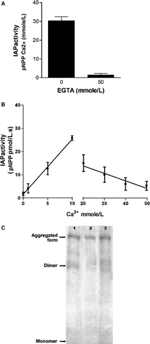
Treatment of IAP with EGTA after calcium exposure (n = 4): IAP was exposed to calcium 50 mmole/L for 5 min to produce inhibition and aggregation. After that, the mixture was exposed to increasing concentration of EGTA (0-50 mmole/L, dissociation constant of chelate Ca2 + -EGTA: 1 × 10− 11 mole/L) to eliminate calcium from the solution. The final concentration of EGTA assures an ionic calcium concentration lower than 7 × 10− 4 mmole/L, without modification of the concentration of magnesium (dissociation constant of chelate Mg2 + -EGTA: 6.3 × 10− 6 mole/L) [Citation13]. Samples were obtained from the solution before and after calcium treatment, and after the successive EGTA additions. IAP activity was measured in all the samples and Western blot was performed with the solutions obtained before calcium addition (, lane 1), after calcium treatment (, lane 2), and after the addition of 50 mmole/L EGTA (, lane 3).
Treatment with EGTA before calcium exposure (n = 4): The aim of this experiment was to verify whether EGTA affects IAP activity per se or by the chelation of magnesium and zinc. IAP was treated with EGTA 50 mmole/L for 5 min to chelate calcium from solution. In this situation, calcium was not added to the solution, as a consequence, the EGTA could act as a chelator of magnesium and zinc. The concentrations of magnesium and zinc in the solution after EGTA addition were 0.126 mmole/L and 2.52 × 10− 9 mmole/L, respectively. Then, the solution was treated with increasing concentrations of calcium, to obtain a final concentration of ionic calcium of 50 mmole/L. IAP activity was measured in all the samples and Western blot was performed with the solutions obtained before EGTA addition (, lane 1), after the treatment with EGTA (, lane 2), and after the addition of 50 mmole/L of calcium (, lane 3).
Figure 1. A: IAP activity before and after EGTA addition. B: IAP activity with different calcium concentrations obtained after calcium addition to IAP that was previously treated with EGTA 50 mmole/L. C: Western blot of IAP before (lane 1) and after EGTA addition (lane 2) and after 50 mmole/L calcium addition (lane 3). Data are expressed as mean ± SD (n = 4).
Statistical analyses
Between groups, comparisons were statistically analyzed using the Student's t test for continuous variables. Regression lines were calculated using the linear least-squares method. Results are expressed as mean ± SD (number of experiments). For both tests, differences with p < 0.05 were regarded as significant.
Results
Isoelectric point measurement
The isoelectric point of IAP increased significantly in the presence of calcium 50 mmole/L. Without calcium: 4.35 ± 0.02 (n = 4), in the presence of calcium: 4.85 ± 0.06 (n = 4), Student's t test, p < 0.05.
2D-electrophoresis
The presence of calcium 50 mmole/L caused the isoelectric focusing of IAP at higher pI and increased the aggregation of IAP. displays the values of isoelectric point (pI) and molecular mass of the dimer and the aggregated form. Values of the molecular mass were not different from those obtained in the previous work [Citation4].
Table I. Molecular mass and isoelectric point obtained by 2D-electrophoresis for the dimer and the aggregated form with and without calcium addition.
Treatment of IAP with EGTA before calcium exposure
The treatment with EGTA 50 mmole/L abolished IAP activity (). However, the addition of calcium to attain 50 mmole/L of free calcium reproduced the biphasic behaviour of the enzyme to the presence of calcium (). The activity of IAP increased as a linear function of the concentration of Ca2 + in the solution (r: 0.96, p < 0.05). reached a maximum at 10-20 mmole/L Ca2 + , and then, the activity of IAP decreased as a function of Ca2 + concentrations (r: − 0.66, p < 0.05). The activity of IAP was plotted as a function of free calcium (the ionic calcium that is not chelate by EGTA). This result assured that EGTA did not produce modification of IAP activity by chelation of magnesium and/or zinc.
Western blot of IAP that was exposed to EGTA displays no differences respect to the enzyme that was not exposed to EGTA (, lane 2 and 1). On the other hand, the addition of calcium to attain calcium concentration of 50 mmole/L increased the aggregated form to the detriment of the dimer (, lane 3).
Treatment of IAP with EGTA after calcium exposure
As it was expected, the activity of IAP decreased after the treatment with calcium 50 mmole/L (). The activity remained at this value after the increasing concentration of EGTA (). The addition of EGTA produces a gradual modification of calcium concentration from 50 to 7 × 10− 4 mmole/L. To make understandable, the activity was plotted as a function of calcium that is free after EGTA addition. Total calcium in the solution remained at 50 mmole/L.
Figure 2. A: IAP activity before and after calcium addition. B: IAP activity as a function of calcium concentration. Calcium concentrations of the horizontal axis are obtained after different additions of EGTA to the solution that started with 50 mmole Ca/L. C: Western blot of IAP before (lane 1) and after Ca2 + addition (lane 2) and after EGTA addition (lane 3) to obtain 0 mmole/L free calcium. Data are expressed as mean ± SD (n = 4).
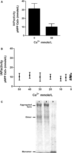
As revealed by Western blot, the treatment with calcium produced an increase of the aggregated form of IAP (, lane 2). The treatment with EGTA after calcium exposure produced a decrease in the aggregated form and an increase in the monomer (, lane 3).
Discussion
Calcium binds to IAP and biphasically modifies the enzymatic activity [Citation4]. As IAP is related to the transcellular transport of calcium at the small intestine, the potential effect of calcium in its own process of absorption is relevant. Some authors have reported contradictory effect of calcium on the activity of IAP [Citation14,Citation15]. While in the first of them IAP activity decreased as an inverse function of calcium concentration, when calcium was below 10 mmole/L [Citation14], the second work demonstrated an increase in activity as a direct function of calcium concentration [Citation15]. The inhibitory or stimulatory mechanism was not investigated in those works. Even though the biphasic effect of calcium on IAP was demonstrated in our laboratory, with a higher activity at 10-20 mmole/L, the sensitivity of IAP to calcium is not always the same. Other unknown factors could interact with calcium to produce the final effect. It is suspected that the state of fasting, the amount of calcium in the diet and the age of animals could be involved in different responses to purified IAP to calcium. This behaviour of IAP in front of calcium deserves further investigation, and could be the cause of contradictory works about the effect of calcium on IAP.
This work demonstrates that calcium modifies the isoelectric point of the enzyme. IAP was separated by others into two regions, each composed of three or four bands. The more acidic (anodic) were focused in the pI range of 4.65-4.73; the more basic (cathodic) were focused in the range 4.85-4.95. The more-cathodically focused band was not always visible [Citation7]. Concentration of calcium in that experiment was not reported. The modification of isolectric point caused by the presence of calcium indicates that calcium binds to IAP by stable bounds, probably not by electrostatic bonds to chemical groups that can change its charge, as it happens during the migration of a protein in a pH gradient.
In the presence of calcium, pI increased simultaneously with the increase of the amount of the aggregated form. However, both forms: dimer and aggregated have the same pI in the presence of 50 mmole/L Ca2 + .
The experiments described in this paper and in the previous work [Citation4] were carried out with purified IAP. Purity of IAP was demonstrated by PAGE and Western blot [Citation4], where a unique band was observed at molecular weight mass of 168 kDa. As a consequence, the aggregated form observed in the presence of calcium is probably a homopolimer of IAP. A band of 60 kDa was observed in polyacrylamide gel electrophoresis in the presence of sodium dodecyl sulphate, and bands of 140 kDa and 443 kDa in the absence of sodium dodecyl sulphate [Citation16]. The bands were detected with Coomasie brilland blue instead of Western blot. However, the molecular weight are coincident with this and the previous work. That work did not point at calcium as a determinant of the different molecular mass. Although we could not assure that other proteins of the same molecular mass were not co-purified with IAP, the same isoelectric point for the dimer and the aggregated form reinforce the conclusion that the aggregated form is a homopolimer. Taken into consideration that the protein is not contaminated with other proteins, the same pI in the dimer and in the aggregated form indicates that the binding of calcium does not cause important changes in the primary structure of the protein and/or in the ionization of the acidic residues.
As small amounts of aggregated form appear even before the addition of calcium, it is suspected that the process is partially independent of calcium and that calcium could accelerate the process.
Western blot of IAP revealed that the treatment with EGTA after calcium addition produces des-aggregation, decrease in the amount of aggregated form and an increase in the amount of dimer and monomer. The treatment with EGTA described in this paper produce chelation of calcium and zinc, leaving a small free calcium and negligible concentration of zinc in the solution, without modification of the concentration of magnesium. As the activity of IAP was not recovered with the decrease in calcium concentration by addition of EGTA, and the amount of monomer increase, we could hypothesize that calcium 50 mmole/L could replace magnesium and/or zinc in the dimer. From other point of view, as the amount of monomer increased after chelation of calcium, we can conclude that calcium not only participate in the formation of aggregated form but also in the stability of the dimer. The lack of activity after chelation of calcium with EGTA indicates that the process of inhibition is irreversible, and the pre-treatment of IAP with EGTA indicates that the inhibition is not due to the effect of EGTA on magnesium or zinc.
The treatment with EGTA previous to the addition of calcium abolishes IAP activity, probably by chelation of magnesium and/or zinc, whose concentrations are insignificant after EGTA treatment in the absence of calcium. But, when calcium is added, the higher stability of the calcium-EGTA complex, frees magnesium from EGTA, and IAP activity is restored, showing a biphasic effect of calcium on the activity of IAP. The chelate Ca2 + -EGTA is less stable than Zn2 + -EGTA (stability constant: 1.26 × 10− 13 mole/L). The addition of calcium to the solution is not sufficient to free zinc from the Zn2 + -EGTA chelate. As a consequence one could conclude that EGTA did not chelate the zinc that is in the structure of IAP. Other works have proved that chelation of zinc that is in the structure of the enzyme is dependent of the presence of substrates [Citation17].
The impact that the aggregation of IAP and the biphasic effect of calcium on its enzymatic activity could have in vivo is unknown. In vivo experiments with rats where activity of IAP is measured in the presence of calcium, simultaneously with calcium transport and the expression of other proteins involved in the process are being carried out.
Acknowledgements
We thank Ariana Foresto for the revision of the manuscript and Dr. Digno Alloatti for providing animals for the experiments. This work was partially funded by CONICET (Grant PIP 5018).
Declaration of interest: The authors report no conflicts of interest. The authors alone are responsible for the content and writing of the paper.
References
- M Besman, JE Coleman. Isozymes of bovine intestinal alkaline phosphatase. J Biol Chem 1985;260 (20):11190–11193.
- AW Norman. Vitamin D metabolism and calcium absorption. Am J Med 1979;67 (6):989–998.
- E Mornet, E Stura, AS Lia-Baldini, T Stigbrand, A Ménez, MH Le Du. Structural evidence for a functional role of human tissue nonspecific alkaline phosphatase in bone mineralization. J Biol Chem 2001;276 (33):31171–31178.
- LR Brun, ML Brance, A Rigalli, RC Puche. Effect of calcium on rat intestinal alkaline phosphatase activity and molecular aggregation. J Enzyme Inhib Med Chem 2006;21 (6):757–763.
- AD Beinlich, LR Brun, A Rigalli, RC Puche. Intestinal absorption of disodium monofluorophosphate in the rat as affected by concurrent administration of calcium. Arzneimittelforschung 2003;53 (8):584–589.
- K Furusaki. Alkaline phosphatase of the rat small intestine–purification of the enzyme and its physiological significance. Hokkaido Igaku Zasshi 1983;58 (3):250–264.
- WC Griffiths, PD Camara, M Rosner, R Lev, EM Brooks. Prevalence and properties of the intestinal alkaline phosphatase identified in serum by cellulose acetate electrophoresis. Clin Chem 1992;38 (4):507–511.
- MM Bradford. A rapid and sensitive method for the quantitation of microgram quantities of protein utilizing the principle of protein-dye binding. Anal Biochem 1976;72:248–254.
- OA Bessey, OH Lowry, MJ Brock. A method for rapid determination of alkaline phosphatase with five cubic milimeters of serum. J Biol Chem 1946;164:321–329.
- UK Laemmli. Cleavage of structural protein during the assembly of the head of bacteriophage T4. Nature 1970;227:680–685.
- PH O'Farell. High resolution two-dimensional electrophoresis of proteins. 1975;250 (10):4007–4021.
- O Salinovich, RC Montelaro. Reversible staining and peptide mapping of proteins transferred to nitrocellulose after separation by sodium dodecylsulfate-polyacrylamide gel electrophoresis. Anal Biochem 1986;156 (2):341–347.
- RMC Dawson, et al. Data for biochemical research. 3rd ed. New York, NY: Oxford University Press; 1986. p 404–405.
- G Deliconstantinos, L Kopeikina-tsiboukidou, S Tsakiris. Perturbations of rat intestinal brush border membranes induced by Ca2+ and vitamin D3 are detected using steady-state fluorescence polarization and alkaline phosphatase as membrane probes. Biochem Pharmacol 1986;35 (10):1633–1637.
- SJ Birge, RC Avioli. Intestinal phosphate transport and alkaline phosphate activity in the chick. Am J Physiol 1981;240 (4):E384–E390.
- K Suzuki, Y Yoshimura, Y Hisada, A Matsumoto. Sensitivity of intestinal alkaline phosphatase to L-homoarginine and its regulation by subunit-subunit interaction. Jpn J Pharmacol 1994;64 (2):97–102.
- SJ Pike, RG Duggleby. Resistance to inactivation by EGTA of the enzyme-substrate and enzyme-phosphate complexes of alkaline phosphatase. Biochem J 1987;244 (3):781–785.