
Abstract
Encapsulation is a well-established method of biomaterial protection, controlled release, and efficient delivery. Here we evaluated encapsulation of monoclonal antibody M75 directed to tumor biomarker carbonic anhydrase IX (CA IX) into alginate microbeads (SA-beads) or microcapsules made of sodium alginate, cellulose sulfate, and poly(methylene-co-guanidine) (PMCG). M75 antibody release was quantified using ELISA and its binding properties were assessed by immunodetection methods. SA-beads showed rapid M75 antibody release in the first hour, followed by steady release during the whole experiment of 7 days. In contrast, the M75 release from PMCG capsules was gradual, reaching the maximum concentration on the 7th day. The release was more efficient at pH 6.8 compared to pH 7.4. The released antibody could recognize CA IX, and target the CA IX-positive cells in 3D spheroids. In conclusion, SA-beads and PMCG microcapsules can be considered as promising antibody reservoirs for targeting of cancer cells.
Introduction
Encapsulation of biologically active materials represents relatively simple but considerably effective way how to immobilize drugs, live mammalian and bacterial cells, and other biophamaceuticals. Moreover, it provides structuration as well as protection and controlled release of the encapsulated materialCitation1.
Out of many applications of encapsulations for therapy, one of the most studied systems is the encapsulation of insulin-producing cells for transplantation and treatment of diabetes Type ICitation1–3. Sodium alginate is a widely used biopolymer implemented in diverse medical applications (wound healing, cartilage repair, bone regeneration, and drug delivery)Citation4 and is considered as one of the most suitable biomaterials for encapsulation. Besides alginate microbeads formed by ionotropic cross-linking using divalent cations, other encapsulation systems for pancreatic islet transplantation have been studied and evaluatedCitation2,Citation5. One of them is the so-called PMCG microcapsule, which is based on polyelectrolyte complexation involving polyanions sodium alginate and cellulose sulfate, the polycation poly(methylene-co-guanidine), calcium gelling and sodium non-gelling ions, respectivelyCitation6–8.
In recent years, encapsulation has also markedly enhanced both local delivery and the protection of several drugs and other pharmaceuticals (enzymes, antibiotics, food supplements, etc.); however, little is known about encapsulation of therapeutic antibodies. Antibody-based therapies are suitable for a wide range of applications in cancer, inflammation, immune-mediated disorders, and wound healingCitation9. Unlike drugs and other pharmaceuticals, monoclonal antibodies (MAb) possess relatively high specificity and their application commonly leads to minimal side effects. When conjugated to other therapeutic agents (inhibitors, toxins, etc.) and radioisotopes, administration of MAbs can increase the efficacy of both diagnostic and therapeutic targeting.
In the case of antibody encapsulation for diagnostics and targeted therapy in a microsphere (i.e. microbead or microcapsule), the MAb has to pass through the microsphere membrane in a controlled way in order to achieve an efficient therapeutic effect. There are several factors, which affect the MAb release, e.g. size of microsphere, drug loading, MAb–polymer network interaction, polymer network molecular weight cutoff and porosity, microsphere morphology, encapsulation efficiency, and properties of MAbCitation10. For optimal targeting of cancer cells, encapsulated MAb must be able to reach tumor tissue in sufficient amounts and, moreover, it must be delivered and released preferentially within or near to tumor tissue. This can be achieved by optimizing size and surface charge of the encapsulation systemCitation11. The efficacy may also be improved by the antibody release in response to the tumor microenvironment (e.g. low pH and partial oxygen pressure), or to external forces (electric pulses, magnetic field, ultrasound, heat, and light)Citation12. On the other hand, the selectivity of the system depends on the specificity of the therapeutic antibody and its target.
More than 20 years ago, MAb M75 directed against human carbonic anhydrase IX protein (CA IX; previously named MN) was described, characterized and its staining pattern in human cell lines and tissues was evaluatedCitation13. CA IX is a transmembrane protein, which consists of a large extracellular part (proteoglycan-like region followed by catalytic domain) exposed at the surface of the cells, single-pass transmembrane region and a short intracytoplasmic tailCitation14. The tumor-associated expression pattern of CA IX is mainly caused by the strong transcriptional activation of its gene (CA9) by hypoxia through the hypoxia-inducible transcription factor (HIF)Citation15. So far, CA IX has been extensively studied due to its strong hypoxic regulation, cancer association, and functional implication in tumor biology. Numerous studies have supported its promising position as one of the most prominent markers of tumor hypoxia with the predictive and prognostic values. As CA IX functions as a highly active enzyme with the catalytic domain exposed on the outer side of the plasma membrane, it can be efficiently targeted by CA IX-inhibitors (sulfonamides, their derivatives and related compounds) or antibodiesCitation16,Citation17. In summary, the expression pattern of CA IX is tightly associated with a broad variety of human tumors, while it is significantly expressed in only few normal tissues.
In this study, the anti-CA IX antibody M75 was encapsulated into SA-beads and PMCG microcapsules and these microspeheres were investigated as potential M75 reservoirs for its long-term release and targeting of CA IX-expressing cancer cells. The M75 antibody is directed to a linear epitope in the proteoglycan-like region of CA IX and recognizes both native and denatured antigenCitation18. Therefore, it is suitable for use in a variety of techniques such as ELISA, Western blotting, FACS, immunofluorescence, and immunohistochemistry. Moreover, favorable tumor uptake, biodistribution data and pharmacokinetics of 125I-labeled M75 antibody were obtained after its intravenous administration into nude mice with HT-29 xenograftsCitation19. The SA-beads and PMCG microcapsules were characterized for their size and structural and morphological properties. The release of M75 antibody in complete DMEM medium at pH values 6.8 and 7.4 from both types of microspheres was determined by Mouse IgG total Ready-SET-Go ELISA kit. The binding specificity of the encapsulated M75 antibody was investigated via immunoblotting and immunofluorescence. Finally, the incorporation of the released M75 antibody was demonstrated through the uptake into multicellular spheroids from cancer cells.
Materials and methods
Reagents and equipment
Sodium alginate (SA-HV, high viscosity, MW = 235 kDa, Mn = 118 kDa, G content = 41 mol %; all quantities were determined in our laboratory) obtained from ISP Alginates (Girvan, Ayrshire, UK) was purified by protocol described by de Vos et al.Citation20 Ultrapure alginate (UP LVM, G content = 41 mol %) was from Novamatrix (Sandvika, Norway). Sodium cellulose sulfate (CS, MW = 760 kDa determined following the work of Kishino et al.Citation21 and degree of substitution ∼2.0; all quantities were determined in our laboratory) was from Acros Organics (Morris Plains, NJ). Poly(methylene-co-guanidine) hydrochloride (PMCG) obtained from Scientific Polymer Products Inc. (Ontario, NY) as 35% aqueous solution was lyophilized. d-Mannitol was from Amresco (Solon, OH), Tween 20 and 3-(N-morpholino)propanesulfonic acid (MOPS) from Sigma-Aldrich (St. Louis, MO). All inorganic salts (NaCl, CaCl2 2H2O, BaCl2 2H2O) were of analytical grade.
Preparation of microspheres with encapsulated M75 antibody
The encapsulation of M75 to either SA-beads or PMCG microcapsules was done under sterile conditions in a laminar airflow cabinet (BH-EN 2000 S/D, Faster S.r.l., Cornaredo, Italy). An in-house made air-stripping nozzle, using a needle with internal diameter of 0.4 mm, was employed for formation of droplets of polyanion solutions at the flow rate of 0.7 ml/min. Polyanion solutions were sterilized by filtration through 0.45 μm and 0.22 μm syringe filters (TPP, Switzerland), gelling solutions and saline were sterilized by filtration through 0.22 μm bottle filter (Sarstedt, Germany).
Encapsulation of M75 antibody in alginate microbeads (SA-beads) followed the protocol for encapsulation of human islets transplanted to nudeCitation22 and Balb/CCitation23 mice. 2.7 ml of solution of 1.9% UP LVM prepared in 0.3 M d-Mannitol in ultrapure water and the pH adjusted to 7.4 was mixed with 0.3 ml of M75 solution in phosphate-buffered saline (PBS) at the concentration of 3.48 mg/ml providing final concentration of UP LVM equal to 1.8% and the concentration of M75 equal to 0.348 mg/ml. The droplets of UP LVM solution containing M75 antibody were air-stripped to 200 ml of gelling solution consisting of 50 mM CaCl2, 1 mM BaCl2, 0.05% Tween 20, 0.14 M d-Mannitol, and 10 mM MOPS dissolved in ultrapure water and of final pH value equal to 7.4. The gelling time was 15 min after the last droplet entered the gelling solution in order to achieve a saturation gelling for SA-beads. Then the SA-beads were washed three times with 200 ml of saline and immediately used for assessing the release of M75 antibody.
The protocol for encapsulation of M75 in the PMCG microcapsules was derived from that developed originallyCitation8 and used for immobilization of whole cellsCitation24,Citation25 and enzymesCitation26, and for transplantation of empty microcapsulesCitation7 and encapsulated allogeneic isletsCitation27 to non-human primates. Polyanion solution was prepared by dissolving 1.0% SA-HV and 1.0% CS in saline solution and by adjusting the pH to 7.4. 0.3 ml of saline solution of M75 at the concentration of 3.48 mg/ml was added to 2.7 ml of polyanion solution to provide the final concentration of used polyanions equal to 0.9% SA-HV and 0.9% CS, and the concentration of M75 equal to 0.348 mg/ml. This solution was air-stripped to the gelling solution consisting of 1.2% PMCG, 1.0% CaCl2, and 0.9% NaCl dissolved in ultrapure water with the pH value adjusted to 7.4, which was flowing at the rate of 55 ml/min in a multi-loop reactorCitation28 to provide gelling time to each microcapsule of ∼40 s. Microcapsules were collected at the exit of the reactor in 150 ml of saline for 60 s to quench the polyelectrolyte complexation. Microcapsules were washed three times with 200 ml of saline using a stainless steel sieve for separation of microcapsules from solutions in all steps. Consequently microcapsules were coated with 0.1% CS in saline for 10 min, washed three times with 200 ml of saline and, identically to SA-beads, were immediately used for assessing the release of M75 antibody.
Release of encapsulated M75 antibody
A quasi-static setting was used for determination of released antibody. Freshly prepared SA-beads and PMCG microcapsules in a volume of 0.5 ml were placed into 8.5 ml of cell culture medium with different pH of either 6.8 or 7.4 in humidified atmosphere and under conditions as the cell cultures. 0.5 ml aliquots of culture medium were removed at various time-points 1 h, 24 h, 48 h, 120 h, and 168 h (7 d) post-encapsulation for determination of released antibody and 0.5 ml of fresh medium was added to keep the total volume constant during the entire experiment. The data are reported as the amount of antibody in ng/ml of media released per 0.5 ml of microspheres and as the total release of M75 corresponding to theoretical value encapsulated in 0.5 ml of microspheres. Antibody release as well as its binding properties were assessed using ELISA, immunoblotting, immunofluorescence, and immunohistochemistry.
Determination of optical and mechanical properties
Prepared microspheres were visualized by optical microscopy (Microscope Kapa 2000, Kvant, s.r.o. Bratislava, Slovakia) equipped with a CCD camera (CC-63KW1P, Mintron, Malaysia) using a digital imaging ImageForge 1.1 software (Prover, Bratislava, Slovakia).
The compression resistance of microspheres was tested on a Texture Analyzer TA-2Xi (Stable Micro Systems, Godalming, UK). The software Texture Expert version 1.16 was used for data collection and evaluation. The equipment consists of a mobile probe that moves vertically from the distance of 1 mm above the stand and compresses the microsphere at a constant velocity of 0.5 mm/s. Microspheres were placed on a stand under the probe in saline and the excess saline was gently removed using a Kimwipe tissue. The microspheres were compressed until bursting occurred, which determines the rupture strength of a microsphere at given conditions of analysis. The force expressed in grams was recorded as a function of the compression distance for 25 microspheres per batch.
Cell cultures
Canine MDCK epithelial cells (ATCC® No. CCL-34™) as well as HeLa, SiHa, and C-33 A cells derived from human cervical carcinoma (CCL-2™, HTB-35™, and HTB-31™) were cultured in Dulbecco’s modified Eagle’s medium (DMEM) with 10% fetal calf serum (FCS; BioWhitteker, Verviers, Belgium) and 40 μg/ml gentamicin (Lek, Ljubljana, Slovenia) in a humidified atmosphere with 5% CO2 at 37 °C.
Immunoblotting
Prior to lysis, MDCK and HeLa cells were rinsed with ice cold PBS and disrupted in RIPA lysis buffer containing 0.1% sodium deoxycholate and 1% Triton-X 100 (Sigma-Aldrich) in PBS, and 1× complete protease inhibitor cocktail (Roche, Mannheim, Germany) for 30 min on ice, collected and clarified. Protein concentrations were determined by bicinchoninic acid assay (Pierce, Rockford, IL) according to the manufacturer’s instructions. Total protein extracts (50 μg/lane) were separated by SDS-PAGE under reducing conditions and blotted onto polyvinylidene fluoride membranes (Immobilon; Millipore, Billerica, MA). Membrane was treated for 1 h in blocking buffer (5% non-fat dry milk, 0.2% NP-40 in PBS) and then incubated for 1 h with undiluted medium containing released M75 antibody. After washing step, the membrane was treated for 1 h with secondary anti-mouse antibody conjugated with horseradish peroxidase (1:5000; Sigma-Aldrich). After an additional washing step, the membrane was developed with the ECL detection system.
ELISA procedures
Measurement of mouse IgG total protein level in both cultivation medium and solutions/buffers for microsphere preparation was determined by commercially available Mouse IgG total Ready-SET-Go ELISA kit (eBioscience, Inc., San Diego, CA) according to the manufacturer's instruction.
Immunofluorescence
MDCK cells grown on glass cover-slips were fixed with ice-cold methanol for 5 min at −20 °C. The cells were rinsed twice with PBS and nonspecific binding was blocked with 1% bovine serum albumin (BSA) in PBS for 30 min at 37 °C. Then the cells were sequentially incubated with cultivation medium with released M75 antibody for 1 h at 37 °C, washed three times with PBS–BSA for 10 min, incubated with Alexa Fluor 488-conjugated anti-mouse secondary antibody (Invitrogen, Carlsbad, CA) diluted 1:1000 in 1% BSA for 1 h at 37 °C and washed again as before. Finally, the nuclei were stained with DAPI (1:36 000, Sigma) and cover-slips were mounted with mounting medium (Calbiochem, Darmstadt, Germany). All samples were analyzed by Zeiss LSM 510 Meta confocal microscope, scanned in multitrack mode and deconvoluted by Huygens software (Scientific Volume Imaging, Hilversum, the Netherlands).
Formation of spheroids in hanging drops
Spheroids from SiHa cells were performed in 20 μl hanging drops containing 600 cells each during 3–4 d incubation in a humidified atmosphere on the lid of a 100 mm Petri dish. 10 ml of PBS was added into the dish to prevent drying of the drops. Resulting cell aggregates were carefully transferred to a 100 mm Petri dish with a non-adherent surface (bacteriological Petri dish) and cultivated in suspension for additional 10 d. The medium was changed on every 3rd day. For the last 24 h of spheroid cultivation, cultivation medium was removed and replaced by pooled medium with released M75 antibody from SA-beads as well as PMCG microcapsules at the end of experiment for medium at pH = 6.8. Cultivation medium without encapsulated antibody served as a negative control. The spheroids were then quickly fixed in Carnoy’s fluid (absolute ethanol/chloroform/glacial acetic acid, 6:3:1) to preserve the integrity and embedded in paraffin.
Immunohistochemistry
SiHa spheroids were embedded in paraffin according to the standard histological procedure. Immunohistochemistry was performed using DakoCytomation EnVision®+ System-HRP (DAB) according to the manufacturer’s instructions. Sections (4 μm) were incubated with secondary anti-mouse antibody overnight at 4 °C. In case of control (spheroids analyzed for expression pattern CA IX) incubation with M75 antibody (hybridoma medium diluted 1:100) for 1 h at room temperature was performed before secondary antibody. Staining was visualized with DAB solution for 1 min with 3,3′-diaminobenzidine as a chromogenic substrate. Finally, the sections were counterstained with Mayer’s hematoxylin and mounted in Aquamount (Merck, Darmstadt, Germany). The stained sections were examined using a Leica DM4500B microscope and images were captured by a Leica DFC480 camera.
Results
Characterization of SA-beads and PMCG microcapsules with encapsulated M75 anti-carbonic anhydrase IX antibody
MAb M75 specific for the human CA IX was encapsulated in either SA-beads or PMCG microcapsules according to the standard protocols at the final antibody concentration in microspheres of 0.348 mg/ml. Aliquots from all solutions and washing buffers were taken for monitoring of eventual loss of the M75 antibody during the encapsulation experiment. ELISA was performed to quantify the amount of immunoglobulins in each step of the encapsulation process including washing solutions and to calculate the loss of antibody for each volume of all solutions.
In general, negligible loss of M75 antibody was observed during the process of both microbeads and microcapsules formation. The gelling solution was the only step in SA-beads preparation where the antibody release was determined and quantified. 21.8 ng/ml was determined in a volume of gelling solution equal to 200 ml, which results in the total loss of more than 4000 ng representing less than 0.5% mass of the total immobilized antibody. In case of washing solutions, the amount of antibody was below the detection capacity of this assay (1.56 ng/ml). The same was true for the washing solutions used for the preparation of PMCG microcapsules where the comparable release of antibody as in case of SA-beads was determined equal to 29.9 ng/ml in a volume 150 ml that results in almost 4500 ng of antibody. Thus, the overall antibody encapsulation efficiency was around 99.5%.
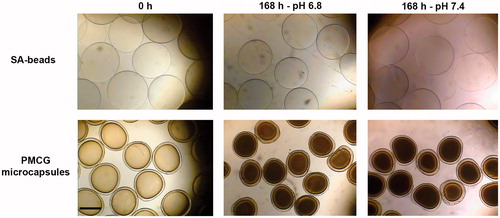
For determination of optical properties, we used optical microscope to measure the size of SA-beads and PMCG microcapsules as well as membrane thickness of PMCG microcapsules at the beginning (after microsphere preparation) and at the end of the incubation time in medium with pH of either 6.8 or 7.4. illustrates representative images for both types of microspheres. PMCG microcapsules size decreased from 0.69 ± 0.02 mm after preparation to 0.59 ± 0.03 mm in cultivation medium pH 6.8 or 0.60 ± 0.03 mm in medium pH 7.4. The membrane thickness determined immediately after preparation (0.049 ± 0.010 mm) remained unchanged after incubation in medium pH 6.8 (0.049 ± 0.014 mm) or slightly increased after incubation in medium pH 7.4 (0.057 ± 0.012 mm). The effect of culture media on PMCG microcapsules was also revealed by a darker core of microcapsules compared to that for microcapsules after preparation. The average size of SA-beads after preparation was 0.72 ± 0.02 mm and after 7 d of incubation in cultivation medium pH 6.8 and 7.4 was 0.80 ± 0.04 mm and 0.86 ± 0.04 mm, respectively.
Figure 1. Optical microscopy images for SA-beads (upper panel) and PMCG microcapsules (lower panel) after encapsulation of M75 antibody (0 h) and after 7 d (168 h) of incubation in medium with pH 6.8 and 7.4. The scale bar is equal to 0.5 mm.
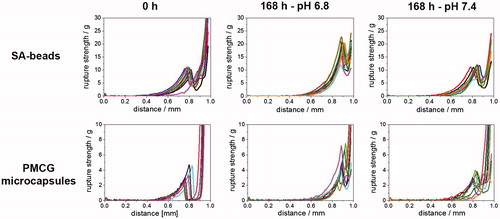
The resistance of PMCG microcapsules to rupture during compression test was 2.8 ± 1.2 g after preparation and, considering high standard deviation for these measurements, remained almost unchanged after 7 d of incubation in medium pH 6.8 (2.9 ± 1.3 g) and pH 7.4 (4.0 ± 2.3 g). The mechanical resistance of SA-beads was equal to 9.8 ± 3.6 g after preparation, 17.1 ± 5.4 g and 10.5 ± 2.2 g for SA-beads incubated for 7 d in medium pH 6.8 and pH 7.4, respectively. The compression curves for SA-beads and PMCG microcapsules are shown in .
Figure 2. Compression curves expressed as compression force versus distance between probe and stand of Texture analyzer for SA-beads (upper panel) and PMCG microcapsules (lower panel) after encapsulation of M75 antibody (0 h) and after 7 d (168 h) of incubation in medium with pH 6.8 and 7.4.
Quantification of the anti-CA IX antibody release
Immediately after encapsulation of anti-CA IX antibody, both SA-beads as well as PMCG microcapsules (each in a volume 0.5 ml) were transferred into cultivation flasks containing 8 ml of cultivation medium with pH value of either 6.8 or 7.4. Different pH conditions were selected to assess similar conditions as occur within tumor tissue mimicking by pH 6.8 extracellular acidic and hypoxic microenvironment. As we were interested in the quantity as well as the kinetic of the M75 antibody released from SA-beads or PMCG microcapsules, medium samples were collected at exact time-points and analyzed.
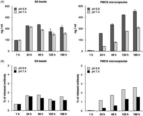
For the quantitative detection of the total antibody released into cultivation medium, we used commercially available mouse IgG ELISA kit. Taking into account the antibody concentration determined from all time-points, similar range for release of M75 was observed for SA-beads and PMCG microcapsules (). However, some differences can be noted. In case of SA-beads, initially (after 1 h) higher concentration of IgG of around 150 ng/ml (under both pH conditions) was measured, which was followed by relatively constant M75 concentration at the level of around 300 ng/ml during the entire experiment. Slightly lower release of M75 was observed at pH 7.4. On the other hand, in case of PMCG microcapsules, the amount of released antibody was continuously increasing with a distinct difference between pH of media revealing a significantly lower release at pH 7.4. The data shown in represent the concentration of the released M75 from 0.5 ml of either SA-beads or PMCG microcapsules that is determined in 0.5 ml of media withdrawn from a total volume of 8.5 ml media at given time points. With respect to 3 ml of initial volume of microspheres and initial loading of M75 antibody 0.348 mg/ml, the amount of 300 ng/ml corresponds to approximately 2% of initially encapsulated antibody. reveals the recalculated release of M75 from 0.5 ml of microspheres demonstrating that during 7 d of the release experiment only a few percent of antibody was liberated from the microspheres, indicating that its release can continue for a substantially prolonged period.
Figure 3. Quantification of encapsulated mouse immunoglobulins released into cultivation medium in quasi-static experiment. A: 0.5 ml aliquot of freshly prepared SA-beads and PMCG microcapsules were transferred into cultivation medium (either pH 6.8 or pH 7.4) and 0.5 ml of medium were taken out in time intervals of 1 h, 24 h, 48 h, 120 h, and 168 h. Withdrawn 0.5 ml medium was replaced by fresh one. B: Total release of M75 antibody in percent corresponding to theoretical content of antibody encapsulated in 0.5 ml of microspheres. IgG release was determined by commercially available ELISA kit for total mouse IgG detection and quantification.
Binding specificity of the released M75 antibody
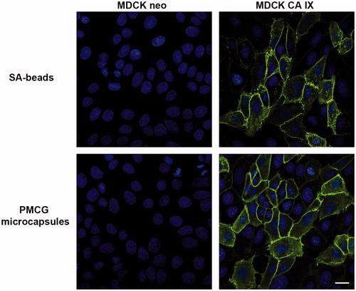
We then examined the ability of the released M75 antibody to recognize its antigen via immunoblotting and immunofluorescence. shows representative immunoreaction of the culture medium with M75 antibody released from the SA-beads with protein lysates prepared from HeLa cells derived from the cervical carcinoma as well as MDCK kidney epithelial cells with the constitutive expression of CA IX. Positive reaction is characterized by detection of twin band corresponding to 54/58 kDa in case of HeLa and MDCK CA IX cells, while no reaction was observed in the control MDCK neo cells. In addition, a positive membrane-specific staining for CA IX was detected via immunofluorescence in MDCK CA IX cells (). These data clearly reveal that there is no obvious difference in the M75 reactivity after encapsulation into either SA-beads or PMCG microcapsules, suggesting that the encapsulation procedure did not affect the integrity of the antibodyCitation17.
Figure 4. Immunoblotting of CA IX expression in different cell cultures. Protein lysates from CA IX-expressing (HeLa and MDCK CA IX) cells as well as negative cells (MDCK neo) were subjected to analysis using cultivation medium with the released M75 antibody. Characteristic twin band (54/58 kDa) appeared only in lines where proteins from CA IX-positive cells were loaded.
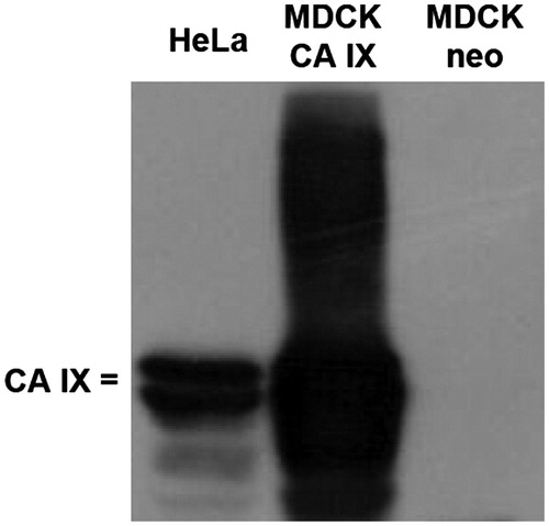
Figure 5. Immunofluorescence analysis of CA IX localization. MDCK CA IX as well as MDCK neo cells were stained with cultivation medium obtained after encapsulation of M75 antibody into both SA-beads as well as PMCG microcapsules followed by Alexa Fluor 488-conjugated secondary antibody. The positive CA IX-specific immunoreaction (green color) was localized at the cell membrane of the MDCK CA IX cells. Empty-vector transfected MDCK cells (MDCK neo) were negative. Cell nuclei were stained using DAPI (blue color). The scale bar is equal to 20 μm.
Incorporation of the released M75 antibody into multicellular spheroid
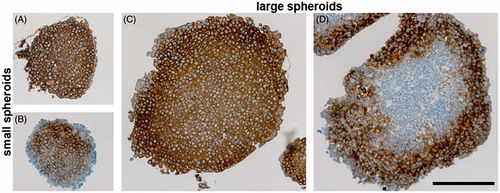
We then investigated whether three-dimensional multicellular spheroids could demonstrate the uptake of the antibody released from the SA-beads and PMCG microcapsules. Spheroids represent a well-known model in cancer biology, allowing for study of cancer cells under conditions more similar to in vivo intratumoral situation. In our study, spheroids were prepared from SiHa human cells derived from cervical carcinoma, which are characterized by relatively high expression of CA IX in hypoxia and/or high cell density. Thus, CA IX-specific staining signal was clearly visible in the membranes of almost all cells across the whole spheroid section processed by immunohistochemistry and visualized with externally added M75 antibody followed by the peroxidase-conjugated secondary anti-mouse antibody (). To confirm the ability of the released antibody to recognize its antigen within three-dimensional spheroids, a medium pooled from both SA-beads and PMCG microcapsules at pH 6.8 at the end of the release experiment (, 168 h) was added to the spheroids from SiHa cells for the last 24 h of their cultivation. shows that the encapsulated M75 antibody released to the cultivation medium was successfully incorporated into both small and large SiHa spheroids as detected by staining with the peroxidase-conjugated secondary anti-mouse antibody. The ability to detect CA IX expression across the multicellular spheroids further supports the view that the antibody encapsulation can be utilized for the protection, delivery, and administration of the monoclonal anti-CA IX antibody for tumor targeting.
Figure 6. Immunohistochemical analysis of CA IX expression in either small (A and B) or large (C and D) spheroids from SiHa cells. A and C: Staining pattern of CA IX in spheroids cultivated without encapsulated antibody (positive control). Standard immunohistochemical staining of CA IX was performed with both primary (hybridoma medium diluted 1:100) as well as secondary antibody. CA IX-specific staining (brown color) is localized across the spheroid. B and D: Uptake of the encapsulated M75 antibody into SiHa spheroids that was determined by incubation of paraffin sections only in secondary anti-mouse antibody. All sections were counterstained with Mayer’s hematoxylin (blue nuclei). The scale bar is equal to 200 μm.
Discussion
Over the last years, there is an increased need to develop new therapeutic tools for detection, prevention, and treatment in oncology. Microencapsulation may improve the use of conventional medicine by offering the protection and sustained delivery of biologically active molecules in combination with controlled release that is considered as particularly valuable for future successful therapies.
Encapsulation as a technology has shown significant improvement in several aspects of therapy. There are basically two concepts of encapsulation for immunotherapeutic purposes, i.e. either encapsulation of antibody or encapsulation of antibody-producing cells. Encapsulation of MAb-producing cells was described by Pelegrin et al. who successfully demonstrated subsequent release of MAb directed against human thyroglobulinCitation29,Citation30. Moreover, encapsulation of hybridoma cells producing antibodies directed to mouse retrovirus neutralization resulted in disease preventionCitation30. More recently, the in vitro and in vivo release of immunostimulatory MAb from hybridoma cells encapsulated in alginate-poly-L-lysine alginate microcapsules was demonstratedCitation31.
Encapsulation of antibody-producing cells provides an effective way how to ensure long-term delivery of antibodies. The design of this system must be versatile and strict enough to enable bidirectional transfer of nutrients for cells and release of antibody through the semipermeable membrane. Additionally, microspheres have to effectively immobilize encapsulated cells and protect them from the destruction by the host immune system. A number of cell encapsulation strategies using various semipermeable membranes in terms of geometry and chemistry have been under developmentCitation2,Citation5. However, the functional encapsulated cell-based therapy for clinics is still not accessible mainly due to unresolved issues of biocompatibility and cell survivalCitation3. Taking into account all these facts, the encapsulation of antibody represents easier and safer approach compared to the encapsulation of antibody-producing cells. This approach provides the principal information on relation between the polymer matrix and antibody properties and release, and may also represent an intermediate step before the effective antibody-producing cell therapy becomes available. Such systems are expected to mediate delivery and administration of antibody on the time-scale from days to weeks that is already viewed as highly beneficial in cases when frequent administration is needed for patients’ compliance. Moreover, this approach can be useful for local administration of the encapsulated therapeutic antibody that requires much lower doses of antibodies than the systemic one therefore reducing both side effects and treatment costs. Indeed, therapeutic potential of the local administration of the hydrogel-encapsulated antibody was recently demonstrated using an anti-angiogenic drug avastin in the mouse model of metastatic colorectal carcinomaCitation32.
There are several possibilities how to encapsulate the antibody. The primary requirement is that encapsulated antibody can pass through the microsphere membrane and recognize its antigen. The membrane should act as a barrier both preventing the antibody destruction by the immune system and controlling the continual antibody release. While SA-beads are typically permeable for IgG (150 kDa) and proteins up to 350 kDaCitation33–35 with approximate viscosity diameter of ∼13 nmCitation36, PMCG microcapsules exhibit reduced permeability with the molecular weight cutoff for proteins in the range of 70 kDa corresponding to viscosity diameter of ∼7 nm (manuscript in preparation). Additional difference is mainly in the residual charge of these ionically cross-linked microspheres, which is negative in case of SA-beads and both negative and positive in case of PMCG microcapsules. These electrostatic interactions together with other types of interactions (hydrogen bonds, polar, hydrophobic) occurring in this type of polymer networks may play a significant role in diffusion of solutes especially in case when solutes are highly interacting such as biological moleculeCitation37.
The size of our PMCG microcapsules and SA-beads was in the submillimeter range (), which is typically used for encapsulation of cells. A slight change in the size after 168 h of storing microspheres in DMEM containing FCS resulted from predominantly electrostatic interactions of the microsphere network with electrolytes, amino acids, and proteins contained in media. This effect leads to slight swelling of the ionotropic network between alginate chains and divalent ions of SA-beads. On the other hand, PMCG microcapsules in media slightly contracted suggesting that additional complexation rather than weakening of the polyelectrolyte network takes place. In addition, the core of PMCG microcapsules became darker due to complexation of residual polyelectrolytes in the core by the media components, which has been observed, in different extent, for explanted PMCG microcapsulesCitation6,Citation7. The different media pH of 6.8 versus 7.4, slightly altering the ionization of weak carboxylic groups (sodium alginate; ionization increases upon increased pH) and weak imine groups (poly(methylene-co-guanidine; ionization decreases upon increased pH) is not manifested by a change in size and membrane thickness of microspheres. The resistance to rapture appearing as the maximum in the curves of compression strength versus distance (i.e. deformation) () is not significantly different in microspheres after preparation and 7-d storage, respectively, in the cultivation media at both pH values. Generally both microspheres survived the experimental conditions, which is in agreement with numerous in vivo applications of these types of microspheres.
Only a negligible amount of antibody was lost during the immobilization of M75 in both types of microspheres. This already indicates that SA-beads as well as PMCG microcapsules can effectively entrap M75. These microspheres possess significantly different molecular weight cutoff values with SA-beads and PMCG microcapsules being and not being permeable, respectively, to solutes of the size corresponding to the size of M75 ∼ 11 nm. Thus, the molecular weight cutoff of these microspheres is not their dominant characteristics for retaining and releasing of M75 antibody, whereas the electrostatic interactions between microspheres and M75 appear to play a significant role. Since the isoelectric point pI of the M75 antibody is equal to 8.5 (Supplementary ), at the physiological pH M75 is of cationic nature and electrostatically interacts with negative charges of the hydrogel networks of SA-beads and PMCG microcapsules.
Negatively charged carboxylate groups on sodium alginate chains are expected to interact with cationically charged M75 and significantly slow down its release from SA-beads. The situation appears to be similar to studies of alginate beads used for the controlled delivery of VEGFCitation38. VEGF exists in dimeric form of molecular weight of ∼40 kDaCitation39 that is significantly lower than the molecular weight cutoff for alginate beads. At the lowest loading of beads with VEGF concentration of 0.2 μg/ml, the loading efficiency was 67%, the retention value was 93% and there was no release of VEGF during 6 d of experiment in PBS. This behavior was ascribed to electrostatic interactions between alginate and VEGF, which pI is 8.5Citation40 as is that of M75. Since M75 is of higher molecular weight than VEGF, SA-beads provide effective retention even though the loading by M75 is higher and the release experiments are performed in complete media with serum present, which resulted in increased release of VEGFCitation38. The static experiment revealed by the shows only about 2% release of M75 seen in the first 24 h of experiment followed by approximately constant concentration. None or only a minor tendency for lowering the content of M75 determined at higher pH values shows that the conditions for electrostatic interactions between pH 6.8 and 7.4 do not change significantly. The most significant release up to 24 h may be associated with an altered environment for the SA-beads after their immersing to complete media and a modest screening of interactions between alginate and M75. In principle, the slow antibody release rate is required for the long-term targeting of CA IX and preventing the tumor progression. Nevertheless, the release rate of M75 can be increased by decreasing the concentration of alginate or decreasing the cross-link density of the alginate network.
PMCG microcapsules exhibit different behavior compared to SA-beads. The membrane of PMCG microcapsule, consisting of sodium alginate and sodium cellulose sulfate, after complexation with polycation poly(methylene-co-guanidine) gain residual negative charge by which microcapsules electrostatically interact with M75. Because of lower molecular weight cutoff of PMCG microcapsules, in addition to electrostatic interactions, the retention of M75 is influenced by obstruction of hydrogel to antibody diffusion. For example, glucose oxidase prepared from Aspergillus niger, an enzyme of pI ∼4.7 and molecular weight of 160 kDCitation44, immobilized in PMCG microcapsules using a slightly modified protocol compared to this workCitation26, showed only 43% encapsulation efficiency with the principal loss of glucose oxidase during the first gelling step in the loop reactor during membrane and microcapsule formation. The principal difference between the immobilization of glucose oxidase and M75 in PMCG microcapsules is the anionic and cationic nature of proteins, respectively, at used pH values of culture media. Albeit molecular weight cutoff value for PMCG microcapsules should completely prevent the release of M75, our data show a slight and continuous release during 7 d of experiment. This may be caused by an inherent heterogeneity of pores of the hydrogels formed by polyelectrolyte complexation and transient stability of interactions between M75 and the hydrogel network.
For PMCG microcapsules the effect of pH is more pronounced than for SA-beads. The release of M75 is small but the difference between culture media seems to be obvious () with a lower release at the physiological pH compared to slightly acidic pH, unlike in case of SA-beads. It is primarily assumed that weakening of interactions between cationically charged M75 and polyanionic components of PMCG microcapsule is responsible for this observation. Incubation of microspheres with the encapsulated antibody in medium with pH 6.8 was done to mimic acidosis, which together with hypoxia frequently occur in microenvironment of solid tumorsCitation41,Citation42. They both induce several crucial changes in tumor cells resulting in acquisition of malignant tumor phenotype characterized by invasion, metastasis, and treatment resistanceCitation43. Therefore, several strategies were performed to establish pH-sensitive nanoparticles (nanocarriers) with drugs for delivery into tumor tissuesCitation11. The fact that M75 is preferentially released under lower pH conditions favors the application of encapsulated anti-CA IX antibodies for tumor diagnostics and therapy. While tumor microenvironment is more acidic than the surrounding healthy tissue, pH sensitivity of antibody release could be perceived as an important advantage. The modulation of M75 release by pH, specifically a more pronounced release at the pH value that is characteristic for tumor environment as seen for PMCG microcapsules, thus represents a direction that should be further explored.
The second goal of our study was to demonstrate that following encapsulation process and release, the M75 antibody still possesses binding specificity and is able to recognize its antigen, i.e. tumor biomarker CA IX. This was confirmed by immunoblotting () and immunofluorescence (). In addition, the released antibody was incorporated into 3D spheroids from SiHa cells ().
The obtained data demonstrate that both types of microspheres function as the long-term reservoirs for the release of M75. This in vitro study does not allow for a straightforward decision, which of the microspheres is more preferable system for the long-term delivery of M75 for anti-cancer purposes. Since these delivery systems are expected to show the efficacy in vivo, one of the crucial factors determining the release of bioactive substances is biocompatibility of microspheres, which is a highly complex issueCitation45 that requires further studies.
Conclusions
In summary, both SA-beads and PMCG microcapsules show high effectiveness in immobilization of the M75 antibody. The immobilization efficiency and release are primarily based on electrostatic interactions between the positively charged antibody and polyanions used for preparation of microcapsules. Released M75 retains its biological activity, recognizes and binds to its antigen CA-IX expressed by tumor cells. These findings stimulate the follow-up work devoted to immobilization and release of this antibody, which will include the ways to control the rate of release and parallel in vivo tests to investigate the correlation between the in vitro and in vivo data. Additionally, these data might be of interest in cell therapies through the polyelectrolyte complex-based microspheres, where the residual charge may drastically influence both availability of nutrients and diffusion of therapeutic compounds.
Declaration of interest
This work was supported by grants from the Slovak Scientific Grant Agency (VEGA 2/0122/16 and VEGA 2/0108/16), the Slovak Research and Development Agency (APVV-0658-11 and APVV-14-0858), and the project MEYS – NPS I – LO1413 for the Regional Center for Applied Molecular Oncology (RECAMO).
IENZ_1177523_Supplemental_information.pdf
Download PDF (141.5 KB)References
- Tomaro-Duchesneau C, Saha S, Malhotra M, et al. Microencapsulation for the therapeutic delivery of drugs, live mammalian and bacterial cells, and other biopharmaceutics: current status and future directions. J Pharmacol 2013;2013:103527
- Scharp DW, Marchetti P. Encapsulated islets for diabetes therapy: history, current progress, and critical issues requiring solution. Adv Drug Deliv Rev 2014;67-68:35–73.
- Weir GC. Islet encapsulation: advances and obstacles. Diabetologia 2013;56:1458–61.
- Sun J, Tan H. Alginate-based biomaterials for regenerative medicine applications. Materials 2014;6:1285–309.
- Lacik I. Current status on immunoprotection of transplanted islets: focus on islet microencapsulation. Micro Nanosyst 2013;5:168–76.
- Wang T, Lacik I, Brissova M, et al. An encapsulation system for the immunoisolation of pancreatic islets. Nat Biotechnol 1997;15:358–62.
- Qi M, Lacik I, Kollarikova G, et al. A recommended laparoscopic procedure for implantation of microcapsules in the peritoneal cavity of non-human primates. J Surg Res 2011;168:e117–23.
- Lacik I, Brissova M, Anilkumar AV, et al. New capsule with tailored properties for the encapsulation of living cells. J Biomed Mater Res 1998;39:52–60.
- Nelson AL, Dhimolea E, Reichert JM. Development trends for human monoclonal antibody therapeutics. Nat Rev Drug Discov 2010;9:767–74.
- Marquette S, Peerboom C, Yates A, et al. Encapsulation of immunoglobulin G by solid-in-oil-in-water: effect of process parameters on microsphere properties. Eur J Pharm Biopharm 2014;86:393–403.
- Jain RK, Stylianopoulos T. Delivering nanomedicine to solid tumors. Nat Rev Clin Oncol 2010;7:653–64.
- Wagner E. Programmed drug delivery: nanosystems for tumor targeting. Exp Opin Biol Ther 2007;7:587–93.
- Pastorekova S, Zavadova Z, Kostal M, et al. A novel quasi-viral agent, MaTu, is a two-component system. Virology 1992;187:620–6.
- Pastorek J, Pastorekova S, Callebaut I, et al. Cloning and characterization of MN, a human tumor-associated protein with a domain homologous to carbonic anhydrase and a putative helix-loop-helix DNA binding segment. Oncogene 1994;9:2877–88.
- Wykoff CC, Beasley NJ, Watson PH, et al. Hypoxia-inducible expression of tumor-associated carbonic anhydrases. Cancer Res 2000;60:7075–83.
- Supuran CT. Carbonic anhydrases: novel therapeutic applications for inhibitors and activators. Nat Rev Drug Discov 2008;7:168–81.
- Zatovicova M, Jelenska L, Hulikova A, et al. Carbonic anhydrase IX as an anticancer therapy target: preclinical evaluation of internalizing monoclonal antibody directed to catalytic domain. Curr Pharm Des 2010;16:3255–63.
- Zavada J, Zavadova Z, Pastorek J, et al. Human tumour-associated cell adhesion protein MN/CA IX: identification of M75 epitope and of the region mediating cell adhesion. Br J Cancer 2000;82:1808–13.
- Chrastina A, Zavada J, Parkkila S, et al. Biodistribution and pharmacokinetics of 125I-labeled monoclonal antibody M75 specific for carbonic anhydrase IX, an intrinsic marker of hypoxia, in nude mice xenografted with human colorectal carcinoma. Int J Cancer 2003;105:873–81.
- De Vos P, De Haan BJ, Wolters GH, et al. Improved biocompatibility but limited graft survival after purification of alginate for microencapsulation of pancreatic islets. Diabetologia 1997;40:262–70.
- Kishino K, Kawai T, Nose T, et al. Dilute solution properties of sodium cellulose disulphate. Eur Polym J 1981;17:623–30.
- Qi M, Strand BL, Morch Y, et al. Encapsulation of human islets in novel inhomogeneous alginate-Ca2+/Ba2+ microbeads: in vitro and in vivo function. Artif Cells Blood Subst Immobil Biotechnol 2008;36:403–20.
- Qi M, Morch Y, Lacik I, et al. Survival of human islets in microbeads containing high guluronic acid alginate crosslinked with Ca2+ and Ba2+. Xenotransplantation 2012;19:355–64.
- Bucko M, Vikartovska A, Lacik I, et al. Immobilization of a whole-cell epoxide-hydrolyzing biocatalyst in sodium alginate–cellulose sulfate–poly(methylene-co-guanidine) capsules using a controlled encapsulation process. Enzyme Microb Technol 2005;36:118–26.
- Bucko M, Schenkmayerova A, Gemeiner P, et al. Continuous testing system for Baeyer-Villiger biooxidation using recombinant Escherichia coli expressing cyclohexanone monooxygenase encapsulated in polyelectrolyte complex capsules. Enzyme Microb Technol 2011;49:284–8.
- Vikartovska A, Bucko M, Mislovicova D, et al. Improvement of the stability of glucose oxidase via encapsulation in sodium alginate–cellulose sulfate–poly(methylene-co-guanidine) capsules. Enzyme Microb Technol 2007;41:748–55.
- Qi M, McGarrigle JJ, Wang Y, et al. Transplantation of pancreatic islets immobilized in alginate-based microcapsules: from animal studies to clinical trials. Micro Nanosyst 2013;5:186–93.
- Anilkumar AV, Lacik I, Wang TG. A novel reactor for making uniform capsules. Biotechnol Bioeng 2001;75:581–9.
- Pelegrin M, Marin M, Noel D, et al. Systemic long-term delivery of antibodies in immunocompetent animals using cellulose sulphate capsules containing antibody-producing cells. Gene Ther 1998;5:828–34.
- Pelegrin M, Marin M, Oates A, et al. Immunotherapy of a viral disease by in vivo production of therapeutic monoclonal antibodies. Human Gene Ther 2000;11:1407–15.
- Dubrot J, Portero A, Orive G, et al. Delivery of immunostimulatory monoclonal antibodies by encapsulated hybridoma cells. Cancer Immunol Immunother 2010;59:1621–31.
- Lee AL, Ng VW, Gao S, et al. Injectable biodegradable hydrogels from vitamin D-functionalized polycarbonates for the delivery of avastin with enhanced therapeutic efficiency against metastatic colorectal cancer. Biomacromolecules 2015;16:465–75.
- Morch YA, Donati I, Strand BL, Skjak-Braek G. Effect of Ca2+, Ba2+, and Sr2+ on alginate microbeads. Biomacromolecules 2006;7:1471–80.
- Rokstad AM, Brekke OL, Steinkjer B, et al. Alginate microbeads are complement compatible, in contrast to polycation containing microcapsules, as revealed in a human whole blood model. Acta Biomater 2011;7:2566–78.
- Vaithilingam V, Kollarikova G, Qi M, et al. Effect of prolonged gelling time on the intrinsic properties of barium alginate microcapsules and its biocompatibility. J Microencapsul 2011;28:499–507.
- Brissova M, Petro M, Lacik I, et al. Evaluation of microcapsule permeability via inverse size exclusion chromatography. Anal Biochem 1996;242:104–11.
- de Vos P, Bucko M, Gemeiner P, et al. Multiscale requirements for bioencapsulation in medicine and biotechnology. Biomaterials 2009;30:2559–70.
- Gu F, Amsden B, Neufeld R. Sustained delivery of vascular endothelial growth factor with alginate beads. J Control Release 2004;96:463–72.
- Ferrara N, Houck KA, Jakeman LB, et al. The vascular endothelial growth factor family of polypeptides. J Cell Biochem 1991;47:211–18.
- Goolcharran C, Cleland JL, Keck R, et al. Comparison of the rates of deamidation, diketopiperazine formation and oxidation in recombinant human vascular endothelial growth factor and model peptides. AAPS PharmSci 2000;2:E5.
- Helmlinger G, Yuan F, Dellian M, Jain RK. Interstitial pH and pO2 gradients in solid tumors in vivo: high-resolution measurements reveal a lack of correlation. Nat Med 1997;3:177–82.
- Harris AL. Hypoxia – a key regulatory factor in tumour growth. Nat Rev Cancer 2002;2:38–47.
- Sedlakova O, Svastova E, Takacova M, et al. Carbonic anhydrase IX, a hypoxia-induced catalytic component of the pH regulating machinery in tumors. Front Physiol 2014;4:400. doi:10.3389/fphys.2013.00400.
- Available from: http://www.brenda-enzymes.org/enzyme.php?ecno=1.1.3.4 [last accessed 20 May 2015].
- Rokstad AM, Lacik I, de Vos P, Strand BL. Advances in biocompatibility and physico-chemical characterization of microspheres for cell encapsulation. Adv Drug Deliv Rev 2014;67–68:111–30.