
Abstract
Glycation-induced cataractogenesis and visual impairment is a major ophthalmic concern of altered sugar homeostasis in humans as well as animals. Searching antiglycating agents from natural sources is widely acknowledged as it can be made bioavailable through diet. The present study was designed to understand the positional suitability of hydroxylation in the flavonoid scaffold for maneuvering it as an anticataract agent. Six naturally occurring monohydroxylated flavonoids rataining hydroxylation at 3, 5, 6, 7, 2′ and 4′ carbon position were evaluated for their effect on glycation induced lens opacity, protein aggregation, carbonyl group formation and nontryptophan fluorescence. The selected flavonoids also evaluated for their aldose reductase inhibition: a key enzyme implicated in cataractogenesis. The results of this study clearly demonstrated the stereo-specificity of hydroxyl substitution and focused the significance of 7-hydroxy substitution as a lead scaffold. Overall, the test flavonoids demonstrated considerable anti-cataract activities in context with studied parameters.
Introduction
Diabetes and aging have been considered as major risk factors for the causation of cataract-induced visual impairment leading to blindnessCitation1. According to international diabetes atlas's report 2012Citation2, it has been estimated that currently over 366 million people worldwide have diabetes and speculated to increase up to 552 million in 2030. Cataracts that are basically aggregates of lens proteins arise as a consequence of dysfunctional protein–protein or protein–water interactions leading to cloudiness or opacification of the lens proteinsCitation3. Cataract has remained a leading cause of blindness all over the world and especially embracing more population in developing countries like India, a capital of diabetes. In the mainstream of diabetic complications, sugar-induced cataractogenesis has been described as one of the significant reasons for accelerating the visual impairments in diabetes-suffering populationCitation4. Crystallins constitute the major structural proteins of the lens that share around 90% of the total soluble lens protein. Crystallins of lens consists of three distinct families such as α-, β- and γ, whose overall structural assembly in concert with short-range interactions imparts stability, which is believed to maintain lens transparencyCitation5.
In diabetic mellitus, the excess levels of glucose forms covalent adducts with several proteins of plasma and many other structural and functional proteins, including lens crystallins, leading to formation of glycated proteins. This nonenzymatic reaction of covalent linkage of sugars with proteins is called glycationCitation6. More specifically, carbonyl group of sugars and a free amino group of proteins reacts to generate the initial unstable glycation products, which upon further rearrangement leads to formation of first stable complex called Amadori products. The reaction being unstable, consequently through series of intermediates transforms the Amadori products into advanced glycation endproducts (AGEs). In many experimental settings, either the amount of AGEs or their fluorescence intensity is measured, as the AGEs are actually involved in formation of protein cross-link adductsCitation6,Citation7. Glycation-driven generation of AGEs may trigger conformational changes in the lens proteins by altering the surface charge of the proteins, which perhaps may adversely affect protein–protein and/or protein–water interactions leading to visual impairment owing to decrease in the transparency of the eye lensCitation7,Citation8. Moreover, glycation of lens proteins is also detrimental to α-crystallin chaperone activity: a mechanism of restoring the natural conformation of lens proteinsCitation9. The formation of lens protein aggregates either by glycation or through other mechanisms capable of scattering light is the initiation phase of cataractogenesisCitation9. The primary function of eye lens is to focus light on the retina by maintaining the prerequisite refractive characteristics. Lens is a perfect architecture meant for serving these functions owing to its biconvex shape, almost perfect transparency and high refractive index. However, the glycation of lens proteins alters dynamic state of the these proteins and leads to formation of high-molecular-weight (HMW) protein aggregates which insults the normal refractive characteristics and ultimately impair the normal vision. There are reports which state that the glycation triggers the formation of HMW α-crystallin aggregates by altering the dynamic state of the proteins and adversely affect their chaperone activityCitation10,Citation11. Therefore, measuring the size of the protein aggregates can be a useful marker of diagnosis and disease progression in both aging and glycation-induced cataractogenesis.
Accumulation of sorbitol (a sugar alcohol and a product of polyol pathways) in the lens tissue has been described as one of the primary cause for the development of diabetic cataractCitation12. Conversion of glucose into sorbitol is an important intermediate reaction of polyol pathway catalyzed by an enzyme called aldose reductase (AR). In humans having normal metabolism of glucose, not more than 3% of glucose enters the polyol pathway; however, in diabetes mellitus, more than 30% of glucose is metabolized via polyol pathway, this in turn generates a sufficient amount of sorbitol in various tissues including lensCitation13. The excess amount of sorbitol generated causes osmotic swelling followed by disruption of fiber cells leading to development of cataract and accelerates other diabetic complicationsCitation1,Citation12,Citation13. Therefore, searching novel AR inhibitors from the dietary sources has remained a major thrust area of developing novel antidiabetic agents.
Flavonoids are the most abundant polyphenolic secondary metabolites of the plants; till date, over 5000 flavonoids have been reported from the plant sources and series of health-promoting flavonoids have been identified in the human dietary ingredients. A discourse on flavonoids and health benefits is constantly updated in the mainstream of functional foods and neutraceuticals owing to the multifunctional/multitargeted biological attributes of this therapeutically important group of phytochemicalsCitation14. Flavonoid fractions from variety of medicinal herbs, fruits and vegetables including green/black tea, Ginkgo biloba, grape seeds Emilia sonchifolia, Vitex negundo and broccoli have demonstrated significant anticataract activity in series of animal model studiesCitation15.
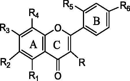
In the present studies, we have attempted to investigate the effect of a panel of six monohydroxylated flavonoids () in relation to their antiglycation activity, lens opacity, tryptophan fluorescence, aldose reductase inhibition (both experimental and in silico) and their effect on formation of glycation induced protein aggregates. A discourse on possible structure activity relationship in context with positional suitability of hydroxylation is also described so as to focus the compatibility of flavonoid scaffold for designing novel and effective anticataract agents.
Figure 1. Structure of the selected flavonoids: 3-Hydroxy flavones (R-OH, R1, R2, R3, R4, R5, R6 -H); 5-Hydroxy flavone (R1-OH, R, R2, R3, R4, R5, R6-H); 6-Hydroxyflavone (R2-OH, R, R1, R3, R4, R5, R6-H); 7-Hydroxy flavone (R3 - OH, R, R1, R2, R4, R5, R6 - H,); 2′- Hydroxy flavone (R5-OH, R, R1, R2, R3, R4, R6-H,); 4′-Hydroxy flavone (R6-OH, R, R1, R2, R3, R4, R5-H).
Materials and methods
Kreb Ringer Bicarbonate (KRB) buffer, Aminoguanidine hydrochloride, NADPH, 2, 4, dinitrophenyl hydrazine (2, 4-DNPH), penicillin, streptomycin, trichloro acetic acid (TCA), urea etc. were purchased from the Himedia Laboratories Pvt. Ltd. Mumbai (MS), India. The flavone, 3-hydroxy, 5-hydroxy, 6-hydroxy, 7-hydroxy, 2′-hydroxy and 4′-hydroxy substituted flavonoids were purchased from Sigma-Aldrich Co., St. Louis, MO. All other chemicals, solvents and reagents were of AR grade and were obtained from commercial sources.
Lens organ culture study
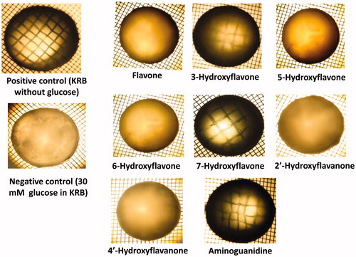
Fresh goat eyeballs (from 6–8-month-old animals) were obtained from the local slaughterhouse at Nanded city and were immediately transferred to a container containing KRB buffer (pH 7.5) and transported to the laboratory in cold conditions using ice packs. The eye balls were dissected using posterior approach for removing the intact lensesCitation16,Citation17. The isolated lenses were incubated individually in KRB buffer (having a baseline glucose concentration of 10 mM and osmolality of 255–295 mOsm) with the addition of 30 mM glucose (a supraphysiological concentration for inducing glycation) and 50 μM individual flavonoids. A set of positive (lens in KRB buffer without further addition of 30 mM glucose) and negative control (lens in KRB + 30 mM glucose) along with a reference compound (aminoguanidine 50 μM) were also arranged simultaneously for comparison purpose. The media of positive control were replaced with plane KRB, while the media of treated groups were replaced with fresh KRB buffer added with 30 mM glucose after every 48 h. Minimum four lenses (from two animals) were used in every set. The lenses in various sets were incubated in CO2 incubator (with 95% air, 5% CO2 and at 37 °C temperature). The antibiotics penicillin and streptomycin were added to the KRB buffer to prevent microbial contamination. After 15 days of incubation, the lenses were observed for the development of generalized opacity, haziness, disruption and other morphological changes. The digitized images of lenses in various sets were captured using Olympus make SZ 61TR zoom trinocular microscope.
Extraction and estimation of crude lens proteins: profiling of soluble and insoluble fractions
After 15 days of incubation, the 10% lens homogenates were prepared according to method described elsewhereCitation15. In brief, the individual lens tissues from different sets were homogenized in a buffer containing, 0.025 M Tris, 0.5 mM EDTA, 0.1 M NaCl and 0.01% sodium azide (pH 8). Lens homogenates were subjected for centrifugation at 10 000 g at 4 °C for 30 min. Crude lens soluble protein present in supernatant was collected and used for further analysis. Total proteins (TP) and total lens soluble proteins (TSP) were estimated by Lowry methodCitation18. The amount of proteins present was calculated as milligram/gram of lens tissues.
Estimation of protein carbonyl groups: a marker of glycation
The protein carbonyl groups were estimated as per previously described methodCitation19. The protocol involves 1-h incubation of lens proteins with equal volume of 0.1% of 2,4-DNPH in 2N HCl at room temperature. After incubation, proteins were precipitated by the addition of 20% trichloro acetic acid (TCA) and washed three times with ethanol/ethyl acetate (1:1) mixture. After final wash, the protein precipitate was solubilized in 133 mM Tris, 13 mM EDTA buffer, pH 7.4 containing 8 M urea and absorbance was read at 365 nm. Concentration of protein carbonyl groups formed in the samples were calculated by using molar extinction coefficient (ɛ365 nm = 21 mM − 1 cm − 1).
Measurement of nontryptophan AGE fluorescence
AGEs formed in the reaction mixture were detected by measuring the fluorescence intensity of TSPCitation17. Protein samples (TSPs of various sets) were calibrated at a concentration of 0.15 mg/ml in phosphate buffer (pH 7.4), the samples were excited at 370 nm, and subsequently, the fluorescence emission was recorded between 400–500 nm using a Jasco make spectrofluorometer (FP-8300).
Size measurements of glycation-induced lens protein aggregates using nanoparticle tracking and analysis (NTA) LM 20 system
After 15 days of incubation, the size of the glycated lens protein aggregates were measured using NTA LM 20 system. The operational settings were carried out as per the manufacturer manual. The glycated protein samples treated with various flavonoids were diluted in ultrapure water to achieve the particle concentration of 107–109/ml. The calibrated samples were injected into LM 20 sample laser module (Sample holder) by sterile syringesCitation20. The particles present in the liquid sample at standard temperature and viscosity possess the Brownian motion; the NTA software (Salisbury, United Kingdom) calculates the size of particles by tracking and analyzing motion of individual particles under laser light illumination. The NTA 2.3 software measure diffraction of laser light by a particles and processed and generated the data. The data are generated in terms of mean (average particle size measured) and mode (most frequently observed). The values obtained by processing the sample were recorded and the modal value (most frequent particle size) was taken as a size of lens protein aggregates. In NTA, Stokes–Einstein equation is the basis for calculation of particle sizeCitation21.
Preparation of aldose reductase (AR)
The isolation and semipurifed AR preparation was carried out as per the previously described protocolCitation22. In brief, the fresh eye balls from young goat were quickly removed and homogenized in 100 mM potassium phosphate buffer (pH 6.2). The homogenate was centrifuged at 15 000 rpm for 30 min at 4 °C. The pellet was discarded and the clear supernatant was stored at 4 °C and used as source of AR enzyme.
Determination of aldose reductase inhibition using selected flavonoids
The AR inhibition assay was carried out according to the method described earlierCitation22. The reaction mixture contained 1 ml of 50 μM potassium phosphate buffer (pH 6.2), 10 μM DL-glyceraldehyde, 0.1 μM NADPH, 0.4 mM lithium sulfate, 5 μM 2-mercaptoethanol and semipurified AR enzyme. The reaction cocktail was incubated at room temperature for 10 min. The reaction was triggered by addition of NADPH and the fall in the extinction was recorded spectrophotometrically at 340 nm. Series of concentrations of selected flavonoids were added to reaction mixture and appropriate blanks were used for corrections. AR activity without inhibitor was considered as 100%, the amount of individual flavonoids required to achieve 50% AR inhibition were considered as IC50 values. Quercetin was used as reference AR inhibitor.
In silico investigation
In silico molecular docking investigation was performed using the formerly well-established protocol reported elsewhereCitation23,Citation24 that involved following steps. Target enzyme's (AR) 3D coordinates were obtained from PDB databaseCitation25 with PDB ID 2DUX. This protein coordinate file was crystallized in complex with inhibitor zopolrestatCitation26 in monomeric state of the enzyme. Before proceeding to actual virtual screening of flavonoids, the coordinate file was edited appropriately to remove non proteinous parts as per the protocol for preparing receptor in AutoDock. The zopolrestat molecule was separately saved to independent file in order to test the validity of system. The target system was generated in conventional pdbqt format.
Initially, the 2D representations of selected flavanoids were drawn in ChemDraw ultra software (CambridgeSoft, Cambridge, MA). SMILES notations of flavanoids were listed in separate file that was subsequently loaded on FROG serverCitation27 for generating the 3D coordinates of the ligands in sdf format. An exceedingly scientifically reputed open access program, AutoDock 4.2 embedded in Python Prescription 0.8 (PyRx) environment was employed for docking investigationCitation28,Citation29. The ligands in sdf format were brought into PyRx environment and converted to required pdbqt format following the process of energy minimization.
Conventionally, docking systems are validated for accuracy by redocking the experimentally verified pose back into receptor file and test if software generate pose having best score with orientation similar to experimental binding mode. This fact is measured in terms of RMSD between predicted and experimental pose of ligand. If the RMSD value falls under the acceptable range, say, less than 2 Å, the prediction of binding is assumed to be successfulCitation30. Default parameters were employed for setting grid and further conformational search algorithm (lamarckian genetic algorithm) was used with default values in all docking runs. The RMSD reading of 0.63 Å was obtained for pose with promising binding free energy value. Structural inspection of docked zopolrestat-AR complex revealed that all the functional groups essential for biological activity were predicted to be oriented as required; and thus, predicted binding mode is observed nearly the same as compared to the crystallographic mode of binding. This observation thereby confirms the successful validation of docking system. Finally, the PyMol version 0.99 (www.pymol.org) was used for rendering the illustrations and 2D interactions of ligands with residues from active site of AR were analyzed with help of LigPlot+ Citation31,Citation32.
Statistical analysis
The experimental results summarized for various parameters are expressed as the mean of n = 4, ± SD. The significance of the difference from the respective controls for each experimental test condition was assayed by using Student’s t-test for each experiment. A p value < 0.05 was considered as a significant difference.
Results and discussion
The basic aim of the study was to assess the efficacy of the selected set of monohydroxylated flavonoids and their positional effect of hydroxylation in concert with the anticataract-related activities in glycation-induced ovine lens model. The representative digitized images of the lenses treated with selected flavonoids in lens organ culture studies are shown in . The glycation-induced lens which was devoid of any treatment resulted in disintegration of the overall morphology of the lens; however, it is clear from the representative images digitized after 15 days of incubation, the 7-hydroxy flavone has maintained the transparency and overall morphology of the lens significantly as compared to negative control and all other test flavonoids. It is interesting to note that the 7-hydroxy treated lens transparency seems to be at par with that of the aminoguanidine: a known antiglycating agent. The 3-hydroxy flavone also demonstrated considerable effect in ameliorating the overall morphology of the glycated lens. All other flavonoids were found effective in maintaining the structural integrity of the lenses; however, they were not found to have significant impact on maintaining lens transparency. Moreover, several structural deformities were apparent in lenses treated with these flavonoids. In the current state of the art, the explanation regarding the underlying mechanism of monohydroxylated flavonoid driven lens transparency is lacking; however, in case of galactosemic rats, the oral treatment of flavonoid quercetin ameliorated the disturbances of eye lens electrolytes and significantly normalized the lens protein levels. The results of this experimental settings concluded that inclusion of flavonoid quercetin contributed significantly towards lens transparency by maintaining the characteristic osmotic ion equilibrium and protein levels of the lensCitation33. Perhaps, the structural compatibility 7- and 3-hydroxy flavonoids might be involved in maintaining osmotic ion equilibrium and protein levels of the lens.
Figure 2. Digitized representative images of the bovine lenses showing effect of selected flavonoids (50 μM) on glycation-induced opacity, haziness, disruption and other morphological changes. The images of lenses were digitized after 15 days of incubation. Minimum four lenses (from two animals) were used in each set.
The profile of TSP and its percentage in glycated and other control sets is summarized in . The significant amount of soluble protein was found in the samples treated with 7-hydroxy (94%) and 3-hydroxy (84%) flavonoid as compared with the glycated sample devoid of any treatment (57%). The 5-hydroxy (82%) flavonoid also contained considerable amount of soluble lens protein. All other samples were found to have soluble proteins in a range of 72–76%. Homogenization of lens tissue in buffer or water and subsequent centrifugation yields water-soluble (WS) fraction and a water-insoluble (WIS) fraction. Usually, the WIS protein fractions tend to increase with agingCitation9. As compare to WS portion, the WIS fraction contains a relatively more amount of modified and cross-linked crystallins, along with cell membranes of lens fibers and cytoskeleton proteinsCitation34. Therefore, presence of more amount of WS fraction is a prognostic marker of lens transparency.
Table 1. Profile of protein concentrations, amount of protein carbonyls and size of protein aggregates in glycation-induced lenses treated with different flavonoids.
The results of the amount of carbonyl group formation owing to glycation of the lens proteins (), clearly shows that less amount of carbonyls were generated in the samples treated with 7-hydroxy flavone (176 nmoles/mg protein) as compared to glycated sample devoid of any treatment (880 nmoles/mg protein). On the other hand, significant amount of carbonyls were detected in samples treated with 2′-hydroxy flavanone (740 nmoles/mg protein), 4′-hydroxy flavanone (609 nmoles/mg protein), followed by 5-hydroxy flavones (506 nmoles/mg protein), indicating that these flavonoids could not inhibit the process of glycation significantly. Glycation is a glyco-oxidative process, wherein the carbonyl groups are generated on protein side chains. In proteins, especially the side chains of Pro, Lys, Arg, and Thr are more prone for sugar mediated oxidation which leaves the carbonyl groups at the oxidized site. Owing to the chemical stability of carbonyls, it is easy to store and detect their identityCitation35. The generation of more amounts of glycation-induced carbonyls is a biomarker of extensive glyco-oxidative damage to the lens proteins. Nevertheless, carbonyls may also contribute protein cross-linking leading to protein dysfunctionCitation16.
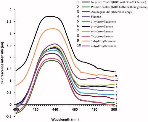
The results of intensity of AGEs formed in the glycated lenses treated with test flavonoids are summarized in . It is clear that once again the sample treated with 7-hydroxy flavone has lesser amount of AGEs generated as compared to the glycated lens devoid of any treatment and even when compared with aminoguinidine: a standard antiglycating agent. The lens treated with 3-hydroxy flavone also demonstrated significant reduction in the intensity of AGEs fluorescence as compared to other test flavonoids. Remaining flavonoids were observed to have moderate effect on inhibition of AGEs formation. Protein glycation-driven formation of AGEs has been considered as one of the major causes leading to series of diabetic complications including cataractogenesisCitation36. AGEs also reported to play an important role in the progression of diabetic retinopathy leading to dysfunction and death of retinal cellsCitation37. Although not monohydroxylated, but the flavonoids containing more than two hydroxyl groups, such as quercetin, luteolin, rutin and rutin metabolites, have been reported to inhibit the formation of fluorescent and nonfluorescent AGEsCitation38,Citation39; however, the literature in relation to mechanism of flavonoid mediated inhibition of AGEs is evolving.
Figure 3. Effect of selected flavonoids (50 μM) on glycation induced AGEs fluorescence of lens proteins. The result summarized is the representative image of individual four experiments performed with four lenses (derived from two animals).
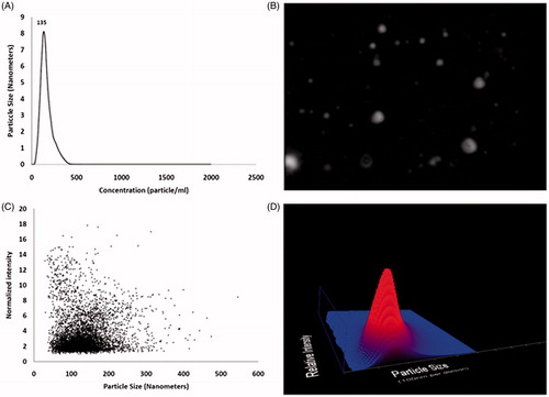
The results of the glycation-induced protein aggregates size measurements using NTA LM20 system are summarized in the . The NTA system offers direct visualization, sizing and counting of the protein aggregates ()Citation20. Among the tested flavonoids the 7-hydroxy flavone (135 nm), 3-hydroxy flavone (146 nm) and 5-hydroxy flavone (161 nm) showed minimum size of glycation-induced lens protein aggregates as compared to glycated lens devoid of any treatment (300 nm). It is interesting to note that with exception of 2′- (223 nm) and 4′-hydroxy flavanone (287 nm), the size of the flavonoids treated proteins aggregates (100–175 nm) was considerably smaller than the aminoguanidine-treated sample (201 nm). It is well-established fact that glycation reaction induces formation of protein aggregatesCitation40. The glycation induced lens crystallins aggregate, causes the lens to increasingly scatter light on the retina instead of focusing light on it and causing the lens to lose its transparency gradually and results in the loss of visual acuityCitation3. The genesis of protein aggregates has been believed to be a consequence of destabilized partially unfolded polypeptides chains. In recent years, inhibition of protein aggregation pathway has been identified as a complementary target for designing novel anticataract drugsCitation3. The plant-derived bioactive flavonoids like (-) epigallactocatechin 3-galate and curcumin successfully inhibited protein aggregate formation and also disaggregated the preformed protein aggregatesCitation41,Citation42; however, the concrete molecular underpinnings of flavonoids-mediated disaggregation of protein aggregates is not known. It is hypothesized that the protein aggregation inhibitors may act chain terminators by blocking the addition of proteins to the growing aggregate; disaggregate the preformed species, or conferring stability to the refolded-protein conformations (native state) of crystallinsCitation3.
Figure 4. Representative image panel of effect of 7-hydroxy flavone (50 μM) on glycation induced lens crystalline aggregate formation. The effect of selected flavonoids (50 μM) on glycation induced lens crystalline aggregate formation was analyzed by measuring the sizes of lens protein aggregates using NTA LM 20 analysis system. (A) Plot representing particle size (nm) and concentration (particle/ml). Value 135 is a mode size (in nm) of observed nano particles. (B) Still frame from video of nano sizing experiment (refer on line supplementary video). (C) Plot representing particle size (nm) and normalized intensity. (D) A 3D plot of particle size (10 nm per division) and relative intensity. The results summarized are the mean value of four lenses (derived from two animals) each used in individual four experiments.
The AR inhibitory activities of selected flavonoids are summarized in . It is clear that 7-hydroxy flavone (IC50, 8.0 μM) has demonstrated significant AR inhibition as compared to other test flavonoids which showed AR inhibition IC50 values in a range of 42–104 μM. It is well established that hyperglycemia activates the polyol pathway. The up-regulated activity of AR leads to production of high levels of sorbitol (a sugar alcohol) which adversely affects the tonicity of the lens, leading to the swelling and disruption of the lens fiber cells. Altogether the collective effect of sorbitol-driven alterations in the lens environment leads to oxidative damage of the lens tissue leading to cataractCitation1. Therefore, developing novel AR inhibitors has remained a significant hope for the amelioration of diabetic complications including cataractogenesis. While defining the structure–activity relationship of flavoinds in concert with AR inhibition, it has been described that the flavones and flavonols having the 7-hydroxyl moiety exhibits strong AR inhibition. Also, the presence of 2–3 double bond (present in flavones and lacking in flavanones) contributes for effective inhibition of ARCitation43. The results of the present investigation are in agreement with the previously described structural requirements, as the 7-hydroxy flavone has demonstrated significant AR inhibition as compared to the 2′ and 4′-hydroxy flavanones which lacks the 2–3 double bonds.
Table 2. Summary of AR inhibition potential of selected flavonoids.
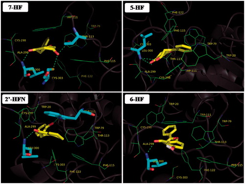
The docking experiments carried out to understand the interactions of the selected flavonoids with AR are summarized in . The docking data clearly demonstrates that the selected flavonoids possess both polar and nonpolar interactions with AR enzyme. The binding-free energies described in may be useful in gaining insights into the protein–ligand interactionsCitation44. The binding efficiency (commonly expressed in terms of tight or loose binding) of ligand to active site of proteins can be inferred using the number and length of hydrogen bonds. Generally, lower the hydrogen bonding distance, tighter is the bindingCitation45. Two flavonoid, 5HF and 7-HF demonstrates promising binding free energy values of −9.32 and −9.54 kcal/mol, respectively, as compared to remaining compounds. These two compounds are observed to make multiple polar interactions with active site residues with shorter hydrogen-bonding distances. These in silico observations might explain their comparable in vitro AR inhibitory potential in contrast to remaining flavonoids (IC50 values of about 42 and 8 μM for 5HF and 7-HF, respectively). The table also features the list of residues from AR that are involved in hydrophobic or van der Waal's contact with the flavonoid compounds. Empirical and modeling studies have demonstrated that nonpolar interactions like hydrophobic interactions and van der Waal's contacts are first line of forces that orient the ligand molecule in appropriate position followed by forming successful polar interactionsCitation46. Thus, such interactions might play a critical role in defining stability of enzyme-inhibitor complexes.
Table 3. In silico data obtained from virtual screening and structural analysis of flavonoids in aldose reductase.
Every flavonoid compound tested for AR inhibitory activity in present study shows the presence of conserved hydrogen bond with backbone nitrogen atom of residue Leu-300 (). Such an interaction is experimentally verified to be an important feature of AR inhibition in a recent crystallographic study involving inhibition of AR by compound JF0064Citation47. Promising flavonoid 7HF is found to be interacting with side chain oxygen of Thr-113. It has been already demonstrated that interaction of inhibitor IDD388 with side chain of Thr-113 is one of the major determinants in AR inhibition mechanismCitation48. Moreover, artificial mutagenesis experiments that result is replacement of alanine in place of threonine at 113th location of AR ended in significant drop of binding-free energy for IDD388 that adversely affected the inhibitory potential of inhibitorCitation49. Additionally, 7HF also appears to be actively engaged in a hydrogen-bonding interaction with side chain sulfhydryl of Cys-303. Recent biochemical and crystallographic study involving inhibition of AR by pyridazinone class of compounds suggest the critical role of polar interaction via side chain of Cys-03Citation50. Many of the test flavonoids (7HF, 6HF and 2'HF) from present investigation were found to be involved in a π–π stacking interaction with the aromatic side chain of Trp-111 (). Such stacking interaction is experimentally verified to stabilize many inhibitors like zopolrestatCitation51 and IDD594Citation26. Considering the observed in vitro AR inhibition data and structural observation from docking study carried out in present investigation along with supporting crystallographic and biochemical data already published, it can be assumed that present flavonoid dependent AR inhibition might have involved residues like Leu-300, Thr-113, Cys-303 and Trp-111.
Figure 5. Docking results of promising flavanoids (yellow sticks) in Aldose reductase (cartoon representation). Residues in hydrophobic contacts are depicted in green color lines. Atoms of residues engaging hydrogen bonds (green dotted lines) with flavonoid molecules are shown as cyan sticks.
Conclusion
The present investigation was designed for understanding the structural requirement of monohydroxylated flavonoids (hydroxylation at 3, 5, 6, 7, 2′ and 4′) in concert with their efficacy in the amelioration of glycation-induced lens opacity, inhibition of protein aggregation, carbonyl group formation, nontryptophan AGE fluorescence and AR. The results obtained clearly focused the significance 7-hydroxy scaffold as a lead molecule for designing novel antiglycating and anticataract agents. In the current state of the art searching the anticataract bioactive compounds like flavonoids are appreciated as they can be made bioavailable through dietary ingredients. Nevertheless, such findings may also help in formulating functional foods and neutraceuticals for the effective management of diabetic complications including cataract.
Declaration of interest
The authors report no conflicts of interest. The authors alone are responsible for the content and writing of this article.
Acknowledgements
Authors are thankful DST-SERB, New Delhi, for financial assistance (F. No. SR/SO/BB-0088/2012) and to Professor Pandit Vidyasagar, Vice Chancellor, SRTM University, for providing facilities and encouragement. KKP thanks DST-SERB for JRF.
References
- Pollreisz A, Schmidt-Erfurth U. Diabetic cataract-pathogenesis, epidemiology and treatment. J Ophthalmol 2010;2010:608751.
- International Diabetes Federation, Diabetes Atlas. 5th ed. Brussels, Belgium: International Diabetes Federation; 2011:1–14.
- Moreau KL, King JA. Protein misfolding and aggregation in cataract disease and prospects for prevention. Trends Mol Med 2012;18:273–82.
- Saraswat M, Suryanarayana P, Reddy PY, et al. Antiglycating potential of Zingiber officinalis and delay of diabetic cataract in rats. Mol Vis 2010;16:1525–37.
- Bloemendal H, de Jong W, Jaenicke R, et al. Ageing and vision: structure, stability and function of lens crystallins. Prog Biophys Mol Biol 2004;86:407–85.
- Luthra M, Balasubramanian D. Nonenzymatic glycation alters protein structure and stability. A study of two eye lens crystallins. J Biol Chem 1993;268:18119–27.
- Chen M, Curtis TM, Stitt AW. Advanced glycation end products and diabetic retinopathy. Curr Med Chem 2013;20:3234–40.
- Ahmed N. Advanced glycation endproducts-role in pathology of diabetic complications. Diabetes Res Clin Pract 2005;67:3–21.
- Sharma KK, Santhoshkumar P. Lens aging: effects of crystallins. Biochim Biophys Acta 2009;1790:1095–108.
- Harding JJ. Viewing molecular mechanisms of ageing through a lens. Ageing Res Rev 2002;1:465–79.
- Swamy MS, Tsai C, Abraham A, Abraham EC. Glycation mediated lens crystallin aggregation and cross-linking by various sugars and sugar phosphates in vitro. Exp Eye Res 1993;56:177–85.
- Obrosova IG, Chung SS, Kador PF. Diabetic cataracts: mechanisms and management. Diabetes Metab Res Rev 2010;26:172–80.
- Murata M, Ohta N, Sakurai S, et al. The role of aldose reductase in sugar cataract formation: aldose reductase plays a key role in lens epithelial cell death (apoptosis). Chem Biol Interact 2001;130–132:617–25.
- Kumar S, Pandey AK. Chemistry and biological activities of flavonoids: an overview. Scientific World J 2013;2013:162750.
- Stefek M. Natural flavonoids as potential multifunctional agents in prevention of diabetic cataract. Interdiscip Toxicol 2011;4:69–77.
- Kumar PA, Kumar MS, Reddy GB. Effect of glycation on alpha-crystallin structure and chaperone-like function. Biochem J 2007;408:251–8.
- Moghaddam MS, Kumar PA, Reddy GB, Ghole VS. Effect of diabecon on sugar-induced lens opacity in organ culture: mechanism of action. J Ethnopharmacol 2005;97:397–403.
- Lowry OH, Rosebrough NJ, Farr AL, Randall RJ. Protein measurement with the folin phenol reagent. J Biol Chem 1951;193:265–75.
- Uchida K, Kanematsu M, Sakai K, et al. Protein-bound acrolein: potential markers for oxidative stress. Proc Natl Acad Sci USA 1998;95:4882–7.
- Filipe V, Hawe A, Jiskoot W. Critical evaluation of nanoparticle tracking analysis (NTA) by NanoSight for the measurement of nanoparticles and protein aggregates. Pharm Res 2010;27:796–810.
- Birla SS, Gaikwad SC, Gade AK, Rai MK. Rapid synthesis of silver nanoparticles from Fusarium oxysporum by optimizing physicocultural conditions. Scientific World J 2013;2013:796018.
- Gacche RN, Dhole NA. Profile of aldose reductase inhibition, anti-cataract and free radical scavenging activity of selected medicinal plants: an attempt to standardize the botanicals for amelioration of diabetes complications. Food Chem Toxicol 2011;49:1806–13.
- Gond DS, Meshram RJ, Jadhav SG, et al. In silico screening of chalcone derivatives as potential inhibitors of dihydrofolate reductase: assessment using molecular docking, paired potential and molecular hydrophobic potential studies. Drug Invent Today 2013;5:182–191.
- Kadam A, Dawane B, Pawar M, et al. Development of novel pyrazolone derivatives as inhibitors of aldose reductase: an eco-friendly one-pot synthesis, experimental screening and in silico analysis. Bioorg Chem 2014;53:67–74.
- Berman HM, Westbrook J, Feng Z, et al. The protein data bank. Nucleic Acids Res 2000;28:235–42.
- Steuber H, Zentgraf M, Gerlach C, et al. Expect the unexpected or caveat for drug designers: multiple structure determinations using aldose reductase crystals treated under varying soaking and co-crystallisation conditions. J Mol Biol 2006A;363:174–87.
- Miteva MA, Guyon F, Tufféry P. Frog2: Efficient 3D conformation ensemble generator for small compounds. Nucleic Acids Res 2010;38:W622–7.
- Huey R, Morris GM, Olson AJ, Goodsell DS. A semiempirical free energy force field with charge-based desolvation. J Comput Chem 2007;28:1145–52.
- Morris GM, Goodsell DS, Halliday RS, et al. Automated docking using a Lamarckian genetic algorithm and an empirical binding free energy function. J Comput Chem 1998;19:1639–1662.
- Gacche RN, Meshram RJ, Shegokar HD, et al. Flavonoids as a scaffold for development of novel anti-angiogenic agents: an experimental and computational enquiry. Arch Biochem Biophys 2015;577-578:35–48.
- Laskowski RA, Swindells MB. LigPlot+: multiple ligand-protein interaction diagrams for drug discovery. J Chem Inf Model 2011;51:2778–86.
- Wallace AC, Laskowski RA, Thornton JM. LIGPLOT: a program to generate schematic diagrams of protein-ligand interactions. Protein Eng 1995;8:127–34.
- Ramana BV, Raju TN, Kumar VV, Reddy PU. Defensive role of quercetin against imbalances of calcium, sodium, and potassium in galactosemic cataract. Biol Trace Elem Res 2007;119:35–41.
- Harrington V, Srivastava OP, Kirk M. Proteomic analysis of water insoluble proteins from normal and cataractous human lenses. Mol Vis 2007;13:1680–94.
- Dalle-Donne I, Rossi R, Giustarini D, et al. Protein carbonyl groups as biomarkers of oxidative stress. Clin Chim Acta 2003;329:23–38.
- Negre-Salvayre A, Salvayre R, Augé N, et al. Hyperglycemia and glycation in diabetic complications. Antioxid Redox Signal 2009;11:3071–109.
- Stitt AW, Curtis TM. Diabetes-related adduct formation and retinopathy. J Ocul Biol Dis Infor 2011;4:10–18.
- Cervantes-Laurean D, Schramm DD, Jacobson EL, et al. Inhibition of advanced glycation end product formation on collagen by rutin and its metabolites. J Nutr Biochem 2006;17:531–40.
- Wu CH, Yen GC. Inhibitory effect of naturally occurring flavonoids on the formation of advanced glycation endproducts. J Agric Food Chem 2005;53:3167–73.
- Adrover M, Mariño L, Sanchis P, et al. Mechanistic insights in glycation-induced protein aggregation. Biomacromolecules 2014;15:3449–62.
- Ferreira N, Saraiva MJ, Almeida MR. Natural polyphenols inhibit different steps of the process of transthyretin (TTR) amyloid fibril formation. FEBS Lett 2011;585:2424–30.
- Meng F, Abedini A, Plesner A, et al. The flavanol (-)-epigallocatechin 3-gallate inhibits amyloid formation by islet amyloid polypeptide, disaggregates amyloid fibrils, and protects cultured cells against IAPP-induced toxicity. Biochemistry 2010;49:8127–33.
- Matsuda H, Morikawa T, Toguchida I, Yoshikawa M. Structural requirements of flavonoids and related compounds for aldose reductase inhibitory activity. Chem Pharm Bull (Tokyo) 2002;50:788–95.
- Meyer AG, Sawyer SL, Ellington AD, Wilke CO. Analyzing machupo virus-receptor binding by molecular dynamics simulations. Peer J 2014;2:e266.
- Osajima T, Suzuki M, Neya S, Hoshino T. Computational and statistical study on the molecular interaction between antigen and antibody. J Mol Graph Model 2014;53:128–39.
- Weng C, Fu Y, Jiang H, et al. Binding interaction between a queen pheromone component HOB and pheromone binding protein ASP1 of Apis cerana. Int J Biol Macromol 2015;72:430–6.
- Cousido-Siah A, Ruiz FX, Mitschler A, et al. Identification of a novel polyfluorinated compound as a lead to inhibit the human enzymes aldose reductase and AKR1B10: structure determination of both ternary complexes and implications for drug design. Acta Crystallogr D Biol Crystallogr 2014;70:889–903.
- Podjarny A, Cachau RE, Schneider T, et al. Subatomic and atomic crystallographic studies of aldose reductase: implications for inhibitor binding. Cell Mol Life Sci 2004;61:763–73.
- Koch C, Heine A, Klebe G. Tracing the detail: how mutations affect binding modes and thermodynamic signatures of closely related aldose reductase inhibitors. J Mol Biol 2011;406:700–12.
- Steuber H, Zentgraf M, Podjarny A, et al. High-resolution crystal structure of aldose reductase complexed with the novel sulfonyl-pyridazinone inhibitor exhibiting an alternative active site anchoring group. J Mol Biol 2006B;356:45–56.
- Wilson DK, Tarle I, Petrash JM, Quiocho FA. Refined 1.8 A structure of human aldose reductase complexed with the potent inhibitor zopolrestat. Proc Natl Acad Sci USA 1993;90:9847–51.