
Abstract
Human dipeptidyl peptidase III (hDPP III), a zinc-metallopeptidase of the family M49, is an activator of the Keap1-Nrf2 cytoprotective pathway involved in defense against oxidative stress. Pathophysiological roles of DPP III have not been elucidated yet, partly due to the lack of specific inhibitors. We showed that substrate analog H-Tyr-Phe-NHOH is a strong competitive inhibitor of hDPP III, while H-Tyr-Gly-NHOH expresses much weaker inhibition. To investigate the effects of amino acid substitutions in inhibitor P1 position, we synthesized three new dipeptidyl hydroxamates and examined their influence on the activity of hDPP III and DPP III from the human gut symbiont Bacteroides thetaiotaomicron. The extent of inhibition of hDPP III, but not of bacterial enzyme, was dependent on the amino acid in P1. H-Phe-Phe-NHOH is recognized as one of the strongest inhibitors of hDPP III (Ki = 0.028 μM), and H-Phe-Leu-NHOH discriminated between human and bacterial ortholog of the M49 family.
Introduction
Dipeptidyl peptidase III (DPP III; EC 3.4.14.4) is a zinc-exopeptidase which cleaves dipeptides from the N-termini of its peptide substrates, consisting of three to ten amino acidsCitation1,Citation2. This enzyme was discovered in extracts of bovine pituitary gland through the hydrolysis of characteristic substrate Arg-Arg-2-naphthylamideCitation3. DPP III was purified and biochemically characterized from several human and animal tissues, slime mold, Drosophila and the yeast Saccharomyces cerevisiaeCitation2. It was shown that this cytosolic enzyme, which is broadly distributed in eukaryotic cells and tissues, in vitro hydrolyzes a number of biologically active peptides, including opioid peptides enkephalins and endomorphinsCitation1,Citation4,Citation5. Therefore, its contribution in normal intracellular protein catabolism was assumed and the role in the endogenous pain modulation was suggested. Recently it was revealed that human DPP III participates in the endogenous defense against oxidative stress, being an activator in the Keap1-Nrf2 signaling pathwayCitation6,Citation7. Pathophysiological roles of human DPP III are indicated in cataractogenesisCitation8, malignant growthCitation9,Citation10 and influenza virus infectionCitation11. He et al. reported that DPP III is a member of a six-gene signature that accurately predicts breast cancer patient survivalCitation12. These data indicated human DPP III as potential drug target.
Molecular cloning and sequencing of the rat and human enzymeCitation2,Citation13 revealed a characteristic zinc-binding motif, hexapeptide HELLGH, enabling the recognition of a distinct DPP III familyCitation14, also known as metallopeptidase family M49Citation15.
DPP III was considered to be an exclusively eukaryotic enzyme until 2003, when the complete genome sequences of Porphyromonas gingivalis and Bacteroides thetaiotaomicron appeared in databases, and the presence of DPP III was revealed in their genomes by bioinformatics approachCitation14. These first two prokaryotic orthologs (from P. gingivalis and B. thetaiotaomicron) helped us to define five conserved regions in the peptidase family M49Citation14. We have cloned and biochemically characterized the first bacterial DPP III, from the human gut symbiont B. thetaiotaomicron, revealing similarities but also important differences compared to its human counterpartCitation16. Until now, crystal structures of two M49 family members have been solved, yeast and human DPP IIICitation17,Citation18. The 3-D structure of human DPP III inactive mutant in complex with pentapeptide tynorphinCitation18 revealed the formation of closed enzyme’s active site during the substrate binding and exposed the amino acid residues which form five deep subsites (S2–S3′). Due to the low homology (less than 25% sequence identity) to eukaryotic proteins, not one 3-D structure of prokaryotic member of the M49 family has been resolved yet.
Although a significant knowledge on the molecular enzymology (structure–activity relationship) of the DPP III family accumulated in the last decadeCitation19–22, physiological roles of any of its members have not been elucidated in sufficient detail. This is partly due to the lack of specific inhibitors. Inhibition by non-specific chelating and thiol reagents is a characteristics of DPP III from various speciesCitation2,Citation16,Citation23, but the studies of more specific inhibitors of the DPP III enzymatic activity are rareCitation24. We have shown that two dipeptidyl hydroxamic acids, H-Tyr-Phe-NHOH and H-Tyr-Gly-NHOH act as the competitive inhibitors of human DPP III activityCitation20. Hydroxamate moiety (-NHOH) is capable of chelating the zinc ion at the catalytic site, and therefore it is essential for DPP III inhibition by dipeptidyl hydroxamic acids. Inhibitory potency of dipeptides was earlier investigated for human and rat DPP IIICitation1,Citation4, which has 93.5% protein sequence identity to hDPP III. Among examined dipeptides, the most potent inhibitors of rat brain DPP III were those with aromatic pairs, Tyr-Tyr and Tyr-Phe, with inhibition constants of 5.8 ± 0.5 μM and 8.4 ± 3.6 μM, respectively. Tyr-Leu inhibited the same enzyme with Ki value of 29.6 ± 6.4 μM, and the Ki of Tyr-Leu-NH2 was much larger (495.6 ± 3.8 μM)Citation4. Human erythrocyte DPP III was only slightly inhibited by dipeptides containing at least one aromatic amino acid residue: 50 μM Phe-Phe caused ∼6% inhibition, and the extent of inhibition by 50 μM Leu-Trp was ∼21% (Ki ∼120 μM)Citation1.
H-Tyr-Phe-NHOH inhibited hDPP III with the Ki value of 0.15 μM at pH 8.0Citation20. However, we observed a dramatic decrease in the inhibitory potency of H-Tyr-Gly-NHOH (70-fold higher Ki value), indicating that for the strong interactions of DPP III with hydroxamate inhibitor, chelating effect of hydroxamate moiety is not sufficient, but, in addition, interactions between the amino acid side chains of dipeptidyl hidroxamate with substrate binding subsites S1 and S2 are of crucial importance.
To investigate further the consequences of the amino acid side chain substitution in P1 or P2 position of dipeptidyl hydroxamates on their inhibitory potency towards human DPP III, we have synthesized a small series of these compounds: H-Phe-Phe-NHOH, H-Phe-Leu-NHOH and H-Phe-Gly-NHOH. Next, we examined their influence on the activity of human DPP III and bacterial DPP III. The enzyme from the Bacteroides thetaiotaomicron was chosen, because this bacterium is a prominent member of the human intestinal microflora and contributes to the host physiologyCitation25,Citation26.
We report here for the first time two novel, potent inhibitors of DPP III and interesting differences found for the interactions of these dipeptidyl hydroxamate compounds with human and bacterial ortholog of the M49 family.
Methods
Synthesis of the hydroxamate inhibitors
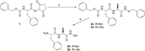
Dipeptidyl hydroxamic acid H-Phe-Phe-NHOH (2b) and H-Phe-Leu-NHOH (3b) have been prepared by the known method as shown in the Scheme 1. The synthesis of the glycine derivative, H-Phe-Gly-NHOH was reported previouslyCitation27. Diprotected derivatives 2a and 3a were obtained by the reaction of the Z-Phe-OSu with H-Phe-NHOBzl or H-Leu-NHOBzl in dry THF. The final compounds were prepared by the hydrogenolysis 2a and 3a over 10% Pd/C in methanol with small quantity CH2Cl2 to improve solubility of Z-protected derivative.
Scheme 1. The synthesis of the hydroxamate inhibitors: (a) H-Phe-NHBzl·CF3COOH (for 2a) or H-Leu-NHBzl·CF3COOH (for 3a), TEA, THF; (b) H2, 10% Pd/C, MeOH.
General: Melting points were determined on Kofler stage and are uncorrected. 1H and 13C NMR spectra were recorded at 300 MHz and 75 MHz, respectively, on a Bruker AV-300 spectrometer (Rheinstetten, Germany) (TMS was used as internal standard). FTIR spectra were recorded on a ABB Bomem MB102 (Québec, Canada). Optical rotations were measured on an Optical Activity AA-10 automatic polarimeter (Ramsey, England) using the wavelength of 589.3 nm. TLC was performed on silica gel coated Merck 60 F254 silica plates (Darmstadt, Germany). RP (reversed phase) HPLC analysis was performed on a Varian ProStar HPLC system (Palo Alto, CA) coupled with UV detector; C-18 semi-preparative (250 × 8 mm, ID 5 μm) column at a flow rate of 1 mL/min was used under isocratic conditions using 40% MeOH in 0.1% aqueous trifluoroacetic acid (TFA). UV detection was performed at 254 nm. HRMS analyses were carried out on a Bruker Ultraflex MALDI TOF/TOF (Bremen, Germany) mass spectrometer. All chemicals were of the best grade commercially available and were used without purification. Solvents were purified according to standard procedures; dry solvents were obtained according to literature methods and stored over molecular sieves.
General procedure for the preparation of protected dipeptidyl hidroxamate 2a and 3a: To a suspension of Z-Phe-OSu (1 mmol) and H-Phe (Leu)-NHOBZl (0.85 mmol) in dry THF (10 mL), TEA (1.25 mmol) was added. The reaction mixture was stirred three days at room temperature. The solvent was removed, CH2Cl2 was added and the solution washed with water, 1% HCl and 5% NaHCO3. Organic layer was separated, dried (Na2SO4) and the solvent evaporated. Recrystallisation (CH2Cl2/light petroleum) gives the title compounds.
Z-Phe-Phe-NHOBzl (2a): Yield 82%; m.p. 209–210 °C; [a]D20 = -23 [c 1, MeOH/CH2Cl2 (4:1)]; IR(KBr) ˜ V max/cm−1: 3279, 3207, 1693, 1649, 1537; 1H NMR (DMSO-d6) δ/ppm: 11.27 (s, 1H, NHhydroxam.), 8.21 (d, 1H, J = 8.1 Hz, NH), 7.44–7.11 (m, 21H, CHarom. and NH), 4.94 (s, 2H, CH2,Z), 4.70, 4.62 (2d, 2 × 1H, 2×J = 10.9, CH2,Bzl), 4.37 (q, 1H, J = 7.5, CH*Phe,), 4.27 (dt, 1H, J = 3.7, J = 10.5, CH*Phe), 2.98–2.78 (m, 3H, CH2,Phe, CHA,Phe), 2.68 (dd, 1H, J = 10.5, J = 13.6, CHB,Phe); 13C NMR (DMSO-d6) δ/ppm: 171.2, 167.4, 155.6 (C=O), 137.9, 137.1, 135.7 (Carom.), 129.2, 129.1, 128.8, 128.2, 128.1, 128.0, 127.6, 127.3, 126.4, 126.2 (CHarom.), 76.8 (CH2, Bzl), 65.1 (CH2,Z), 55.9 (CH*Phe), 51.7 (CH*Phe), 37.8 (CH2,β,Phe), 37.5 (CH2,β,Phe); HRMS (m/z) calculated for C33H33N3O5 [M + Na]+ 574.2318, found. 574.2309.
Z-Phe-Leu-NHOBzl (3a): Yield 72%; m.p. 169–170 °C; [a]D20 = -34 [c 1, MeOH/CH2Cl2 (4:1)]; IR(KBr) ˜ Vmax/cm−1: 3420, 3288, 3201, 1701, 1669, 1648, 1537; 1H NMR (DMSO-d6) δ/ppm: 11.28 (s, 1H, NHhydroxam.), 8.09 (d, 1H, J = 8.1 Hz, NH), 7.39–7.14 (m, 16H, CHarom. and NH), 4.94, 4.78 (2s, 2 × 2H, CH2,Bzl and CH2,Z), 4.37–4.25, 4.25–4.14 (2m, 2 × 1H, CH*Phe, CH*Leu), 2.99 (dd, 1H, J = 3.5, J = 13.6, CHA,Phe), 2.73 (dd, 1H, J = 10.7, J = 13.6, CHB,Phe), 1.61–1.31 (m, 3H, CH2,β,Leu and CHγ,Leu), 0.87, 0.83 (2d, 2 × 3H, 2× J = 6.2, CH3,δ,Leu); 13C NMR (DMSO-d6) δ/ppm: 171.2, 168.5, 155.7 (C=O), 137.9, 137.0, 135.8 (Carom.), 129.2, 128.8, 128.2, 127.9, 127.6, 127.3, 126.2 (CHarom.), 76.7 (CH2,Bzl), 65.1 (CH2,Z), 55.9 (CH*Phe), 48.7 (CH*Leu), 41.0 (CH2,β,Leu), 37.3 (CH2,β,Phe), 24.1 (CHγ,Leu), 22.6, 21.9 (CH3,δ,Leu); HRMS (m/z) calculated for C30H35N3O5 [M + Na]+ 540.2474, found. 540.248.
General procedure for the preparation of dipeptidyl hidroxamic acid 2b and 3b: To a solution of Z-Phe-(Phe or Leu)-NHOBzl (0.2 mmol) in MeOH (9 mL) and CH2Cl2 (2 mL-to improve solubility), 10 Pd/C (25 mg) was added. The reaction mixture was hydrogenated at room temperature and 0.55 MPa for 20 h. The catalyst was filtered and the solvent evaporated. The residue was crystallized from diethyl ether. The samples were analyzed by HPLC-MS. Hydroxamic acid 2b was purified by HPLC.
H-Phe-Phe-NHOH·CF3COOH (2b): Yield 22.0%; 1H NMR (DMSO-d6) δ/ppm: 10.73 (s, 1H, OH), 8.89 (d, 1H, J = 8.3 Hz, NH), 8.05 (s, 3H, NH3+), 7.38–7.17 (m, 11H, CHarom. and NH), 4.46–4.44 (m, 1H, CH*Phe,), 4.01 (s br, 1H, CH*Phe), 3.10 (dd, 1H, J = 4.6, J = 14.1, CHA,Phe), 2.97 (dd, 1H, J = 6.6, J = 13.7, CHA’Phe), 2.89 (m, 1H, J = 8.4, J = 14.1, CHB,Phe), 2.86 (dd, 1H, J = 7.9, J = 13.7, CHB’Phe), 2.68 (dd, 1H, J = 10.5, J = 13.6, CHB,Phe); 13C NMR (DMSO-d6)) δ/ppm: 167.7, 166.6 (C=O), 137.1, 134.6 (Carom.), 129.5, 129.0, 128.5, 128.2, 127.1, 126.5 (CHarom.), 53.1 (CH*Phe), 52.0 (CH*Phe), 38.0 (CH2,β,Phe), 37.0 (CH2,β,Phe); HRMS (m/z) calculated for C18H21N3O3 [M + Na]+ 350.1481, found. 350.1486.
H-Phe-Leu-NHOH (3b): Yield 95%; 1H NMR (DMSO-d6) δ/ppm: 10.62 (s br, 1H, OH), 8.80 (s br, 1H, NH), 7.91 (d, 1H, J = 7.7 Hz, NH), 7.30–7.15 (m, 5H, CHarom.), 4.24 (dd, J = 7.7, J = 13.6, 1H, CH*Leu), 3.42 (dd, 1H, J = 4.2, J = 8.7, CH*Phe), 2.94 (dd, 1H, J = 4.1, J = 13.5, CHA,Phe), 2.61 (dd, 1H, J = 8.5, J = 13.5, CHB,Phe), 1.52–1.31 (m, 3H, CH2,β,Leu and CHγ,Leu), 0.87, 0.84 (2d, 2 × 3H, 2×J = 6.4, CH3,δ,Leu); 13C NMR (DMSO-d6) δ/ppm: 173.7, 168.3 (C=O), 138.6 (Carom.), 129.3, 128.0, 126.0 (CHarom.), 55.9 (CH*Phe), 48.3 (CH*Leu), 41.6 (CH2,β,Leu), 40.3 (CH2, β,Phe), 24.0 (CHγ,Leu), 22.7, 21.9 (CH3,δ,Leu); HRMS (m/z) calculated for C15H23N3O3 [M + Na]+ 316.1637, found. 316.1633.
Heterologous expression and purification of human and bacterial DPP III
Both DPP III were heterologously expressed in Escherichia coli and affinity purified as recombinant proteins with C-terminal six-histidine tag. Production of recombinant human DPP III was performed according to Špoljarić et al.Citation28. Gene amplification and expression of DPP III from Bacteroides thetaiotaomicron was described by Vukelić et al.Citation16. Protein purity was confirmed by SDS-PAGE according to LaemmliCitation29. Protein concentrations were determined by the Bradford methodCitation30.
Determination of kinetic parameters
The Km values for hydrolysis of fluorogenic substrate Arg2–2NA were determined for both recombinant His-tagged DPP III under the same conditions (pH 7.4, 20 mM Tris-HCl buffer; 25 °C), from the initial reaction rates as previously describedCitation28. Under the conditions used, the Km values were 6.0 μM and 0.8 μM for human and bacterial enzyme, respectively.
All potential inhibitors were dissolved in 100% DMSO and were further diluted with 20 mM Tris-HCl buffer, pH 7.4 to give a stock solution 0.3 mM to 3 mM. Final concentration of human and bacterial DPP III in reaction mixture (3 ml) was 0.17 nM and 4 nM, respectively.
The control sample contained DMSO in the same % as the inhibitor-treated enzyme sample. Enzyme inhibition studies were performed using the Arg2–2NA as substrate. Dixon and s/v versus i plots were used to establish the type of inhibition exhibited by hydroxamate inhbitor at pH 8.0 and to determine the Ki valuesCitation20. At pH 7.4, the inhibition constant, Ki, was calculated according to the equation:
where, v0 and vi are the initial hydrolysis rates of Arg2–2NA catalyzed by DPP III in the absence and presence of the competitive inhibitor, and Km is the Michaelis constant.
Results and discussion
Generation and purification of bacterial and human DPP III
Purified recombinant His-tagged proteins (B. thetiotaomicron DPP III and human DPP III) were prepared as described in Methods and used for inhibition studies.
Effects of synthesized dipeptidyl hydroxamic acids on the activity of human and bacterial DPP III
We have shown previously that dipeptidyl hydroxamic acids H-Tyr-Phe-NHOH and H-Tyr-Gly-NHOH inhibit human DPP III at pH 8.0 competitively with Ki value of 0.15 μM and 10.5 μM, respectively. The competitive character of this inhibition was demonstrated by Dixon and Cornish–Bowden (s/v versus i) plotsCitation20.
Competitive inhibitors are valuable tools for study of enzyme active sites. By using H-Tyr-Phe-NHOH as an potent inhibitor of the wild-type human DPP III, we have recognized the functional importance of two evolutionary conserved amino acid residues (Tyr318 and Trp300) outside the zinc binding motives H450ELLGH and E507ECRAE, when the data on crystal structure of this zinc-metallopeptidase have not been available yetCitation19,Citation20.
In the present study, at first we examined the effect of H-Phe-Phe-NHOH, H-Phe-Leu-NHOH and H-Phe-Gly-NHOH on the activity of human DPP III at pH 8.0 and 25 °C for comparison with data reported for H-Tyr-Phe-NHOH and H-Tyr-Gly-NHOH. All three newly synthesized dipeptidyl hydroxamic acids inhibited this enzyme, but with different potency (), in competitive manner, as expected. Our preliminary investigation of novel dipeptidyl hydroxamic acids additionally indicated that the most potent inhibitors (Ki ∼ 0.1 μM) of human DPP III are those with a Phe as side chain substituent in P1 position.
Table 1. Inhibition of human DPP III by dipeptidyl hydroxamic acids at pH 8.0Table Footnote*.
Further goal was to examine newly synthesized hydroxamate compounds as potential inhibitors of another member of the same metallopeptidase family – DPP III from the human gut symbiont B. thetiotaomicron (Bt DPP III) at physiological pH (7.4), and to compare the inhibitory potency of these compounds towards bacterial and human enzyme.
Since the type of DPP III inhibition with dipeptidyl hydroxamic acids was a competitive, for determination of inhibition constant Ki at pH 7.4, we applied the equation:
where v0 and vi are the initial hydrolysis rates of Arg2–2NA catalyzed by DPP III in the absence and presence of the inhibitor, and Km is the Michaelis constant. summarizes the results on inhibition of bacterial and human DPP III by newly synthesized dipeptidyl hydroxamic acids, and H-Tyr-Phe-NHOH at pH 7.4. Interestingly, pH value strongly influenced the inhibitory activity of all examined dipeptidyl hydroxamates: the Ki values were 2–3 times lower at pH 7.4, compared to those determined at pH 8.0 ( and ).
Table 2. Inhibition of human and bacterial DPP III by dipeptidyl hydroxamic acids at pH 7.4Table Footnote*.
At pH 7.4, H-Phe-Phe-NHOH and H-Tyr-Phe-NHOH inhibited human DPP III with equal potency (Ki ∼ 30 nM), while this enzyme’s affinity for H-Phe-Leu-NHOH and especially for H-Phe-Gly-NHOH was much lower (20-fold and 150-fold increased Ki value, respectively). However, the differences in inhibitory potency of dipeptidyl hydroxamic acids towards DPP III from B. thetaiotaomicron were much less pronounced. The inhibition constant was about the same for H-Phe-Phe-, H-Phe-Leu- and H-Tyr-Phe-NHOH, and that obtained for H-Phe-Gly-NHOH was about 10-fold higher ().
Furthermore, while the two inhibitors with Phe in P1 position (H-Phe-Phe- and H-Tyr-Phe-NHOH) inhibited human DPP III somewhat stronger (2-fold decrease in the Ki values) than the bacterial enzyme, inhibitory potency of H-Phe-Leu-NHOH was 9-fold higher for Bt DPP III ().
Obtained results revealed that for the strong binding of dipeptidyl hydroxamic acids to the active site of human DPP III, but not of bacterial enzyme, aromatic amino acid residue in P1 position is required, thus indicating the differences in the structural determinants of the S1 subsite of human and bacterial enzyme. Specificity nomenclature for peptidase interactions with substrate/inhibitor is presented in Supplementary Figure 1. Recently, Bezerra et al.Citation18 resolved the crystal structure of human DPP III in complex with pentapeptide tynorphin (Val-Val-Tyr-Pro-Trp), revealing the formation of closed enzyme's active site during the ligand binding which induces a large domain motion. This work defined the amino acid residues forming five deep subsites (S2–S3′) which accommodate the amino acid side chains in the positions P2–P3′ of the peptide substrate.
Figure 1. Multiple sequence alignment (MSA) of human (UniProt entry: Q9NY33) and B. thetaiotaomicron (UniProt: Q8A6N1) protein sequences. MSA was obtained using ClustalO (www.uniprot.org./align). A section of MSA comprising the constituents of the S1 and S2 subsite, marked with 1 and 2, below alignment is presented in the figure. Five evolutionary conserved regions of the M49 family are framed. Identical amino acid constitutents of S1 and S2 subsites are shadowed grey and those which differ are printed in white on black.
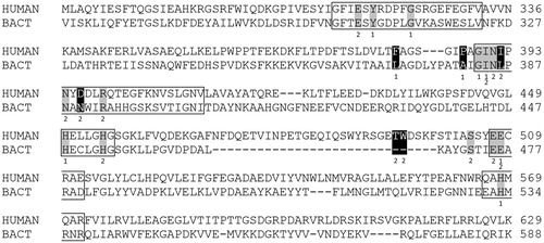
To investigate potential structural basis for observed difference in interaction with dipeptidyl hydroxamic acids, multiple sequence alignment (MSA) of human and Bt DPP III was performed (). MSA enabled us to compare structurally equivalent amino acid residues in human and bacterial DPP III. Dipeptidyl hydroxamic acids could be considered as substrate analogs which occupy the S2 and S1 subsite of DPP III. The S2 subsite is in human enzyme formed by Glu316, Ile390, Asn391, Ile392, Asn394, Asp396, Arg399, His450, Trp495, Asp496, Ser504, Glu507 and Glu508Citation18. Nine amino acid residues constitute the S1 subsite: Tyr318, Glu329, Phe381, Pro387, Gly389, Ile390, His450, Glu508 and His568. These two subsites are mostly formed by the residues from the five evolutionary conserved regions of M49 (DPP III) familyCitation14. Comparison of the primary structures of human and Bt DPP III by MSA revealed only a few changes in amino acid constituents of these enzymes S2 and S1 subsites (, ): two variations in the S2 subsite were rather conservative substitutions (Ile for Leu, Asp for Asn). However, significant difference was found in the constituents of S1 subsite: Phe381 and Pro387 in human DPP III are structurally equivalent to Leu372 and Ala381 in B. thetaiotaomicron enzyme, respectively.
Table 3. Constituents of the substrate binding subsites S2 and S1 in human and bacterial DPP III *.
Our present study revealed more potent inhibition of hDPP III at pH 7.4 than at pH 8.0. For careful interpretation we need knowledge of Ki dependence on broader pH range, and of pKa values of dipeptidyl hydroxamic acids used in our study. It is known that hydroxamic acids are weak acids with pKa values of the N-OH in aqueous solution of the order 7.6 to 10.3Citation31,Citation32. Taking in account literature data on pKa values of hydroxamic acids derivatives, our results may indicate that un-ionized form of dipeptidyl hydroxamic acid binds to human DPP III in order to cause this enzyme’s inhibition. The optimal pH of hDPP III enzyme activity towards Arg2–2NA is in alkaline region (about pH 8.6)Citation1. However, the pH optimum for other substrates (Ala-Ala-2 NA, peptides) is shifted towards neutrality. Therefore, we believe that pKa2 value of ∼ 9.5 of the pH dependence curve for Arg2–2NA hydrolysis reflects the ionization of this substrate. In contrast, pKa1 of ∼7.5 reveals the presence of functional group governing catalysis. Chemical identity of this functional group has not been determined. However, two histidine residues are ligands of the active-site zinc ion in DPP III. Further experimental data are required to be able to speculate whether the same enzymic functional group (with pKa 7.5) is influencing binding of hydroxamate inhibitors to hDPP III.
We showed recently that the tetrahedral coordination of the catalytic zinc ion (two His, one Glu and one water molecule as ligands) in DPP III closely resembles that observed in metalloendopeptidase thermolysinCitation17,Citation18, which has been thoroughly investigated. An equivalent catalytic mechanism has been proposed for DPP III accordingly that involves general base (Glu451 in hDPP III) activation of the zinc-bound water moleculeCitation17.
The pH dependence of the inhibition of thermolysin by the hydroxamic acid inhibitor benzyloxycarbonyl-Gly-Leu-NHOH has been reported by Nishino and PowersCitation33, showing a bell-shaped curve with a minimum (strongest inhibition) at pH 7.0, which coincided with the optimum pH for used enzyme substrate. Izquierdo-Martin and SteinCitation34 investigated extensively pH and temperature dependences and solvent isotope effects for the inhibition of thermolysin by HONH-isobutylmalonyl-Ala-GlyNH2 and determined Ki value of 63 nM (at pH 6.5 and 25 °C). In their mechanistic studies, these authors showed that pH dependence of 1/Ki is bell-shaped with pKa1 = 5.4 and pKa2 = 8.2. Furthermore, a mechanism for the inhibition of thermolysin by the hydroxamic acid inhibitor is formulated in which the neutral, un-ionized form of the hydroxamic acid binds to thermolysin, and not the hydroxamate anion as was suggested earlier. Initially, monodentate complex with inhibitor is formed with displacement of zinc-bound water molecule, and formation of stable bidentate complex is accompanied by ionization of the bound hydroxamic acid. Earlier, the mode of binding of a series of hydroxamic acid derivatives, potent inhibitors of thermolysin has been determined by X-ray crystallographyCitation35, revealing that hydroxamate moiety forms a bidentate complex with the zinc. For another metallopeptidase, carboxypeptidase A (CP A), graphical presentation of the pH dependence of pKi for substrate analog hydroxamic acids derivatives was a curve which exhibits a maximum near neutralityCitation32. Interestingly, weaker binding to enzyme in alkaline pH (alkaline limb of the profile) depended on the pKa of the hydroxamate inhibitor, i.e. binding of the hydroxamate inhibitors fell off at pH values that exceeded the pKa of the hydroxamic acid residue within the inhibitors.
There are only a few studies investigating more specific inhibitors of DPP III. Previously, non-peptidic inhibitors of microbial origin, fluostatins A and B, the compounds containing tetracyclic nucleus with fluorenone moiety were reported to inhibit DPP III from human placenta (Ki value for fluostatin A: 14 μM), but not dipeptidyl peptidases of other typesCitation36. The heptapeptide spinorphin, isolated from bovine spinal cord as an endogenous inhibitor of enkephalin degrading enzymes, inhibited brain dipeptidyl aminopeptidase with the Ki = 0.5 μMCitation37. Truncated form of spinorphin, the synthetic pentapeptide tynorphin, inhibited DPP III from monkey brain with a Ki of 7.5 × 10⁻⁸ MCitation38. However, both spinorphin and tynorphin are cleaved by human serum peptidasesCitation38, and could not be considered as true inhibitors. We earlier investigated and reported the inhibition of human erythrocyte DPP III by cyclobutane derivatives containing amidino-substituted benzimidazole moieties (Ki of 0.2 μM was determined for one of the most potent inhibitors of this series)Citation24. Newly synthesized H-Phe-Phe-NHOH (Ki = 0.028 μM), together with H-Tyr-Phe-NHOH, thus represents up-to-date the strongest inhibitor of human DPP III. These inhibitors might be useful to clarify the roles of DPP III in the cells. Since DPP III is a cytosolic enzyme, the cellular uptake of such inhibitor could be mediated by the peptide transporters PEPT1 and/or PEPT2. These plasma membrane proteins, expressed in a variety of tissues, predominantly occurring in the brush-border membranes in the small intestine and kidney, can translocate various dipeptides, tripeptides and peptide-like drugs across the biological membraneCitation39.
Conclusions
To examine the influence of amino acid side chain substitution in P1 position of dipeptidyl hydroxamates on their inhibitory potency towards the activity of human and bacterial DPP III, a small series of related compounds of the general structure H-Phe-X-NHOH was synthesized. The inhibition constants (Ki values) were determined by competitive inhibition of the hydrolysis of fluorogenic substrate at pH 7.4. The most potent inhibitor of human enzyme, H-Phe-Phe-NHOH (Ki = 0.028 μM) was found to be 20-fold and 160-fold more potent than H-Phe-Leu-NHOH (Ki value 0.65 μM) and H-Phe-Gly-NHOH (Ki = 4.6 μM), respectively. However, the differences in inhibitory potency towards DPP III from the human gut symbiont B. thetiotaomicron were less pronounced, most probably reflecting the subtle variations in the structural determinants of human and bacterial DPP III subsite S1. H-Phe-Leu-NHOH inhibited bacterial enzyme with a Ki value of 0.075 μM, almost nine-fold more potent than the human enzyme. Our study has identified strong inhibitors of human and bacterial DPP III, and revealed that variations within the S1 subsite might be exploited to develop inhibitors which would discriminate between the members of this metallopeptidase family.
Declaration of interest
The authors report that they have no conflicts of interest. We thank the Croatian Ministry of Science, Education and Sport (project 098-1191344-2938) and the Croatian Science Foundation (projects number 7235 “Flexibility, activity and structure correlations in the dipeptidyl peptidase III family”, and IP-11-2013-7387 “Supramolecular Synthesis of Self-Assembled Functional Nanomaterials and Complex Chemical Systems”) for the financial support for this study. We greatly acknowledge the access to equipment in possession of University of Rijeka within the project RISK “Development of University of Rijeka campus laboratory research infrastructure”, financed by European Regional Development Fund (ERDF).
Supplementary material available online.
IENZ_1186021_Supplementary_Material.pdf
Download PDF (207 KB)Acknowledgements
We are grateful to Dr. Jeffrey I. Gordon and Dr. Janaki L. Guruge for the kind gift of genomic DNA isolated from B. thetaiotaomicron. We thank Marija Kozlović, M.S., for the purification of recombinant human DPP III, Dr. Ivanka Jerić for help with purification of hydroxamic acid inhibitor by HPLC, and Dr. Karlo Wittine and Dr. Uroš Anđelković for HRMS analyses.
References
- Abramić M, Zubanović M, Vitale L. Dipeptidyl peptidase III from human erythrocytes. Biol Chem Hoppe-Seyler 1988;369:29–38
- Chen J-M, Barrett AJ. Dipeptidyl-peptidase III. In: Barrett AJ, Rawlings ND, Woessner JF, eds. Handbook of proteolytic enzymes. Vol. 1. Amsterdam, The Netherlands: Elsevier Academic Press; 2004:809–12
- Ellis S, Nuenke JM. Dipeptidyl arylamidase III of the pituitary: purification and characterization. J Biol Chem 1967;242:4623–9
- Lee C-M, Snyder SH. Dipeptidyl-aminopeptidase III of rat brain. Selective affinity for enkephalin and angiotensin. J Biol Chem 1982;257:12043–50
- Baršun M, Jajčanin N, Vukelić B, et al. Human dipeptidyl peptidase III acts as a post-proline-cleaving enzyme on endomorphins. Biol Chem 2007;388:343–8
- Liu Y, Kern JT, Walker JR, et al. A genomic screen for activators of the antioxidant response element. Proc Natl Acad Sci USA 2007;104:5205–10
- Hast BE, Goldfarb D, Mulvaney KM, et al. Proteomic analysis of ubiquitin ligase KEAP1 reveals associated proteins that inhibit NRF2 ubiquitination. Cancer Res 2013;73:2199–210
- Zhang H, Yamamoto Y, Shumiya S, et al. Peptidases play an important role in cataractogenesis: an immunohistochemical study on lenses derived from Shumiya cataract rats. Histochem J 2001;33:511–21
- Šimaga Š, Babić D, Osmak M, et al. Dipeptidyl peptidase III in malignant and non-malignant gynaecological tissue. Eur J Cancer 1998;34:399–405
- Šimaga Š, Babić D, Osmak M, et al. Tumor cytosol dipeptidyl peptidase III activity is increased with histological aggressiveness of ovarian primary carcinomas. Gynecol Oncol 2003;91:194–200
- Meliopoulos VA, Andersen LE, Brooks P, et al. MicroRNA regulation of human protease genes essential for influenza virus replication. PLoS One 2012;7:e37169
- He M, Mangiameli DP, Kachala S, et al. Expression signature developed from a complex series of mouse models accurately predicts human breast cancer survival. Clin Cancer Res 2010;16:249–59
- Fukasawa K, Fukasawa KM, Kanai M, et al. Dipeptidyl peptidase III is a zinc metallo-exopeptidase: molecular cloning and expression. Biochem J 1998;329:275–82
- Abramić M, Špoljarić J, Šimaga Š. Prokaryotic homologs help to define consensus sequences in peptidase family M49. Period Biol 2004;106:161–8
- Rawlings ND, Waller M, Barrett AJ, Bateman A. MEROPS: the database of proteolytic enzymes, their substrates and inhibitors. Nucleic Acids Res 2014;42:D503–9
- Vukelić B, Salopek-Sondi B, Špoljarić J, et al. Reactive cysteine in the active-site motif of Bacteroides thetaiotaomicron dipeptidyl peptidase III is a regulatory residue for enzyme activity. Biol Chem 2012;393:37–46
- Baral PK, Jajčanin-Jozić N, Deller S, et al. The first structure of dipeptidyl-peptidase III provides insight into the catalytic mechanism and mode of substrate binding. J Biol Chem 2008;283:22316–24
- Bezerra GA, Dobrovetsky E, Viertlmayr R, et al. Entropy-driven binding of opioid peptides induces a large domain motion in human dipeptidyl peptidase III. Proc Natl Acad Sci USA 2012;109:6525–30
- Salopek-Sondi B, Vukelić B, Špoljarić J, et al. Functional tyrosine residue in the active center of human dipeptidyl peptidase III. Biol Chem 2008;389:163–7
- Špoljarić J, Salopek-Sondi B, Makarević J, et al. Absolutely conserved tryptophan in M49 family of peptidases contributes to catalysis and binding of competitive inhibitors. Bioorg Chem 2009;37:70–6
- Tomić A, Abramić M, Špoljarić J, et al. Human dipeptidyl peptidase III: insights into ligand binding from a combined experimental and computational approach. J Mol Recognit 2011;24:804–14
- Abramić M, Karačić Z, Šemanjski M, et al. Aspartate 496 from the subsite S2 drives specificity of human dipeptidyl peptidase III. Biol Chem 2015;396:359–66
- Jajčanin-Jozić N, Deller S, Pavkov T, et al. Identification of the reactive cysteine residues in yeast dipeptidyl peptidase III. Biochimie 2010;92:89–96
- Agić D, Hranjec M, Jajčanin N, et al. Novel amidino-substituted benzimidazoles: Synthesis of compounds and inhibition of dipeptidyl peptidase III. Bioorg Chem 2007;35:153–69
- Xu J, Bjursell MK, Himrod J, et al. A genomic view of the human-Bacteroides thetaiotaomicron symbiosis. Science 2003;299:2074–6
- Zocco MA, Ainora ME, Gasbarrini G, Gasbarrini A. Bacteroides thetaiotaomicron in the gut: molecular aspects of their interaction. Dig Liver Dis 2007;39:707–12
- Odake S, Nakahashi K, Morikawa T, et al. Inhibition of urease activity of dipeptidyl hydroxamic acids. Chem Pharm Bull 1992;40:2764–8
- Špoljarić J, Tomić A, Vukelić B, et al. Human dipeptidyl peptidase III: the role of Asn406 in ligand binding and hydrolysis. Croat Chem Acta 2011;84:259–68
- Laemmli UK. Cleavage of structural proteins during the assembly of the head of bacteriophage T4. Nature 1970;227:680–5
- Bradford MM. A rapid and sensitive method for the quantitation of microgram quantities of protein utilizing the principle of protein-dye binding. Anal Biochem 1976;72:248–54
- Codd R. Traversing the coordination chemistry and chemical biology of hydroxamic acids. Coordin Chem Rev 2008;252:1387–408
- Mock WL, Cheng H. Principles of hydroxamate inhibition of metalloproteases: carboxypeptidase A. Biochemistry 2000;39:13945–52
- Nishino N, Powers JC. Peptide hydroxamic acids as inhibitors of thermolysin. Biochemistry 1978;17:2846–50
- Izquierdo-Martin M, Stein RL. Mechanistic studies on the inhibition of thermolysin by a peptide hydroxamic acid. J Am Chem Soc 1992;114:325–31
- Holmes MA, Matthews BW. Binding of hydroxamic acid inhibitors to crystalline thermolysin suggests a pentacoordinate zinc intermediate in catalysis. Biochemistry 1981;20:6912–20
- Akiyama T, Harada S, Kojima F, et al. Fluostatins A and B, new inhibitors of dipeptidyl peptidase III, produced by Streptomyces sp. TA-3391 – I. Taxonomy of producing strain, production, isolation, physico-chemical properties and biological properties. J Antibiot 1998;51:553–9
- Nishimura K, Hazato T. Isolation and identification of an endogenous inhibitor of enkephalin-degrading enzymes from bovine spinal cord. Biochem Biophys Res Commun 1993;194:713–19
- Yamamoto Y, Hashimoto J-I, Shimamura M, et al. Characterization of tynorphin, a potent endogenous inhibitor of dipeptidyl peptidaseIII. Peptides 2000;21:503–8
- Rubio-Aliaga I, Daniel H. Mammalian peptide transporters as targets for drug delivery. Trends Pharmacol Sci 2002;23:434–40
- Schechter I, Berger A. On the size of the active site in proteases. I. Papain. Biochem Biophys Res Commun 1967;27:157–62