
Abstract
The mosquito-borne West Nile virus (WNV) causes a wide range of symptoms ranging from fever to the often fatal viral encephalitis. To date, no vaccine or drug therapy is available. The trypsin-like WNV NS2B-NS3 protease is deemed a plausible drug target and was shown to be inhibited by bovine pancreatic trypsin inhibitor (BPTI), a 58-residue protein isolated from bovine lung. Herein, we report a protein truncation study that resulted in a novel 14-residue cyclic peptide with equipotent inhibitory activity to native BPTI. We believe our truncation strategy can be further applied in the development of peptide-based inhibitors targeting trypsin-like proteases.
Introduction
The West Nile virus (WNV) is a member of the Flaviviridae family of viruses transmitted by infected Culex mosquitoesCitation1,Citation2. Originating in Africa, it has spread to Asia, southern Europe, the Middle East, Canada, and North AmericaCitation3–5. Infection symptoms include fever, chills, diaphoresis, headaches, giddiness, nausea, lethargy, enlarged lymph nodes, muscle aches, arthralgia, and in rare cases, may progress to encephalitis with a fatality rate of 20%Citation6. Between 1999 and 2014, the United States Centers for Disease Control and Prevention reported more than 32 000 human infections in North America resulting in 1765 fatalitiesCitation7. The most recent North American outbreak occurred in 2012, resulting in 1118 infections and 41 deathsCitation8. There is currently no vaccine or drug therapy to treat WNV infections, creating an urgent need for a new antiviral drugCitation6,Citation9.
The WNV possess an 11-kb single-stranded RNA genome which acts as a messenger RNA for protein synthesis and a template for RNA replication in infected mammalian cellsCitation10,Citation11. The viral genome encodes seven nonstructural proteins (NS1, NS2A, NS2B, NS3, NS4A, NS4B, and NS5) where NS3 complexes to its cofactor NS2B to form a trypsin-like serine protease. This enzyme plays a crucial role in hydrolyzing polyprotein precursors into functional proteins responsible for viral replication and is deemed a potential drug targetCitation12,Citation13. However, due to its shallow and very polar nature of the S1 and S2 sites, small molecules may not be well suited and a peptide-based approach may be more suitable for targeting this proteaseCitation14.
Bovine pancreatic trypsin inhibitor (BPTI, aprotinin, Trasylol®), a 58-residue protein trypsin inhibitor isolated from bovine lung tissueCitation15–17, was reported to inhibit the trypsin-like WNV NS2B-NS3 protease with an IC50 of ∼1 μMCitation18. The X-ray co-crystal of BPTI bound to WNV NS2B-NS3 revealed that the protease-bound BPTI adopted a compact, twisted antiparallel β-hairpin with a short α-helix at the C-terminus (; 2IJO.pdb)Citation19. BPTI binds to the NS2B-NS3 protease by an induced-fit mechanism where it docks onto the protease’s active-site, occupying the S2–S2′ specificity pockets, preventing peptide substrate attachment and subsequent hydrolysisCitation19. A closer structure inspection revealed only two regions of BPTI were involved in protease binding (): G12 to Y21 and T32 to R39 (amber and green regions, respectively; herein termed “fragment 1” and “fragment 2”). Intrigued by this, we conducted a systematic truncation study on BPTI to determine the minimal active sequence required for activity against the viral protease by sequentially removing amino acids from each terminus. In this communication, we report the structures and inhibitory activities of 18 truncated BPTI analogs, herein called “m-BPTIs” (miniature-BPTIs), against the WNV NS2B-NS3 protease. In addition, we also rigidified one of these peptides by the introduction of a second disulfide bond to enhance its binding affinity, resulting in a 14-residue cyclic peptide with equipotent activity to the parent 58-residue BPTI. We believe our truncation strategy and inhibitory data will prove useful in the design of peptide-based inhibitors targeting not just viral proteases but also other trypsin-like proteases targeted by BPTI.
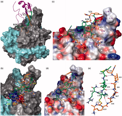
Figure 1. (a) X-ray co-crystal structure of BPTI (ribbon representation) bound to the WNV NS2B-NS3 protease (surface representation). Amber and green ribbons represent G12 to Y21 (fragment 1) and T32 to R39 (fragment 2) of BPTI, respectively. NS2B and NS3 are colored cyan and gray, respectively; (b) Stick model of peptide 1 bound to the protease active site using 2IJO.pdb as template. The C14–C38 disulfide bond is colored yellow. The surface of the acidic residues D75 and S135 and basic residue H51 of the catalytic triad is colored red and blue; (c) Stick model of peptide 3. Fragment 1 and protease residues are labeled black and purple; (d) Stick representation of peptide 3. Fragment 2 and protease residues are labeled black and yellow, respectively; (e) Stick model of cyclic peptide 19. Residues in amber and green represent fragment 1 and 2, respectively. Yellow bonds represent disulfide bridges.
Materials and methods
BPTI and peptides
BPTI from bovine lung was purchased from Sigma-Aldrich (St. Louis, MI). All m-BPTI peptides were custom-made by GL Biochem (Shanghai, China) and were HPLC-purified to >90% purity. All peptides were acetylated and amidated at their N- and C-termini, respectively.
WNV NS2B-NS3 protease inhibition assay
The WNV NS2B-NS3 inhibitory assays were based on published workCitation20 and performed in a pH 8.0 buffer containing Tris-HCl (10 mM), CHAPS (1 mM), and glycerol (20% v/v). The protease (20 nM) and varying concentrations of inhibitor were next added and pre-incubated at 25 °C for 1 h. The reaction was initiated by the addition of the fluorogenic peptide substrate Pyr-RTKR-AMC (Bachem, Switzerland) to make a final concentration of 20 μM. The reaction components were shaken for 5 s, and the reaction progress monitored at 37 °C by measuring the increase in fluorescence (λex355 nm and λem460 nm) every 45 s for 1 h on a SpectraMax Gemini XS plate reader (Molecular Devices, Sunnyvale, CA). Experiments were conducted in duplicates. IC50 values were derived by fitting the initial velocity against the log [inhibitor] with a sigmoidal dose–response curve using GraphPad Prism 5 software (La Jolla, CA). Nona-D-Arg-NH2 peptide (GenScript, Piscataway, NJ) was used as positive control. IC50 graphs can be found in the supplementary notes.
Molecular modeling and visualization software
The WNV NS2B-NS3 protease X-ray co-crystal structure PDB entry 2IJOCitation19 was downloaded from the Protein Data Bank [www.pdb.org] and prepared with the protein preparation wizard in Maestro 9.3 (Schrödinger, New York, NY) using standard settings. This included the addition of hydrogen atoms, bond assignments, removal of water molecules > 7 Å from the ligand, protonation state assignment, optimization of the hydrogen bond network, and restrained minimization using the OPLS2005 force fieldCitation21. Co-crystallized BPTI was used as a template for modeling the conformation and orientation in the binding site of the peptide inhibitors discussed in this work. The inhibitor-protein complex was finally energy-minimized using Macromodel 9.9 (Schrödinger, New York, NY). All residues >9 Å from the ligand were constrained before the complex was subjected to 500 steps of Polak-Ribière Conjugate GradientCitation22 energy minimization using the OPLS2005 force field and GB/SA continuum solvation methodCitation23. Pymol Molecular Graphics System (Schrödinger, New York, NY) was used for model visualization.
Results and discussion
In our assay, the 58-residue native BPTI was found to inhibit the WNV NS2B-NS3 protease with an IC50 of 1.4 ± 0.1 μM, close to the 1 μM reported in an earlier paper by Robin et al.Citation18 From the X-ray co-crystal structure of BPTI bound to the WNV NS2B-NS3 protease (2IJO.pdbCitation19), it was observed that only certain BPTI residues were involved in protease binding: G12 to Y21 and T32 to R39, herein referred as fragment 1 and 2, respectively, found in the antiparallel β-strand region of BPTI (). Based on this, we designed our first truncated 18-residue peptide consisting of fragments 1 and 2 (peptide 1; ; ) and subjected it to the WNV protease inhibition assay using a reported procedureCitation20. Peptide 1 exhibited an IC50 of 14.5 ± 1.1 μM, approximately 10-fold less potent than native BPTI (IC50 of 1.4 ± 0.1 μM). We postulated this to be due to the highly flexible nature of peptide 1 as a result of losing two disulfide bridges (C5–C55 and C30–C51) present in native BPTI. Highly flexible molecules tend to suffer reduced affinities toward their targets due to the high entropic penalties that have to be paid on bindingCitation24. We started truncating peptide 1 by removing G12, yielding peptide 2 with an IC50 of 15.0 ± 0.9 μM, similar to peptide 1 (IC50 of 14.5 ± 1.1 μM). This was expected as the X-ray co-crystal (2IJO.pdb) revealed G12 to be solvent-exposed and uninvolved in intermolecular and intramolecular H-bonding. Further truncation by removing P13 (peptide 3; ) resulted in an unexpected IC50 of 3.8 ± 0.5 μM, approximately 4-fold more potent than peptides 1 and 2. An inspection of the X-ray co-crystal suggested that the P13 pyrrolidine ring of peptides 1 and 2 could be responsible for unfavorable steric interactions to the I155 side-chain of NS3 (). In contrast, peptide 3 without proline 13 was likely to fit better into the protease active site.
Table 1. Inhibitory activities of BPTI and truncated peptides against the WNV NS2B-NS3 protease.
Based on these initial findings, peptide 3 was used as a template for the next series of peptides involving C-terminal truncation of fragment 1 denoted in amber (peptides 4–10; ; ). First, removing Y21 gave peptide 4 with an IC50 of 3.9 ± 0.5 μM which was comparable to peptide 3. Inspection of the X-ray co-crystal structure revealed Y21 to be largely solvent-exposed with only its side-chain hydroxyl in close proximity to the NS3 L31 and L32 nonpolar side-chains (). Hence, its removal was not expected to result in significant IC50 changes. Further truncation of R20 (peptide 5; IC50 of 4.0 ± 0.5 μM) also did not significantly alter its IC50. This was likely due to R20 being solvent-exposed and was not involved in protease binding (). However, a further truncation involving the removal of I19 (peptide 6; IC50 of 16.5 ± 3.4 μM) resulted in an approximate 4-fold decrease in potency compared to peptide 5. We postulated this could be due to the loss of (a) H-bonding between the peptide’s I19 HN to the phenol hydroxyl of Y34 of NS3 and (b) loss of hydrophobic interactions between the peptide’s I19 side-chain and the face of the Y34 aromatic ring of NS3 (). Truncating I18 (peptide 7; IC50 of 36.3 ± 9.7 μM) resulted in a further 2-fold decrease in potency compared to peptide 6. The X-ray co-crystal structure (2IJO.pdb) revealed that although the peptide’s I18 side-chain was solvent-exposed and not involved in protease binding, its backbone NH and C=O were involved in intramolecular H-bonding to the peptide’s Y35 backbone C=O and NH, respectively (). These H-bonds possibly helped rigidify the peptide so that its entropic penalty involved in protease binding was reduced. Removing R17 (peptide 8; IC50 >100 μM) abrogated the peptide’s protease inhibitory activity. We attributed this to the loss of H-bonding between R17’s backbone NH to NS3 A36’s backbone C=O (). Further residue truncations exemplified by peptides 9 and 10 resulted in inactive peptides (IC50s >100 μM). This was expected as they lacked the residues responsible for peptide–protease binding.
Based on these results, we reverted back to the most potent peptide (peptide 3) and removed R39 to yield peptide 11 (). The IC50 was found to be 8.7 ± 1.1 μM, approximately 2-fold less potent than peptide 3 (IC50 3.8 ± 0.5 μM). An inspection of the X-ray co-crystal structure revealed R39’s side-chain to form a salt bridge with NS2B’s D80 and hydrogen bond to the side-chain of NSD3 Q296 () and its removal was the likely cause of the observed reduction in binding affinity. Hence, we concluded that R39 played an important role in protease binding.
In the final series of peptides, we continued employing peptide 3 as a template while truncating fragment 2’s N-terminus (colored green in and ). Removing T32 at the N-terminus yielded peptide 12 with a comparable IC50 of 4.4 ± 0.4 μM (versus 3.8 ± 0.5 μM for 3). Examining the X-ray co-crystal structure revealed T32 to be solvent-exposed and is involved in neither protease nor intramolecular interactions. Next, removal of F33 resulted in peptide 13 whose inhibitory activity was not significantly altered (IC50 4.6 ± 0.4 μM) compared to 12. The X-ray co-crystal structure showed that F33 did not have any contacts with the protease. Likewise, truncating V34 (peptide 14, IC50 5.0 ± 0.5 μM) also did not cause a significant change to its IC50 when compared to 12 and 13. Although the X-ray co-crystal structure revealed V32’s isopropyl side-chain to be in close proximity to the NS3 T132 side-chain, the loss of this poor hydrophobic interaction was unlikely to have caused significant changes in binding affinity. However, further peptide truncation involving the removal of Y35 (peptide 15, IC50 9.2 ± 1.9 μM) resulted in an approximate 2-fold reduction in activity compared to 14. We postulated that this was due to the loss of intramolecular interactions. The side-chain of Y35 was involved in electrostatic interactions with the backbone NH of R20 while its backbone formed two H-bonds to I18 that were likely to play an important role in rigidifying the peptide to enhance its binding affinity toward the target protease. Further peptide truncation by removing G36 and G37 to yield peptides 16 and 17 did not cause significant IC50 changes (9.5 ± 2.0 and 9.7 ± 2.1 μM, respectively) when compared to peptide 15 (IC50 9.2 ± 1.9 μM). Interestingly, the removal of the disulfide bridge by truncating C38 and R39 yielded the inactive linear peptide 18 (IC50 >100 μM), suggesting that the disulfide bridge and R39 play an important role in protease binding, possibly based on the reasons discussed for peptide 11. In summary, the initial N-terminal truncation of fragment 2 involving T32 to V34 (peptides 12–14) did not significantly affect inhibitory potencies with IC50s ranging from 4.4 ± 0.4 to 5.0 ± 0.5 μM and we postulated this to be due to the three residues not being involved in protease binding. Further residue truncation from Y35 onwards (peptides 15 to 18) started to affect inhibitory activities significantly (IC50s ≥ 9.2 μM), suggesting that they were important for bioactivity ().
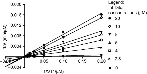
Finally, using the IC50 results obtained from peptides 1–18, we introduced a second disulfide bridge in the bid to enhance the peptide’s rigidity to produce a more potent inhibitor. Based on the X-ray co-crystal structure (2IJO.pdb), we postulated that R20 and F33 were suitable replacement candidates for a disulfide bridge due to their close spatial proximity and their noninvolvement in protease interactions. Hence, the 14-residue cyclic peptide 19 (; ) was synthesized. Its IC50 was found to be 1.5 ± 0.1 μM, comparable to the 58-residue parent BPTI (IC50 1.4 ± 0.1 μM) although 19 was 75% smaller in size, based on the number of residues present. We attributed this to the successful selection of crucial residues required for protease binding and the enhanced rigidity of peptide 19 due to the introduction of a second disulfide bridge between residues I19 and V34. A Lineweaver–Burk plot revealed peptide 19 to be a competitive inhibitor of the WNV NS2B-NS3 protease (.
Figure 2. Lineweaver–Burk plot showing competitive inhibition of the WNV NS2B-NS3 protease by peptide 19 using Pyr-RTKR-AMC as substrate.
In conclusion, we designed a small peptide inhibitor based on the parent protein (BPTI) structure by the sequential removal of amino acids followed by the judicious selection of crucial residues involved in target binding. Further peptide rigidification by the introduction of an additional disulfide bond resulted in a 14-residue cyclic peptide with equipotent inhibitory activity compared to the 58-residue parent protein. We believe this protein miniaturization strategy can potentially be applied to the design of new peptide-based protease inhibitors targeting not only viral proteasesCitation25 but also other trypsin-like proteases such as plasminCitation26 and kallikreinCitation27. In particular, our truncation and cyclization strategy can potentially lead to smaller, novel, and more potent analogs of the 58-residue BPTI (Trasylol®) which is used as a plasmin and kallikrein inhibitor during cardiopulmonary bypass surgeryCitation28.
Declaration of interest
The authors of this paper declare no conflicts of interest.
Additional note
Peptide structures #1 to 19 described in this manuscript have been submitted for patent filing (UK Patent Application No. 1513019.8) on 25 July 2015.
Supplementary material available online.
Acknowledgement
We thank A*STAR Biomedical Research Council for providing financial support.
References
- Colpitts TM, Conway MJ, Montgomery RR, Fikrig E. West Nile virus: biology, transmission, and human infection. Clin Microbiol Rev 2012;25:635–48
- Solomon T. Flavivirus encephalitis. N Engl J Med 2004;351:370–8
- Johnson RT. West Nile virus in the US and abroad. Curr Clin Top Infect Dis 2002;22:52–60
- Gould LH, Fikrig E. West Nile virus: a growing concern? J Clin Invest 2004;113:1102–7
- Kramer LD, Li J, Shi PY. West Nile virus. Lancet Neurol 2007;6:171–81
- Gray TJ, Webb CE. A review of the epidemiological and clinical aspects of West Nile virus. Int J Gen Med 2014;7:193–203
- www.cdc.gov/ncidod/dvbid/westnile/index.htm
- Kaiser J. Public health. Outbreak pattern stymies vaccine work. Science 2012;337:1030
- Diamond MS. Progress on the development of therapeutics against West Nile virus. Antiviral Res 2009;83:214–27
- Mukhopadhyay S, Kuhn RJ, Rossmann MG. A structural perspective of the flavivirus life cycle. Nat Rev Microbiol 2005;3:13–22
- Chappell KJ, Stoermer MJ, Fairlie DP, Young PR. Insights to substrate binding and processing by West Nile virus NS3 protease through combined modeling, protease mutagenesis and kinetic studies. J Biol Chem 2006;281:38448–58
- Chappell KJ, Stoermer MJ, Fairlie DP, Young PR. West Nile virus NS2B/NS3 protease as an antiviral target. Curr Med Chem 2008;15:2771–84
- Radichev I, Shiryaev SA, Aleshin AE, et al. Structure-based mutagenesis identifies important novel determinants of the NS2B cofactor of the West Nile virus two-component NS2B-NS3 proteinase. J Gen Virol 2008;89:636–41
- Poulsen A, Kang C, Keller TH. Drug design for Flavivirus proteases: what are we missing? Curr Pharm Des 2014;20:3422–7
- Kunitz M, Northrup J. Isolation from beef pancreas of crystalline trypsinogen, trypsin, a trypsin inhibitor, and an inhibitor-trypsin compound. J Gen Physiol 1936;19:991–1007
- Kassell B, Radicevic M, Ansfield MJ, Laskowski M. The basic trypsin inhibitor of bovine pancreas. IV. The linear sequence of the 58 amino acids. Biochem Biophys Res Commun 1965;18:255–8
- Ascenzi P, Bocedi A, Bolognesi M, et al. The bovine basic pancreatic trypsin inhibitor (Kunitz inhibitor): a milestone protein. Curr Protein Pept Sci 2003;4:231–51
- Robin G, Chappell K, Stoermer MJ, et al. Structure of West Nile virus NS3 protease: ligand stabilization of the catalytic conformation. J Mol Biol 2009;385:1568–77
- Aleshin AE, Shiryaev SA, Strongin AY, Liddington RC. Structural evidence for regulation and specificity of flaviviral proteases and evolution of the Flaviviridae fold. Protein Sci 2007;16:795–806
- Nall TA, Chappell KJ, Stoermer MJ, et al. Enzymatic characterization and homology model of a catalytically active recombinant West Nile virus NS3 protease. J Biol Chem 2004;279:48535–42
- Kaminski GA, Friesner RA, Tirado-Rives J, Jorgensen WL. Evaluation and reparametrization of the OPLS-AA force field for proteins via comparison with accurate quantum chemical calculations on peptides. J Phys Chem B 2001;105:6474–87
- Polak E, Ribière G. Rev. Francaise Informat Recherche Opérationelle, Série Rouge. Note sur la Convergence de Directions Conjuguée 1969;16:35–43
- Hasel W, Hendrickson TF, Still WC. A rapid approximation to the solvent accessible surface areas of atoms. Tetrahedron Comput Methodol 1988;1:103–16
- Chang CA, Chen W, Gilson MK. Ligand configurational entropy and protein binding. Proc Natl Acad Sci USA 2007;104:1534–9
- Zhirnov OP, Klenk HD, Wright PF. Aprotinin and similar protease inhibitors as drugs against influenza. Antiviral Res 2011;92:27–36
- Takeda-Shitaka M, Kamiya K, Miyata T, et al. Structural studies of the interactions of normal and abnormal human plasmins with bovine basic pancreatic trypsin inhibitor. Chem Pharm Bull 1999;47:322–8
- Chen Z, Bode W. Refined 2.5 Å X-ray crystal structure of the complex formed by porcine kallikrein A and the bovine pancreatic trypsin inhibitor. J Mol Biol 1983;164:283–311
- www.fda.gov/downloads/Drugs/DrugSafety/PostmarketDrugSafetyInformationforPatientsandProviders/UCM142741.pdf