
Abstract
Human carbonic anhydrase IX (CA IX) is overexpressed in the most aggressive and invasive tumors. Therefore, CA IX has become the promising antitumor drug target. Three inhibitors have been shown to selectively and with picomolar affinity inhibit human recombinant CA IX. Their inhibitory potencies were determined for the CA IX, CA II, CA IV and CA XII in Xenopus oocytes and MDA-MB-231 cancer cells. The inhibition IC50 value of microelectrode-monitored intracellular and extracellular acidification reached 15 nM for CA IX, but with no effect on CA II expressed in Xenopus oocytes. Results were confirmed by mass spectrometric gas analysis of lysed oocytes, when an inhibitory effect on CA IX catalytic activity was found after the injection of 1 nM VD11-4-2. Moreover, VD11-4-2 inhibited CA activity in MDA-MB-231 cancer cells at nanomolar concentrations. This combination of high selectivity and potency renders VD11-4-2, an auspicious therapeutic drug for target-specific tumor therapy.
Introduction
The changes in pH regulation and altered cellular metabolism are key features of solid tumors. Due to inadequate blood supply, metabolism in tumor cells is highly affected by expression of many hypoxia-inducible genes. Changes in metabolic pathways occur to survive hypoxic conditions. Known as “Warburg effect”Citation1,Citation2, extensive glycolysis leads to tremendous amounts of lactate and protons, which significantly contribute to acidification of extracellular space. The efflux of lactate and H+ is facilitated by the monocarboxylate transportersCitation3,Citation4. In addition, the active transport for removing H+ from the cell is mediated by H+-ATPase pumps and secondary active Na+/H+ exchangers (NHE), or affected by HCO3− influx via Na+-HCO3− co-transporters (NBC) or Na+-dependent Cl−/HCO3− exchangers (NDAE)Citation5–9. Hypoxic and acidic microenvironment promotes the breakdown of extracellular matrixCitation10 and allows tumor progression.
Carbonic anhydrase (CA) IX plays an important role in tumor acid-base homeostasis under hypoxic conditionsCitation11. CA IX is one of the 12 catalytically active human CA isoformsCitation12. Being transmembrane homodimer, CA IX possesses extracellular activity, catalyzing the reversible hydration of carbon dioxideCitation13,Citation14. Low levels of CA IX have been reported in normal tissues, except in stomachCitation15, whereas the increase in CA IX expression has been found in many aggressive hypoxic cancersCitation16–18. Thus, CA IX has been considered as a promising target in the development of antitumor agents. The design of potent and CA IX-selective inhibitors has been extensively exploredCitation14,Citation19.
To confirm the potency of the compound, in vivo assays are essential in the initial phases of drug discovery. The heterologous protein expression in Xenopus laevis oocytes has emerged as a promising tool in molecular and physiological research due to efficient translation of heterologous RNA into proteinCitation20. The heterologous expression of various CA isoforms in Xenopus has become suitable for the investigation of selective CA inhibition because oocytes do not express any CA themselvesCitation21,Citation22. Therefore, the cRNA of target CA can be injected into the oocyte to obtain the expression of the protein and the selective inhibitory effect can be determined as it has been shown with CA IVCitation23 and CA IXCitation24.
The present study assessed the inhibitory effects of three compounds – VD12-09, VD11-4-2 and EA2-3 – in Xenopus oocytes expressing CA II, CA IV, CA IX or CA XII and in MDA-MB-231 breast cancer cells. CA activity in oocytes was evaluated by measuring pH in the cytosol and at the outer membrane surface and confirmed by mass spectrometry of lysed oocytes. Our results show that VD11-4-2 not only features high selectivity toward CA IX over other tested CA isoforms, but also significantly inhibits extracellular and intracellular CA IX activity with low nanomolar IC50 value in oocytes and in tumor cells.
Materials and methods
Inhibitors
The synthesis, structural and purity characterization of VD11–4-2, VD12-09 and EA2-3 have been previously describedCitation25–27. The compounds were dissolved in 100% dimethyl sulfoxide (DMSO) to prepare 100 mM stock solutions. Series of dilutions of each compound were carried out in DMSO at a final DMSO concentration up to 0.01%.
Constructs, oocytes and injections of cRNA
Xenopus laevis females were purchased from Xenopus Express, Vernassal, France. Segments of ovarian lobules were surgically removed under sterile conditions from frogs anesthetized with 1 g/l of 3-amino-benzoic acid ethylester (MS-222, Sigma-Aldrich, St. Louis, MO), and rendered hypothermic. After disection frogs were kept solitary in a basin for one week to recover. The wound was checked regularly for infections. The procedure was approved by the Landesuntersuchungsamt Rheinland-Pfalz, Koblenz (23 177–07/A07–2-003 §6). Afterwards the oocytes were dispersed by collagenase (Collagenase A, Roche, Mannheim, Germany) treatment and stored over-night in oocyte saline (82.5 mM NaCl, 2.5 mM KCl, 1 mM CaCl2, 1 mM MgCl2, 1 mM Na2HPO4 and 5 mM HEPES, pH 7.8) at 18 °C to recover. A detailed description of the frog dissection and processing of oocytes is given in [29]. Oocytes of the stages V and VI were injected with 1–6 ng of cRNA coding either for human CA II, CA IV, CA IX or CA XII, cloned into the Xenopus oocyte expression vector pGemHeJuel. Measurements were carried out 3–5 days after target cRNA microinjections. For pH measurements, the oocyte saline was titrated to pH 7.4. In bicarbonate-containing saline, 25 mM NaCl was replaced by an equivalent amount of NaHCO3 and the solution was aerated using 5% CO2.
Intracellular and extracellular pH measurements
Single-barreled microelectrodes were used to measure intracellular [H+] ([H+]i) while double-barreled microelectrodes – for extracellular [H+] at outer membrane surface ([H+]s). Their manufacture and application have already been reportedCitation28–30.
Mass-spectrometry measurements
By monitoring the 18O depletion of doubly labeled 13C18O2 through several hydration and dehydration steps of CO2 and HCO3−, with a quadrupole mass spectrometer (Omni Star GSD 320; Pfeiffer Vacuum, Asslar, Germany), the effect of 1–10 nM VD11-4-2 on CA IX activity was determined in oocyte lysate at 25 °C. The rates of catalyzed and non-catalyzed reaction and the activity of CA IX in units (U) were evaluated as previously describedCitation31–34. 1 U is equal to 100% stimulation of the non-catalyzed 18O depletion of 13C18O2.
Cell lines and culture
The human breast adenocarcinoma cell line MDA-MB-231 was purchased from the German Collection of Microorganisms and Cell Cultures DSMZ, Braunschweig, Germany (DSMZ-No. ACC-732). Cells were aliquoted after purchase and used within 6 months. The cell line was verified by the DSMZ with fluorescent nonaplex PCR of short tandem repeat markers. Cells were cultured in Gibco Leibovitz-L15 medium (Life Technologies, Waltham, MA), supplemented with 10% fetal calf serum, 2 mM glucose and 1% penicillin/streptomycin, pH 7.2, either under normoxia (5% CO2 in air) or hypoxia (5% CO2, 1% O2, 94% N2) at 37 °C in humidified cell culture incubators.
pH measurements in MDA-MB-231 breast cancer cells
Changes in intracellular proton concentration in MDA-MB-231 cells were measured with an epifluorescence microscope (BX50WI upright microscope, Olympus Deutschland GmbH, Hamburg, Germany; with Polychrome IV epifluorescence unit, Till Photonics GmbH, Munich, Germany). Cells were loaded with 2′,7′-bis-(carboxyethyl)-5-(and-6)-carboxyfluorescein (BCECF-AM, 2 μM; Molecular Probes, Eugene, OR). The procedure of pH measurements in MDA-MB-231 cells has been described in detail previouslyCitation35.
Calculation and statistics
Results are presented as mean ± SEM. Student’s t-test or, if possible, a paired t-test was used for the determination of significance in differences. As it is shown in figures, * indicates a significance level of p ≤ 0.05, ** indicate a significance level of p ≤ 0.01, *** indicate a significance level of p ≤ 0.001.
Results
VD11-4-2 inhibits intracellular CA IX activity
The inhibitory potency of VD11-4-2 against CA IX and CA II was determined by measuring the rate of rise in intracellular [H+] (Δ[H+]i/Δt) during repeated application of CO2/HCO3−. Acid transients were recorded in native oocytes and oocytes injected with 5 ng CA IX-cRNA or 6 ng CA II-cRNA in the absence and presence of 10 μM of VD11-4-2 with single-barreled pH-sensitive microelectrodes. shows the acid transients for oocytes injected with CA IX-cRNA. Compared with native oocytes, the rate of acidification increased from 25.4 ± 2.9 nM/min to 82.0 ± 8.8 nM/min and to 137.5 ± 14.7 nM/min for CA IX and CA II cRNA injected oocytes, respectively (). No impact of 10 μM VD11-4-2 on Δ[H+]i/Δt in CA II expressing oocytes was detected and Δ[H+]i/Δt was evaluated to be 137.1 ± 17.1 nM/min (). The raw inhibition curve can be found in the Supplementary Material, Figure S1.
Figure 1. Measurements of cytosolic H+ changes in native Xenopus oocytes (B) and oocytes expressing target CA isoform (A, C–H). Cells were injected with 5 ng CA IX-cRNA (A, D, F and H) or 6 ng CA II-cRNA (C, E and G) during application of 5% CO2/25 mM HCO3- (from a nominally CO2-free, HEPES-buffered solution) in the absence of compounds and in the presence of 10 μM VD11-4-2 (C and D), 10 μM VD12-09 (E and F) and 10 μM EA2–3 (G and H). * indicates a significance level of p ≤ 0.05, ** indicate a significance level of p ≤ 0.01, *** indicate a significance level of p ≤ 0.001.
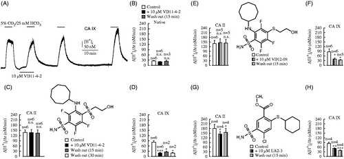
A completely different effect was detected in the CA IX expressing oocytes. The Δ[H+]i/Δt was reduced from 82.0 ± 8.8 nM/min down to 20.3 ± 3.5 nM/min upon administration of 10 μM VD11-4-2 (). This value (20.3 ± 3.5 nM/min) matched the rate of 20.2 ± 1.4 nM/min obtained in native cells (w/o any CAs) treated with the same dose of VD11-4-2 (). Moreover, VD11-4-2 was characterized to be not washable from CA IX after exposure to the inhibitor for 15 and 45 min (Δ[H+]i/Δt were 26.4 ± 2.9 nM/min and 27.7 ± 3.5 nM/min, respectively). Thus, VD11-4-2 was shown to be membrane-permeable inhibitor that completely blocked the CA IX-generated rate of acidification at 10 μM in Xenopus oocytes.
The other two compounds, VD12-09 and EA2-3, exhibited lower potential to inhibit CA IX activity than VD11-4-2 according to [H+]i measurements in oocytes. The raw inhibition curves can be found in the Supplementary Material, Figure S2. The fast rise of [H+]i was not affected by 10 μM of VD12-09 in CA II expressing cells and was inhibited in CA IX expressing cells from 87.5 ± 17.8 nM/min to 40.6 ± 6.9 nM/min (). Even though VD12-09 displayed the selectivity toward CA IX, the CA IX inhibition effect is lower as compared with the one of 10 μM VD11-4-2. In the presence of 10 μM EA2-3, Δ[H+]i/Δt was decreased significantly in both CA II (from 171.2 ± 27.1 nM/min to 143.8 ± 17.2 nM/min) and CA IX (from 84.7 ± 5.9 nM/min to 51.9 ± 3.9 nM/min) expressing oocytes (). Therefore, VD12-09 and EA2-3 are much less effective CA IX inhibitors than VD11-4-2.
Intracellular CA IX inhibition by VD11-4-2 is dose-dependent
In order to find the correlation between the cytosolic CA IX activity and the amount of VD11-4-2 necessary for inhibition, the experiments of treating CA IX expressing oocytes with various VD11-4-2 concentrations and measuring Δ[H+]i/Δt were carried out (repeated four times). The lowest dose of VD11-4-2 was 1 nM. It was increased by 10 fold until 10 μM. Each concentration was tested on two CO2/HCO3− applications (). Average values were determined and used for IC50 value determination.
shows the intracellular IC50 of VD11-4-2 as determined using the sigmoidal dose-response Hill model. Data points were obtained experimentally and represent the values of changes in [H+]i when 1 nM to 10 μM VD11-4-2 was applied on oocytes expressing CA IX. The rate of change in [H+]i from the first pulse in the absence of VD11-4-2 was defined as 100% CA IX activity. Δ[H+]i/Δt during the pulse in the presence of 10 μM VD11-4-2 was evaluated to be 0% due to the complete inhibition of CA IX catalytic activity using 10 μM VD11-4-2 as shown in the .
Figure 2. Dose-dependent inhibition of intracellular CA IX catalytic activity with VD11-4-2 in Xenopus oocytes. (A) Recordings of [H+]i in oocytes injected with 5 ng CA IX-cRNA during application of 5% CO2/25 mM HCO3- (from a nominally CO2-free, HEPES-buffered solution) in the absence and presence of 1 nM to 10 μM of VD11-4-2. (B) The dependence of changes in [H+]i on added VD11-4-2 concentrations. Black data points were obtained experimentally. The solid line was simulated according to the Hill model with variable slope.
![Figure 2. Dose-dependent inhibition of intracellular CA IX catalytic activity with VD11-4-2 in Xenopus oocytes. (A) Recordings of [H+]i in oocytes injected with 5 ng CA IX-cRNA during application of 5% CO2/25 mM HCO3- (from a nominally CO2-free, HEPES-buffered solution) in the absence and presence of 1 nM to 10 μM of VD11-4-2. (B) The dependence of changes in [H+]i on added VD11-4-2 concentrations. Black data points were obtained experimentally. The solid line was simulated according to the Hill model with variable slope.](/cms/asset/3e9afdfa-2462-4c3e-a0d2-86e76a7c0842/ienz_a_1217854_f0002_b.jpg)
Intracellular pH in CA IX expressing oocytes was already affected by 1 nM VD11-4-2 and Δ[H+]i/Δt was decreased by ∼20%. The fast rise of cytosolic [H+] upon the exposure of CO2/HCO3− was reduced when the concentration of VD11-4-2 was increased. IC50 was found to be 15 nM. Thus, VD11-4-2 exhibited the significant inhibitory potential toward the intracellular CA IX in the nanomolar range.
VD11-4-2 inhibits extracellular CA IX activity
Being an important therapeutic target, the mature CA IX is a homodimeric and membrane-anchored protein displaying extracellular catalytic activity. The sensitivity of extracellular CA IX activity to 10 μM of VD11-4-2 was evaluated by recording [H+] at the extracellular membrane surface ([H+]s) and the reference potential (Eref) using double-barreled pH-sensitive microelectrodes (). Changes in Eref were negligible (≤2 mV) during the measurements and are not presented here. A transient decrease in [H+]s was caused upon addition of CO2/HCO3- and a rise of [H+]s was induced upon removal of CO2/HCO3-. The extracellular CA IX activity was assessed by the amplitude of the transient rise of [H+]s (Δ[H+]s) during the removal of CO2/HCO3-.
Figure 3. Measurements of [H+]s in Xenopus oocytes injected with 5 ng CA IX-cRNA (A and C) and native oocytes (B) during application of 5% CO2/25 mM HCO3- (from a nominally CO2-free, HEPES-buffered solution) in the absence and presence of 10 μM VD11-4-2 and after 15 min of washing out. ** indicate a significance level of p ≤ 0.01, *** indicate a significance level of p ≤ 0.001.
![Figure 3. Measurements of [H+]s in Xenopus oocytes injected with 5 ng CA IX-cRNA (A and C) and native oocytes (B) during application of 5% CO2/25 mM HCO3- (from a nominally CO2-free, HEPES-buffered solution) in the absence and presence of 10 μM VD11-4-2 and after 15 min of washing out. ** indicate a significance level of p ≤ 0.01, *** indicate a significance level of p ≤ 0.001.](/cms/asset/6258d655-eb72-4498-808e-911eb595f6ff/ienz_a_1217854_f0003_b.jpg)
In the absence of VD11-4-2, Δ[H+]s was found to be 15.6 ± 0.4 nM in native cells () which was increased to 63.0 ± 3.1 nM in CA IX expressing oocytes (). The impact of VD11-4-2 on the CA-mediated changes in extracellular pH in CA IX expressing oocytes was significant and nearly complete. The amplitude of the acid transient decreased to 22.1 ± 0.7 nM with 10 μM VD11-4-2 (). This Δ[H+]s correlated with the result of treating native cells with the same dose of VD11-4-2 – 14.5 ± 0.5 nM (). Similar to intracellular pH measurements applying VD11-4-2 on CA IX expressing oocytes, the interaction between extracellular CA IX and VD11-4-2 was also shown to be essentially irreversible and Δ[H+]s after 15 min of washing out was 34.0 ± 6.0 nM ().
VD11-4-2 inhibits extracellular CA IX in a concentration-dependent manner
The dependence of extracellular CA IX catalytic activity on added VD11-4-2 concentration was determined. The amplitudes of Δ[H+]s transients after the application of different doses of VD11-4-2 are shown in . The highest concentration of VD11-4-2 was 10 μM, which was decreased by 10 fold down to 0.1 nM. Each concentration was applied during two applications of CO2/HCO3−. The average values were calculated and used for IC50 evaluation.
Figure 4. Dose-dependent inhibition of extracellular CA IX catalytic activity with VD11-4-2 in Xenopus oocytes. (A) Recordings of [H+]s in oocytes injected with 5 ng CA IX-cRNA during application of 5% CO2/25 mM HCO3- (from a nominally CO2-free, HEPES-buffered solution) in the absence and presence of 0.1 nM to 10 μM of VD11-4-2. (B) The dependence of changes in [H+]s as a function of added VD11-4-2 concentrations. The data points were obtained experimentally while the solid line was simulated according to the Hill model with varying slope.
![Figure 4. Dose-dependent inhibition of extracellular CA IX catalytic activity with VD11-4-2 in Xenopus oocytes. (A) Recordings of [H+]s in oocytes injected with 5 ng CA IX-cRNA during application of 5% CO2/25 mM HCO3- (from a nominally CO2-free, HEPES-buffered solution) in the absence and presence of 0.1 nM to 10 μM of VD11-4-2. (B) The dependence of changes in [H+]s as a function of added VD11-4-2 concentrations. The data points were obtained experimentally while the solid line was simulated according to the Hill model with varying slope.](/cms/asset/57ca7055-c089-4c2e-818d-b67b2c949e40/ienz_a_1217854_f0004_b.jpg)
shows the dose-response curve (of changes in extracellular Δ[H+]s as a function) of added VD11-4-2 concentration. When higher concentrations of the inhibitor were used, the amplitude of Δ[H+]s transient decreased. Simulation of the dependence according to Hill model yielded the extracellular IC50 of 25 nM, which correlates with the intracellular IC50 of VD11-4-2 (). Thus, VD11-4-2 exhibits high inhibitory potential toward both the intracellular and extracellular CA IX and is a good lead compound candidate for CA IX inhibition.
VD11-4-2 at 50 nM concentration features lower inhibitory potency to CA IV and CA XII than CA IX
The high selectivity of VD11-4-2 toward CA IX was also confirmed by determining the inhibitory potential of the compound toward CA IV and CA XII. VD11-4-2 was shown to have a relatively minor effect on extracellular pH in CA IV and CA XII cRNA injected oocytes at 50 nM concentration. However, the administration of 10 μM of VD11-4-2 resulted in Δ[H+]s of 54.3 ± 5.7 nM and 26.5 ± 1.4 nM in oocytes expressing CA IV and CA XII, respectively (). Moreover, the effect of VD11-4-2 on CA IV was found to be washable after 15 min, while non-washable for CA XII. These experiments support the conclusion that VD11-4-2 is highly selective for CA IX, but not for CA II, CA IV and CA XII.
Figure 5. Measurements of [H+]s in oocytes injected with 1 ng CA IV-cRNA (A) or 5 ng CA XII-cRNA (B) during application of 5% CO2/25 mM HCO3- (from a nominally CO2-free, HEPES-buffered solution) in the absence and presence of 50 nM and 10 μM VD11-4-2 and after 15 min of washing out. ** indicate a significance level of p ≤ 0.01, *** indicate a significance level of p ≤ 0.001.
![Figure 5. Measurements of [H+]s in oocytes injected with 1 ng CA IV-cRNA (A) or 5 ng CA XII-cRNA (B) during application of 5% CO2/25 mM HCO3- (from a nominally CO2-free, HEPES-buffered solution) in the absence and presence of 50 nM and 10 μM VD11-4-2 and after 15 min of washing out. ** indicate a significance level of p ≤ 0.01, *** indicate a significance level of p ≤ 0.001.](/cms/asset/3436169b-792a-4de1-9c9f-8e7404caee52/ienz_a_1217854_f0005_b.jpg)
CA IX activity is significantly inhibited by the administration of single-digit nanomolar concentration of VD11-4-2 according to mass spectrometry
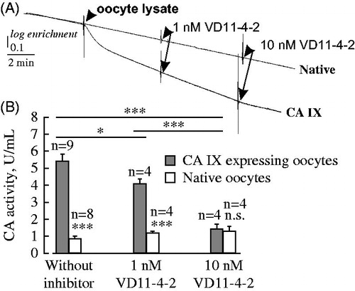
CA IX activity was decreased by 25% after the addition of 1 nM VD11-4-2 to oocyte lysate according to mass-spectrometry measurements (). In the lysate of oocytes expressing CA IX, which was exposed to 10 nM of VD11-4-2, the rate of 18O enrichment did not differ from the rate in the lysate of native cells treated with the same dose of inhibitor. Therefore, the complete inhibition of CA IX activity was identified using 10 nM of VD11-4-2. These results confirm the outcome from intracellular and extracellular pH measurements that VD11-4-2 with nanomolar affinity inhibits CA IX.
Figure 6. Recordings of the degradation of 18O-labeled CO2 (A) and evaluations of CA IX activity (B) in the absence and presence of 1 and 10 nM VD11-4-2 in lysed oocytes as measured by mass spectrometry. The addition of oocyte lysate made from 20 cells and different concentrations of VD11-4-2 are shown by arrows. *** indicate a significance level of p ≤ 0.001.
Inhibition of CA activity by VD11-4-2 in MDA-MB-231 tumor cells
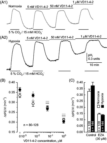
The effect of VD11-4-2 on CA activity was further determined in the human adenocarcinoma cell line MDA-MB-231, which was incubated either under normoxic (21% O2) or hypoxic (1% O2) conditions. CA catalytic activity was determined by the rate of change in intracellular pH (ΔpHi/Δt) during application of 5% CO2/15 mM HCO3- (pH 7.2) in the absence and presence of VD11-4-2 (). In the absence of VD11-4-2, ΔpHi/Δt was significantly increased in hypoxic cells as compared to normoxic cells (p ≤ 0.05), indicating increased CA expression under hypoxic conditions (most likely a hypoxia-induced increase in CA IX expression). In the presence of 5 nM VD11-4-2, a significant decrease in ΔpHi/Δt both in normoxic and hypoxic cells was recorded. 1 μM VD11-4-2 reduced ΔpHi/Δt to around 60% (). Full inhibition of total CA activity with 30 μM ethoxzolamide (EZA) decreased ΔpHi/Δt to ∼40% (). The 20% difference in inhibition between VD11-4-2 and EZA might originate from the additional inhibition of other CA isoforms by EZA. The residual rate of pHi change in the presence of EZA can be attributed to the spontaneous hydration of CO2 in the cell. Furthermore, no significant differences in ΔpHi/Δt could be found between normoxic and hypoxic cells in the presence of 1 μM VD11-4-2, which also indicates inhibition of hypoxia-induced CA IX. Therefore, it can be assumed that VD11-4-2 has comparable inhibitory potency in tumor cells and in oocytes.
Figure 7. Inhibition of CA activity with VD11-4-2 in MDA-MB-231 breast cancer cells. (A) Recordings of pHi in MDA-MB-231 cells, kept under normoxic (21% O2, A1) or hypoxic (1% O2, A2) conditions, during application of 5% CO2/15 mM HCO3- (pHo 7.2) in the absence and presence of 5 nM, 50 nM and 1 μM of VD11-4-2. (B) Rate of change in pHi in normoxic (white) and hypoxic (black) MDA-MB-231 cells, induced by application of 5% CO2/15 mM HCO3-, as a function of VD11-4-2 concentrations. The asterisks refer to the values in the absence of VD11-4-2. (C) Rate of change in pHi in normoxic (white) and hypoxic (black) MDA-MB-231 cells, induced by application of 5% CO2/15 mM HCO3-, in the absence (control) and presence of 30 μM EZA.
Discussion
CA IX has emerged as a promising anticancer target, and it is highly expressed in numerous human cancers and essentially absent in normal tissue. There are many studies which have investigated the affinity of various inhibitors toward CA IX in vitro using enzymatic assaysCitation36–40. High affinity and significant selectivity of three recently discovered inhibitors, VD12-09, VD11-4-2 and EA2-3, toward CA IX in vitro have been previously described using three complementary methods: isothermal titration calorimetry, the fluorescent thermal shift assay and the stopped-flow CO2hydration assayCitation27. VD12-09 bound CA IX with 1.1 nM affinity. Interactions between VD12-09 and other remaining 12 catalytically active human CA isoforms were 25–100 000-fold weaker than with CA IX. VD11-4-2 exhibited a dissociation constant of 50 pM for CA IX and featured significant selectivity toward CA IX. EA2-3 bound CA IX with 330 pM affinity and was highly selective for CA IX. Therefore, there was a crucial demand to confirm VD12-09, VD11-4-2 and EA2-3 as tight binders of CA IX in live biological systems in order to develop these compounds as therapeutics.
In the present study, the catalytic activity of CA IX heterologously expressed in Xenopus oocytes was evaluated in the presence of newly designed VD12-09, VD11-4-2 and EA2-3. The measurements of the rate of cytosolic pH changes and the amplitude of pH changes at the outer membrane surface during addition/removal of 5% CO2/25 mM HCO3− revealed that VD11-4-2 possesses the best balance of affinity and selectivity to CA IX in vivo. IC50 value for intracellular and extracellular CA IX varied in low-nanomolar range whereas intracellular CA II as well as extracellular CA IV and CA XII were not affected significantly by the administration of VD11-4-2 at nanomolar concentrations. Mass spectrometry experiments using oocyte lysate showed the complete block of CA IX catalytic activity upon administration of nanomolar doses of VD11-4-2. Therefore, the present study for the first time confirmed the nanomolar potency of VD11-4-2 toward CA IX in vivo. In addition, we have indicated here that the interactions between VD11-4-2 and CA IX are irreversible after 45 min. These results correlate perfectly with Talibov et al. (2016) investigation, which showed that VD11-4-2 is a tight binder to recombinant CA IX with dissociation rates too slow to be determined by surface plasmon resonanceCitation41. Thus, VD11-4-2 has been shown as a promising lead compound.
The impact of VD12-09 on the catalytic activity of CA IX was lower than VD11-4-2. VD12-09 exhibited high affinity and selectivity to intracellular CA IX compared with negligible effects on intracellular CA II activity. However, VD12-09 did not influence the amplitude of pH changes at the outer oocyte membrane surface. Thus, VD12-09 possibly was not able to accommodate in the active site of the mature membrane-anchored CA IX with extracellular catalytic domain. Furthermore, it may be caused by structural differences in intracellular and extracellular CA IX due to incomplete protein folding and the lack of post-translation modifications in intracellular CA IX structure.
Klier et al. has recently investigated the concentration-dependent inhibition of intracellular and extracellular CA IX catalytic activity in Xenopus oocytes by EZA.Citation24 This CA inhibitor has been used for many years to treat glaucoma, duodenal ulcers and as a diureticCitation42–44. Results have demonstrated the 30 μM EZA induced slow rates of cytosolic pH changes and small amplitudes of pH changes at the outer membrane side in CA IX expressing oocytes, which were analogous to the respective parameters of native oocytes, when only the spontaneous, non-catalyzed reaction of CO2 hydration to H+ and HCO3− occurred. Similar to their studies, the dose response of CA IX inhibition by VD11-4-2 was observed by measuring inner and outer pH in Xenopus oocytes with pH-sensitive microelectrodes and 1 μM of VD11-4-2 resulted in the complete inhibition of CA IX catalytic activity. The amount of VD11-4-2 is 30-fold lower than EZA, which induces the same effect using the same method. Thus, VD11-4-2 is more potent inhibitor against CA IX than clinically used drug EZA.
Both the extracellular and intracellular pH measurements yielded a similar inhibition dose response curve. Since the compounds are expected to be membrane-permeable, it is difficult to be certain that extracellular measurements reflect inhibition of only extracellularly expressed CA IX. However, as previously shownCitation24, around 20% of CA IX in oocytes is located outside at the membrane and 80% inside the oocyte. Thus, the applied inhibitors would primarily inhibit an outside-located CA IX.
The investigation of changes in pH exhibits high potential toward the development of cancer therapyCitation45. The invasion of cancer cells correlates with the increase of intracellular pH as well as the decrease of extracellular pHCitation46. To monitor tumor progression, many derivatives of fluorescein (BCECF, BCPCF, fluorescein) and benzoxanthene (SNAFL, SNAFR, SNARF) have been extensively applied as pH-sensitive fluorescent probes to measure intracellular pHCitation47. The measurements of extracellular pH in solid tumors in vivo have been also carried outCitation48,Citation49. In our study, we could show that already 5 nM of VD11-4-2 significantly inhibit CA activity in MDA-MB-231 breast cancer cells and 1 μM VD11-4-2 does seem to fully inhibit CA IX activity. Therefore, VD11-4-2 appears to be a promising agent for CA IX inhibition.
In summary, we have discovered an inhibitor of CA IX, compound VD11-4-2 exhibiting 15–25 nM IC50 for both intracellularly and extracellularly expressed CA IX in Xenopus oocytes while possessing strong selectivity over CA II, CA IV and CA XII. In addition, the high inhibitory potency of VD11-4-2 against hypoxic CA IX was confirmed in MDA-MB-231 breast cancer cells. Thus, VD11-4-2 features with a strong promise to be further developed as a human anticancer drug targeting CA IX.
Declaration of interest
The authors have no competing interests. This research was funded by the grant [No. TAP LLT-1/2016] from the Research Council of Lithuania.
Supplementary materials available online
IENZ_1217854_Supplementary_Material.pdf
Download PDF (58 KB)Acknowledgements
We thank Hans-Peter Schneider and Sina Ibne Noor for their skillful assistance with the experiments and experimental data analysis.
Related Research Data
References
- Warburg O. On respiratory impairment in cancer cells. New York, NY: Science; 1956;124:269–70
- Vander Heiden MG, Cantley LC, Thompson CB. Understanding the Warburg effect: the metabolic requirements of cell proliferation. New York, NY: Science; 2009;324:1029–33
- Poole RC, Halestrap AP. Transport of lactate and other monocarboxylates across mammalian plasma membranes. Am J Physiol 1993;264:761–82
- Parks SK, Chiche J, Pouyssegur J. pH control mechanisms of tumor survival and growth. J Cell Physiol 2011;226:299–308
- Hulikova A, Harris AL, Vaughan-Jones RD, Swietach P. Regulation of intracellular pH in cancer cell lines under normoxia and hypoxia. J Cell Physiol 2013;228:743–52
- Boron WF. Regulation of intracellular pH. Adv Physiol Educ 2004;28:160–79
- Lee AH, Tannock IF. Heterogeneity of intracellular pH and of mechanisms that regulate intracellular pH in populations of cultured cells. Cancer Res 1998;58:1901–8
- Hulikova A, Vaughan-Jones RD, Swietach P. Dual role of CO2/HCO3(–) buffer in the regulation of intracellular pH of three-dimensional tumor growths. J Biol Chem 2011;286:13815–26
- Martinez-Zaguilan R, Lynch RM, Martinez GM, et al. (+)-ATPases are functionally expressed in plasma membranes of human tumor cells. Am J Physiol 1993;265:1015–29
- Giusti I, D’Ascenzo S, Millimaggi D, et al. Cathepsin B mediates the pH-dependent proinvasive activity of tumor-shed microvesicles. Bioorg Med Chem 2008;10:481–8
- Wykoff CC, Beasley NJP, Watson PH, et al. Hypoxia-inducible expression of tumor-associated carbonic anhydrases. Proc Natl Acad Sci USA 2000;60:7075–83
- Imtaiyaz Hassan M, Shajee B, Waheed A, et al. Structure, function and applications of carbonic anhydrase isozymes. Bioorg Med Chem 2013;21:1570–82
- Alterio V, Hilvo M, Fiore AD, et al. Crystal structure of the catalytic domain of the tumor-associated human carbonic anhydrase IX. Proc Natl Acad Sci 2009;106:16233–8
- Aggarwal M, Boone CD, Kondeti B, McKenna R. Structural annotation of human carbonic anhydrases. J Enzyme Inhib Med Chem 2013;28:267–77
- Pastorekova S, Parkkila S, Parkkila A, et al. Carbonic anhydrase IX, MN/CA IX: analysis of stomach complementary DNA sequence and expression in human and rat alimentary tracts. Gastroenterology 1997;112:398–408
- Pastorekova S, Zatovicova M, Pastorek J. Cancer-associated carbonic anhydrases and their inhibition. Curr Pharm Des 2008;14:685–98
- McDonald PC, Winum J-Y, Supuran CT, Dedhar S. Recent developments in targeting carbonic anhydrase IX for cancer therapeutics. Oncotarget 2012;3:84–97
- Pastorek J, Pastorekova S. Hypoxia-induced carbonic anhydrase IX as a target for cancer therapy: from biology to clinical use. Semin Cancer Biol 2015;31:52–64
- Alterio V, Di Fiore A, D’Ambrosio K, et al. Multiple binding modes of inhibitors to carbonic anhydrases: how to design specific drugs targeting 15 different isoforms? Chem Rev 2012;112:4421–68
- Hilken G, Dimigen J, Iglauer F. Growth of Xenopus laevis under different laboratory rearing conditions. Laboratory Animals 1995;29:152–62
- Becker HM, Deitmer JW. Nonenzymatic proton handling by carbonic anhydrase II during H+-lactate cotransport via monocarboxylate transporter 1. J Biol Chem 2008;283:21655–67
- Nakhoul NL, Davis BA, Romero MF, Boron WF. Effect of expressing the water channel aquaporin-1 on the CO2 permeability of Xenopus oocytes. Am J Physiol 1998;274:543–8
- Schneider H-P, Alt MD, Klier M, et al. GPI-anchored carbonic anhydrase IV displays both intra- and extracellular activity in cRNA-injected oocytes and in mouse neurons. Proc Natl Acad Sci 2013;110:1494–9
- Klier M, Jamali S, Ames S, et al. Catalytic activity of human carbonic anhydrase isoform IX is displayed both extra- and intracellularly. FEBS J 2016;283:191–200
- Čapkauskaitė E, Zakšauskas A, Morkūnaitė V, Matulis D. Selective inhibitors of carbonic anhydrase. PCT/IB2015/056626; 2015/09/01
- Dudutienė V, Zubrienė A, Smirnov A, et al. Functionalization of fluorinated benzenesulfonamides and their inhibitory properties toward carbonic anhydrases. Chem Med Chem 2015;10:662–87
- Dudutienė V, Matulienė J, Smirnov A, et al. Discovery and characterization of novel selective inhibitors of carbonic anhydrase IX. J Med Chem 2014;57:9435–46
- Deitmer JW. Electrogenic sodium-dependent bicarbonate secretion by glial cells of the leech central nervous system. J Gen Physiol 1991;98:637–55
- Becker H. Transport of lactate: characterization of the transporters involved in transport at the plasma membrane by heterologous protein expression in Xenopus Oocytes. In: Hirrlinger J, Waagepetersen HS, eds. Brain Energy Metabolism. New York: Springer; 2014. p. 25–43
- Bröer S, Schneider HP, Bröer A, et al. Characterization of the monocarboxylate transporter 1 expressed in Xenopus laevis oocytes by changes in cytosolic pH. Biochem J 1998;333:167–74
- Becker HM, Hirnet D, Fecher-Trost C, et al. Transport activity of MCT1 expressed in Xenopus oocytes is increased by interaction with carbonic anhydrase. J Biol Chem 2005;280:39882–9
- Silverman DN. Carbonic anhydrase: oxygen-18 exchange catalyzed by an enzyme with rate-contributing proton-transfer steps. Methods Enzymol 1982;87:732–52
- Badger MR, Price GD. Carbonic anhydrase activity associated with the Cyanobacterium synechococcus PCC7942. Plant Physiol 1989;89:51–60
- Sültemeyer DF, Fock HP, Canvin DT. Mass spectrometric measurement of intracellular carbonic anhydrase activity in high and low Ci cells of Chlamydomonas studies using 18O exchange with 13C/18O labeled bicarbonate. Plant Physiol 1990;94:1250–7
- Jamali S, Klier M, Ames S, et al. Hypoxia-induced carbonic anhydrase IX facilitates lactate flux in human breast cancer cells by non-catalytic function. Sci Rep 2015;5:13605
- Sharma A, Tiwari M, Supuran CT. Novel coumarins and benzocoumarins acting as isoform-selective inhibitors against the tumor-associated carbonic anhydrase IX. J Enzyme Inhib Med Chem 2014;29:292–6
- Winum J-Y, Colinas PA, Supuran CT. Glycosidic carbonic anhydrase IX inhibitors: a sweet approach against cancer. Bioorg Med Chem 2013;21:1419–26
- Nocentini A, Ceruso M, Carta F, Supuran CT. 7-Aryl-triazolyl-substituted sulfocoumarins are potent, selective inhibitors of the tumor-associated carbonic anhydrase IX and XII. J Enzyme Inhib Med Chem 2015. [Epub ahead of print]. doi: 10.3109/14756366.2015.1115401
- Yaseen R, Ekinci D, Senturk M, et al. Pyridazinone substituted benzenesulfonamides as potent carbonic anhydrase inhibitors. Bioorg Med Chem Lett 2016;26:1337–41
- Rami M, Dubois L, Parvathaneni N-K, et al. Hypoxia-targeting carbonic anhydrase IX inhibitors by a new series of nitroimidazole-sulfonamides/sulfamides/sulfamates. J Med Chem 2013;56:8512–20
- Talibov VO, Linkuvienė V, Matulis D, Danielson UH. Kinetically selective inhibitors of human carbonic anhydrase isozymes I, II, VII, IX, XII, and XIII. J Med Chem 2016;59:2083–93
- Puscas I, Buzas G. Treatment of duodenal ulcers with ethoxzolamide, an inhibitor of gastric mucosa carbonic anhydrase. Int J Clin Pharmacol Ther Toxicol 1986;24:97–9
- McKenna R, Supuran CT. Carbonic anhydrase inhibitors drug design. Sub-Cell Biochem 2014;75:291–323
- Maren TH, Brechue WF, Bar-Ilan A. Relations among IOP reduction, ocular disposition and pharmacology of the carbonic anhydrase inhibitor ethoxzolamide. Exp Eye Res 1992;55:73–9
- Parks SK, Chiche J, Pouysségur J. Disrupting proton dynamics and energy metabolism for cancer therapy. Nat Rev Cancer 2013;13:611–23
- Damaghi M, Wojtkowiak JW, Gillies RJ. pH sensing and regulation in cancer. Front Physiol 2013;4:370
- Johnson I. The molecular probes handbook: a guide to fluorescent probes and labeling technologies. 11th ed. Carlsbad (CA):Life Technologies Corporation; 2010
- Hight MR, Nolting DD, McKinley ET, et al. Multispectral fluorescence imaging to assess pH in biological specimens. J Biomed Opt 2011;16:016007
- Robey IF, Baggett BK, Kirkpatrick ND, et al. Bicarbonate increases tumor pH and inhibits spontaneous metastases. Cancer Res 2009;69:2260–8