
Abstract
In this article, the derivatives of 3-quinoline carboxylic acid were studied as inhibitors of protein kinase CK2. Forty-three new compounds were synthesized. Among them 22 compounds inhibiting CK2 with IC50 in the range from 0.65 to 18.2 μM were identified. The most active inhibitors were found among tetrazolo-quinoline-4-carboxylic acid and 2-aminoquinoline-3-carboxylic acid derivatives.
Introduction
CK2 is a ubiquitous, highly pleiotropic, and constitutively active Ser/Thr protein kinase with tetrameric structure (two catalytic α and/or α′ and two regulatory β subunits). This enzyme has approximately 400 physiological substrates including growth and transcription factors, cell cycle, apoptosis, and stress response regulatorsCitation1–3. The overexpression of CK2 is associated with the development of autoimmune and CNS diseasesCitation4, cardiac hypertrophyCitation5, inflammationCitation6, cancersCitation7, etc. Also CK2 is known to be used by many viruses for phosphorylation of their own proteinsCitation8.
The development of novel effective and selective CK2 inhibitors, which can be stable in physiological fluids and able to penetrate cell membranes, requires a few steps of chemical optimization. Among identified to date various CK2 inhibitors, the only one compound CX-4945 (Silmitasertib)Citation9 has entered the second phase of clinical trials, as anticancer drugCitation10,Citation11. Thus, development of new effective inhibitors of CK2 is still actual.
Experimental
Chemistry
The 2-chloroquinoline-3-carbaldehydes 2 were synthesized from acetanilides 1 (Scheme 1) with the excess of Vilsmeier complex by O. Meth-Cohn methodCitation12. The key intermediates are 2-chloroquinoline-3-carboxylic acids 3, which were obtained by means of oxidation with silver nitrate in alkaline mediumCitation13.
Scheme 1. Synthesis of 3-quinoline carboxylic acids.
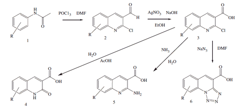
A series of 2-oxo-1,2-dihydroquinoline-3-carboxylic acids 4 were obtained after hydrolysis of chlorine in position 2 of quinoline by treatment with boiling acetic acid with addition of water. Alternatively, chlorine in position 2 was replaced with amino group in aqueous ammonia at 150 °C, 2-aminoquinoline-3-carboxylic acids 5 were isolated with 30–50% yield after fractional crystallization. A series of tetrazolo[1,5-a]quinoline-4-carboxylic acids 6 were synthesized by reaction of intermediates 3 with sodium azide in dimethylformamide (DMF) at 100 °CCitation14.
The bromine in benzene ring of 2-oxo-1,2-dihydroquinoline-3-carboxylic acid 4 and tetrazolo[1,5-a]quinoline-4-carboxylic acid 5 was replaced with phenylboronic acid residues by modificated Suzuki reaction (Scheme 2).
Scheme 2. Synthesis of phenyl substituted 3-quinoline carboxylic acids.
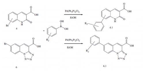
Under reaction conditions, bromine atom in 6 and 7 positions of 2-oxo-1,2-dihydroquinoline-3-carboxylic acid 4 and tetrazolo[1,5-a]quinoline-4-carboxylic acid 5 has been more replaceable, and products of interaction of 8-bromo tetrazolo[1,5-a]quinoline-4-carboxylic acid 6j () with phenylboronic acids were not formed. The 1-acetyl-1,2,3,4-tetrahydroquinoline 8 was a starting compound for the synthesis of 5-oxo-2,3-dihydro-1H,5H-pyrido[3,2,1-ij]quinoline-6-carboxylic acid derivatives 11, 13, and 14 (Scheme 3). This compound was used in reaction with Vilsmeier complex with obtaining 5-oxo-2,3-dihydro-1H,5H-pyrido[3,2,1-ij]quinoline-6-carbaldehyde 9, which was treated with hydroxylamine hydrochloride in phosphorus oxychloride at 100 °C thus yielding 5-oxo-2,3-dihydro-1H,5H-pyrido[3,2,1-ij]quinoline-6-carbonitrile 10 (Scheme 3). The last one was bromated to position 9 by N-bromosuccinimide action in DMF at room temperature. Obtained compounds 10 and 12 were hydrolyzed in process of prolonged boiling with ethanol solution of sodium hydroxide. Obtained 9-bromo-5-oxo-2,3-dihydro-1H,5H-pyrido[3,2,1-ij]quinoline-6-carboxylic acid 14 was added to the modificated Suzuki reaction with phenylboronic acids generating 14а and 14b derivatives ().
Scheme 3. Synthesis of 5-oxo-2,3-dihydro-1H,5H-pyrido[3,2,1-ij]quinoline-6-carboxylic acid derivatives.
![Scheme 3. Synthesis of 5-oxo-2,3-dihydro-1H,5H-pyrido[3,2,1-ij]quinoline-6-carboxylic acid derivatives.](/cms/asset/6f573c61-b721-42e2-95cd-4c490daea4e9/ienz_a_1222584_sch0003.gif)
Molecular docking
Software packages of Autodock 4.2.6Citation15 and Dock 4.0Citation16 were used for flexible docking. The crystal structure of human protein kinase CK2 was obtained from the Brookhaven Protein Data Bank (PDB ID: 3R0T)Citation17–20.
AutoDock
Water molecules, ions, and ligand were removed from the PDB file of receptor.
Ligands were prepared by Vega ZZ (command line)Citation21 and MGL Tools 1.5.6Citation15. To carry out calculation with the aid of Autodock program the incoming formats of receptor and ligands data were converted into PDBQT-format with Vega ZZ in AUTODOCK force field. This format contains the coordinates of the atoms and partial charges. Hydrogen atoms were removed from nonpolar atoms. The receptor was prepared using MGL Tools and AutoGrid.
For docking calculation we have used the following parameters: translation step – 2 Å, quaternion step – 50°, torsion step – 50°. Torsional degrees of freedom and coefficient were 2 and 0.274, respectively. Cluster tolerance – 2 Å. External grid energy – 1000, max initial energy – 0, max number of retries – 10 000. Number of individuals in population – 300, maximum number of energy evaluations – 850 000, maximum number of generations – 27 000, number of top individuals, which survived to the next generation – 1, rate of gene mutation – 0.02, rate of crossover – 0.8, mode of crossover – arithmetic. Alpha parameter of Cauchy distribution was 0, Beta parameter Cauchy distribution – 1. The number of iterations of Lamarckian genetic algorithm was 50 for each ligand.
Dock
The receptor molecule has been minimized in water with GROMACS molecular dynamics simulation packageCitation22 (GROMACS force field, steepest descent algorithm, 1000 steps, em_tolerance = 100, em_step = 0.001). Active site spheres were calculated with DOCK sphgen software. The spheres with the positions outside the ATP-binding site were deleted manually. Connolly MSCitation23 and Grid programs from DOCK package were used to generate receptor Connolly surface and energy grids. Surface and grid calculations were performed with parameter settings as inCitation24, except for grid spacing that was set to 0.3.
Calculations of ligand geometry were performed using YFF force field developed by the authorsCitation25, partial atomic charges of the ligands were calculated with Kirchhoff methodCitation26,Citation27.
For receptor–ligand flexible docking the DOCK program has been used. DOCK input parameters were set as inCitation24 with some exceptions to increase the calculations accuracy: the minimum of heavy atoms in the anchor was set to 6, the maximum number of orientations was set to 1000, and the “all atoms” model has been chosen. DOCK employs an incremental reconstruction algorithm, where rigid anchor fragments are identified first. At the next step, the selected fragment is placed into the active site of the receptor using a sphere-matching procedure. The complete ligand is constructed by adding the remaining components step by step. At each step of reconstruction a specified number of optimal partial solutions are selected for the next extension step. Solutions are scored using energy score (sum of van der Waals and electrostatic components).
Visual analysis
For visual inspection of obtained with molecular docking protein kinase–ligand complexes Discovery Studio Visualizer 4.0Citation28 was used.
Biology
Compounds were tested using in vitro kinase assayCitation29. Each test was carried out in triplicate in 30 μL reaction volume which contains 6 μg of peptide substrate RRRDDDSDDD (New England Biolabs); 10 units of recombinant human CK2 holoenzyme (New England Biolabs); 50 μM ATP and 0.05–0.1 μCi γ-labeled 32P ATP with final specific activity of labeled ATP 3000 mCi/mmol; CK2 buffer (20 mM Tris-HCl, pH 7.5; 50 mM KCl; 10 mM MgCl2) and inhibitor in varying concentrations. Incubation time was 20 min at 30 °C. The reaction was stopped by adding an equal volume of 10% o-phosphoric acid and the reaction mixture was loaded onto 20-mm discs of phosphocellulose paper (Whatman). Disks were washed three times with 1% o-phosphoric acid solution, air-dried at room temperature, and counted by the Cherenkov method in a beta-counter (LKB). As negative control an equal volume of dimethyl sulfoxide (DMSO) was added to the reaction mixture. Inhibition percentage was calculated as ratio of substrate-incorporated radioactivity in the presence of inhibitor to the radioactivity incorporated in control reactions, i.e. in the absence of inhibitor. Serial dilutions of inhibitor stock solution were used to determine its IC50 concentration. The IC50 values represent means of triplicate experiments with SEM never exceeding 15%.
Results and discussion
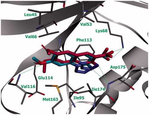
Earlier, we found protein kinase CK2 inhibitors among the derivatives of 3-carboxy-4(1H)-quinoloneCitation30. Recently, we have identified hit compound among the derivatives of 3-carboxy quinolin-2-one-7-bromo-2-oxo-1,2-dihydroquinoline-3-carboxylic acid 93 () which decreased activity of CK2 on 22% at concentration of 33 μMCitation31,Citation32. In this article, we have optimized the structure of this compound with the aim of improvement of CK2 inhibitory activity.
Figure 1. Schematic representation of the complex of compound 93 with active site of protein kinase CK2 (green line indicates amino acid residues involved in hydrophobic contacts; dotted lines indicate hydrogen bonds).
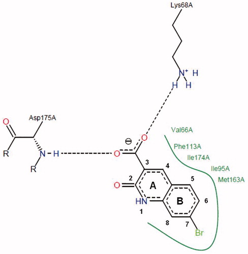
Accordingly to the molecular docking results, the interaction between ligand and ATP-binding pocket of CK2 occur due to hydrophobic contacts with five amino acid residues of protein kinase (Ile95, Phe113, Ile174, Val66, and Met163) and two hydrogen bonds, which are formed between carboxylic group of the compound and Lys68 and Asp175 amino acid residues. It should be noted that the interaction with Lys68 is observed among many low molecular weight inhibitors of human protein kinase CK2Citation33. Taking into account the key interactions of compound 93 in CK2 active site, the several ways for structural optimization of quinoline-3-carboxylic acid derivatives were proposed:
To study the impact of substituents of benzene ring B () on formation of hydrogen bonds and hydrophobic interactions with CK2 active site, we have synthesized derivatives with methoxy group and hydrophobic substituents in position 5, 6, 7, and 8 of quinolin-2(1H)-one cycle.
To determine the influence of carbonyl group on hydrogen bonds formation we have substituted it with chlorine atom or amino group in position 2 of quinoline cycle A.
To investigate influence of cyclic systems on compound activity via enhancing of hydrophobic effect, we have synthesized derivatives with additional hydrophobic cycle in position 5–6, 7–8 and aliphatic cycle in 1–8 position with fixed amide bond of cycle A.
Also, it was interesting to determine the influence of another heterocycles in compounds structure on stabilization of complex “inhibitor-CK2” via π–π stacking with Phe113. Thus, the derivatives with condensed tetrazole cycle in 1 and 2 positions of quinoline cycle have been obtained.
Table 1. Structure of 2-chloro- and 2-aminoquinoline-3-carboxylic acid derivatives and the data related to their in vitro screening.
As a result of our modifications, 43 novel derivatives of 3-quinoline carboxylic acid have been synthesized and tested in vitro. Their chemical structures and inhibitory activity toward CK2 (IC50 values) are shown in .
Table 2. Structure of 2-oxo-1,2-dihydroquinoline-3-carboxylic acid derivatives and the data related to their in vitro screening.
Table 3. Structure of tetrazolo[1,5-a]quinoline-4-carboxylic acid derivatives and the data related to their in vitro screening.
Accordingly to the results of structure–activity relationship (SAR) analysis, a series of regularities can be observed. Inhibitors’ activity is greatly influenced by the replacement of substituents in position 2 of quinoline heterocycle, which can be shown by comparing groups of compounds 3b–4b–5b–6b, 3k–4k–5k–6k, 5c–93 (see ) –6j. In order to explain this relationship, we performed molecular docking of novel 3-carboxyquinoline derivatives into ATP-binding pocket of human CK2. Accordingly to computer modeling results, the ligands quinoline heterocycle forms hydrophobic contacts with amino acid residues Val66, Ile95, Met163, and Ile174; stacking with Phe113 is observed here as well. The substituent in position 2 of quinoline heterocycle is located in ribose pocket of CK2 and forms a series of hydrophobic contacts: compounds with tetrazole cycle (6b, 6k, and 6j) have strong hydrophobic contacts with amino acid residues Ile95, Phe113, and Ile174; compounds with chlorine atom (3b, 3k) and oxygen atom (4b, 4k, and 93) do not have hydrophobic interaction with Ile95. Besides ligands with oxygen atom or tetrazole cycle form hydrogen bond with Asp175. Thus, the most active inhibitor among above-mentioned compounds is tetrazole derivative (compound 6b, IC50 = 0.8 μM). It can be explain by the fact that this ligand has more hydrophobic contacts in ATP-binding site of CK2.
For ligands with NH2- group 5a–5k, we were expecting the same binding mode as described above, but both docking programs AutoDock and Dock proposed reverse position (). The carboxylic and amino groups of these compounds were oriented toward the CK2 hinge region (). The quinoline heterocycle of compounds 5b, 5k, and 5c has hydrophobic interactions with Val66, Ile95, Met163, Ile174, π–π stacking with Phe113 and additional contact with Val53. Carboxylic acid residue of ligands forms H-bond with Val116, while -NH2 is involved in the formation of hydrogen bond with Glu114. Opposite to this, the empirical data of inhibitory activity for compounds 5b, 5k, and 5c confirmed substituent effects in the ring B on compounds activity, as well as for 3b, 3k, and 6j. Also, we have found that the introduction of –NH2 into the second position of 3-carboxyquinolines, instead C=O, leads to considerable increase of CK2 inhibition activity (pair of compounds 4k–5k, 93–5c, and 4b–5b).
Figure 2. Compound 5k (violet) in ATP-binding site of CK2 (the complex was obtained with molecular docking, hydrogen bonds are indicated by green dotted lines).
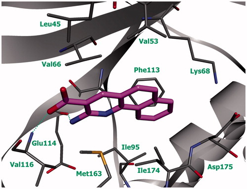
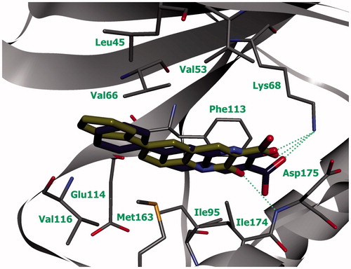
Compounds with 7-methoxy substituent of quinoline are more active than compounds with 6-methoxy or 6,7-dimethoxy substituents that can be shown by comparing the groups of compounds 3a–3b–3c and 6a–6b–6c. This regularity can be explained based on the complexes of pairs of compounds 6a–6b () and 6b–6c (). Carboxyl group of compounds is located in the area with a positive electrostatic potential (near Lys68), which attracts negatively charged groups of compounds and influences on position of ligands in the active site. In this area, the hydrogen bond is formed between oxygen atom of the ligand’s carboxyl group and nitrogen atom of Lys68 side chain and hydrogen bond occurs between tetrazole cycle and nitrogen atom of Asp175 main chain.
Figure 3. Compounds 6а (green) and 6b (blue) in ATP-binding site of CK2 (the complex was obtained by molecular docking, hydrogen bonds are indicated by green dotted lines).
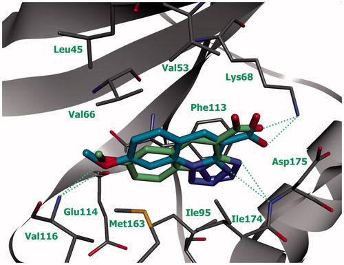
Figure 4. Compounds 6b (blue) and 6с (purple) in ATP-binding site of CK2 (the complex was obtained by molecular docking, hydrogen bonds are indicated by green dotted lines).
Oxygen atom of the methoxy group of compounds 6a and 6b forms hydrogen bond with nitrogen atom of Val116. As both 6- and 7-substituted quinolines have the same hydrogen bonds, higher inhibitory activity of the last one can be explained by tighter hydrophobic interaction with Val66 (). In its turn, the absence of expected inhibitory activity of dimethoxy substituted derivative 6с can be caused by the fact that the introduction of the second methoxy group results in the repulsion of methoxy group in position 6 of quinoline from Leu45, whereupon compound is buried deeper into enzyme’s active site and loses the hydrogen bonds with CK2 hinge region ().
In a way similar to methoxy substituted quinoline derivatives, 7-bromine substituted compounds exhibit better inhibitory activity toward CK2, compared to 6-bromine substituted: 5c–5d and 6f–6j. It is interesting that compounds 4.1a and 4.1b, with bulk substituent (Ph) in position 6- and 7 of quinolone, show the opposite regularity. This can be explained by the binding modes of these compounds with CK2 active site. As we can see from the , substituent in position 6 of quinolone (compound 4.1a) is located in hydrophobic region II while the Ph in position 7 of quinolone (compound 4.1b) should be directed toward hinge region, but its size prevents it. As a result, 4.1b changes its location in active site that leads to weakening of hydrophobic contacts with Leu45 and Val66, and breakdown of π–π stacking with Phe113.
Figure 5. Compounds 4.1а (yellow) and 4.1b (pale gray) in ATP-binding site of CK2 (the complex was obtained by molecular docking, hydrogen bonds are indicated by green dotted lines).
Introduction of oxygen atom between phenyl substituent and 2-quinolone heterocycle negatively effects compounds activity, as was shown by pair of compounds 4d–4.1a and 6d–6.1f. In this case, the compounds with phenoxy in position 6 lost their inhibitory activity.
The positive effect on inhibitory activity of compounds through increasing hydrophobicity of ligands can be seen in the pair of compounds 6.1a–6.1b–6.1c. Thus, the introduction of additional substituents into the phenyl cycle leads to increased interactions with Leu45 and contributes to the formation of additional hydrophobic contacts with imidazole cycle of His115. Also, halogen bonds between oxygen atom of Asn118 and halogen atoms of ligand are likely to occur.
Additional aliphatic cycle (–CH2–CH2–CH2–N–) decreases the activity of compounds which was proved by pairs 4.1а–14а, 4.1f–14b. The quinolones with additional cycle have lost the hydrogen bond with Asp175 due to inability to locate in the depth of the active site. Also, their hydrophobic interaction with Phe113 was weakened.
The best inhibitory activity due to hydrophobic substituent is shown by benzo[h]quinoline-3-carboxylic acid derivatives 3–6k. At the same time, their benzo[f]quinoline-2-carboxylic acid analogs (compounds 3 l, 4 l, and 6 l) are inactive at all. The lack of activity can be explained by different position of compounds in the CK2 active site. The complexes of compounds 3k, 4k, and 6k with enzyme have shown that quinoline heterocycle is located in the depth of active site and forms strong hydrophobic interaction with Val53, Val66, Ile95, and Ile174. The presence of additional cycle in position 5–6 prevents similar position of compounds 3 l, 4 l, and 6 l. These ligands are located closer to the exit from the active site and consequently their hydrophobic contacts with Val66 and Ile95 are weakened.
Thus, taking into account the results obtained by SAR analysis and molecular modeling data, it is possible to conclude that the key interactions, which cause the increase of inhibitory activity of studied 3-quinoline carboxylic acid derivatives are hydrophobic contacts.
Conclusion
Modifications of the 3-quinoline carboxylic acid moiety have led to the identification of promising protein kinase CK2 inhibitors. The most active compounds with IC50 in submicromolar range were found among the tetrazolo-quinoline-4-carboxylic acid and 2-amino-quinoline-3-carboxylic acid derivatives. We suppose that the last ones are the most perspective for further chemical optimization and biological investigations.
Experimental section
Starting materials and solvents were purchased from commercial suppliers and were used without further purification. 1HNMR spectra were recorded on Varian VXR 400 spectrometer at 400 MHz (Agilent Technologies, Santa Clara, CA). Chemical shifts are described as parts per million (δ) downfield from an internal standard of tetramethylsilane, and spin multiplicities are given as s (singlet), d (doublet), dd (double doublet), t (triplet), q (quartet), or m (multiplet). High-performance liquid chromatography-mass spectra (HPLC-MS) analysis was performed using the Agilent 1100 LC/MSD SL (Agilent Technologies) separations module and Mass Quad G1956B mass detector with electrospray ionization (Agilent Technologies) (+ve or − ve ion mode as indicated) and with HPLC performed using Zorbax SB-C18 (Agilent Technologies), Rapid Resolution HT Cartridge 4.6 30 mm 1.8-m (Agilent Technologies P/N:823975-902) i.d. column, at 40 °C with gradient elution of 0–100% CH3CN (with 1 mL/L HCOOH): H2O (with 1 mL/L HCOOH) at a flow rate of 3 mL/min and a run time of 2.8 min. Compounds were detected at 215 nm using Diode Array G1315B detector. Melting points were determined on a micro hot stage apparatus and are uncorrected.
General procedure synthesis of 2-chloroquinoline-3-carboxylic acids 3а–l
To stirring suspension of corresponding 2-chloroquinoline-3-carbaldehyde 2a–l [9, 16] (0.01 mol) in 60 mL EtOH was added warm AgNO3 solution 2.71 g (0.016 mol) in 30 mL EtOH. Solution of NaOH 2 g (0.05 mol) in 30 mL 80% aqueous EtOH was added dropwise with intensive stirring during 15 min at room temperature. Reaction mixture was stirred 12 h and filtered through CELIT pad. The solvent was removed by rotary evaporation. Water was added to completely solved Na+ salt of 3, than acidified 15% aqueous HCl solution to pH 1. Solid products 3a–l were filtered, washed with water (2 × 20 mL) and then dried in a vacuum oven at 60 °C.
2-chloro-6-methoxyquinoline-3-carboxylic acid 3а. Yield = 89%. Mp. = 259 °C; white powder. LCMS m/z 238, 240 [M + H+], Rf = 0.75 min. 1HNMR (DMSO-d6) δ 3.91 (s, 3H, –OCH3), 7.52–7.64 (m, 2H), 7.91 (d, 1Н, CН, J = 8.79 Hz), 8.79 (s, 1Н, CН).
2-chloro-7-methoxyquinoline-3-carboxylic acid 3b. Yield = 83%. Mp. = 203 °C; white powder. LCMS m/z 238, 240 [M + H+], Rf = 0.77 min. 1HNMR (DMSO-d6) δ 3.95 (s, 3H, –OCH3), 7.29–7.45 (m, 2H), 8.07 (d, 1Н, CН, J = 8.79 Hz), 8.86 (s, 1Н, CН), 13.55 (br. s., 1Н, COOН).
2-chloro-6,7-dimethoxyquinoline-3-carboxylic acid 3c. Yield = 87%. Mp. = 236 °C; yellow powder. LCMS m/z 268, 270 [M + H+], Rf = 0.81 min. 1HNMR (DMSO-d6) δ 3.93 (s, 3H, –OCH3), 3.97 (s, 3H, –OCH3), 7.39 (s, 1H, CH), 7.53 (s, 1H, CH), 8.71 (s, 1Н, CН).
2-chloro-6-phenoxyquinoline-3-carboxylic acid 3d. Yield = 92%. Mp. = 184 °C; light-yellow powder. LCMS m/z 300, 302 [M + H+], Rf = 0.88 min. 1HNMR (DMSO-d6) δ 7.16 (d, 2Н, C2Н2, J = 7.81 Hz), 7.24 (t, 1Н, CН, J = 7.08 Hz), 7.47 (t, 2Н, C2Н2, J = 6.84 Hz), 7.60 (s, 1H, CH), 7.66 (d, 1Н, CН, J = 8.79 Hz), 8.03 (d, 1Н, CН, J = 8.30 Hz), 8.81 (s, 1H, CH), 13.77 (br. s., 1Н, COOH).
7-bromo-2-chloro-6-methoxy-3-carboxylic acid 3e. Yield = 94%. Mp. = 264 °C; yellow powder. LCMS m/z 321 [M + H+], Rf = 0.93 min. 1HNMR (DMSO-d6) δ 4.00 (s, 3H, –OCH3), 7.71(s, 1H, CH), 8.28 (s, 1H, CH), 8.81 (s, 1H, CH), 13.75 (br. s., 1Н, COOН).
2-chlorobenzo[h]quinoline-3-carboxylic acid 3k. Yield = 83%. Mp. = 228 °C; beige powder. LCMS m/z 258, 260 [M + H+], Rf = 0.89 min. 1HNMR (DMSO-d6) δ 7.82 (m, 2 H, C2Н2), 8.04 (m, 3 H, C3Н3), 8.95 (s, 1 H, CН), 9.00 (d, J = 7.32 Hz, 1 H, CН), 13.85 (s, 1 H, COОН).
3-chlorоbenzo[f]quinoline-2-carboxylic acid 3 l. Yield = 89%. Mp. = 233 °C; white powder. LCMS m/z 258, 260 [M + H+], Rf = 0.97 min. 1HNMR (DMSO-d6) δ 7.75–7.85 (m, 2 H, C2Н2), 7.89 (d, 1 H, CН, J = 9.16 Hz), 8.11 (d, 1 H, CН, J = 7.32 Hz), 8.28 (d, 1 H, CН, J = 9.16 Hz), 8.90 (d, 1 H, CН, J = 7.94 Hz), 9.59 (s, 1 H, CН).
General procedure synthesis of 2-oxo-1,2-dihydroquinoline-3-carboxylic acids 4a–l
Suspension of corresponding 2-chloroquinoline-3-carboxylic acid 3 (1 mmol) in glacial acetic acid (25 mL) and water (1.5 mL) was boiled with stirring during 12–24 h. The reaction was monitored by NMR. After cooling, water (100 mL) was added and solid products 4a–l were filtered, washed with water (2 × 20 mL) and then dried in a vacuum oven at 60 °C.
6-methoxy-2-oxo-1,2-dihydroquinoline-3-carboxylic acid 4а. Yield = 89%. Mp. = 289 °C; yellow powder. LCMS m/z 220 [M + H+], Rf = 0.67 min. 1HNMR (DMSO-d6) δ 3.84 (s, 3H, –OCH3), 7.41–7.53 (m, 2H, C2H2), 7.59 (s, 1Н, CН), 8.92 (s, 1Н, CН), 13.08 (br. s., 1Н, NН), 14.94 (br. s., 1Н, COOН).
7-methoxy-2-oxo-1,2-dihydroquinoline-3-carboxylic acid 4b. Yield = 73%. Mp. = 293 °C; beige powder. LCMS m/z 220 [M + H+], Rf = 0.69 min. 1HNMR (DMSO-d6) δ 3.90 (s, 3H, –OCH3), 6.97 (s, 1Н, CН), 7.03 (d, 1Н, CН, J = 8.3 Hz), 7.96 (d, 1Н, CН, J = 8.79 Hz), 8.87 (s, 1Н, CН), 12.93 (br. s., 1Н, NН), 14.59 (br. s., 1Н, COOН).
6,7-dimethoxy-2-oxo-1,2-dihydroquinoline-3-carboxylic acid 4с. Yield = 92%. Mp. = 287 °C; yellow powder. LCMS m/z 250 [M + H+], Rf = 0.72 min. 1HNMR (DMSO-d6) δ 3.84 (s, 3H, –OCH3), 3.91 (s, 3H, –OCH3), 7.00 (s, 1H, CH), 7.53 (s, 1Н, CН), 8.81 (s, 1Н, CН), 12.93 (br. s., 1Н, NН), 14.85 (br. s., 1Н, COOН).
2-oxo-6-phenoxy-1,2-dihydroquinoline-3-carboxylic acid 4d. Yield = 97%. Mp. = 272 °C; yellow powder. LCMS m/z 282 [M + H+], Rf = 0.76 min. 1HNMR (DMSO-d6) δ 7.04 (d, 2Н, C2Н2, J = 7.32 Hz), 7.17 (t, 1Н, CН, J = 7.08 Hz), 7.41 (t, 2Н, C2Н2, J = 7.08 Hz), 7.50–7.60 (m, 2H, C2H2), 7.71 (s, 1Н, CН), 8.92 (s, 1Н, CН), 13.47 (br. s., 1Н, NH), 14.08 (br. s., 1Н, COOH).
2-oxo-1,2-dihydrobenzo[h]quinoline-3-carboxylic acid 4k. Yield = 65%. Mp. = 228 °C; yellow powder. LCMS m/z 240 [M + H+], Rf = 0.82 min. 1HNMR (DMSO-d6) δ 7.77 (m, 3 H, C3Н3), 7.94 (d, J = 9.16 Hz, 1 H, CН), 8.05 (d, J = 7.94 Hz, 1 H, CН), 9.00 (d, J = 7.94 Hz, 1 H, CН), 9.03 (s, 1 H, CН), 13.49 (s, 1 H, ОН).
3-oxo-3,4-dihydrobenzo[f]quinoline-2-carboxylic acid 4 l. Yield = 74%. Mp. = 228 °C; yellow powder. LCMS m/z 240 [M + H+], Rf = 0.79 min. 1HNMR (DMSO-d6) δ 7.65 (m, 2 H, C2Н2), 7.78 (t, J = 7.63 Hz, 1 H, CН), 8.04 (d, J = 8.55 Hz, 1 H, CН), 8.28 (d, J = 9.16 Hz, 1 H, CН), 8.67 (d, J = 8.55 Hz, 1 H, CН), 9.57 (s, 1 H, CН).
General procedure synthesis of 2-aminoquinoline-3-carboxylic acids 5а–5 l
Suspension of corresponding 2-chloroquinoline-3-carboxylic acids 3 (1 mmol) in 26% aqueous NH3 (5 mL) was heated in stainless steel autoclave at 150 °C during 4 h. After reaction mixture cooling, clear solution was acidified by 5% aqueous HCl solution to pH-4. Solid products 5a–l were filtered and recrystallized from IPA–DMF mixture.
2-amino-6-methoxyquinoline-3-carboxylic acid 5а. Yield = 36%. Mp. = 283 °C; yellow powder. LCMS m/z 219 [M + H+], Rf = 0.56 min. 1HNMR (DMSO-d6) δ 3.83 (s, 3H, –OCH3), 7.47 (m, 2H, C2H2), 7.61 (s, 1H, CH), 8.93 (s, 1H, CH), 15.01 (br. s., 1Н, COOН).
2-amino-7-methoxyquinoline-3-carboxylic acid 5b. Yield = 49%. Mp. = 269 °C; white powder. LCMS m/z 219 [M + H+], Rf = 0.58 min. 1HNMR (DMSO-d6) δ 3.86 (s, 3H, –OCH3), 6.85 (d, 2H, C2H2, J = 6.1 Hz), 6.89 (s, 1H, CH), 7.69 (d, 2H, C2H2, J = 6.1 Hz), 8.61 (s, 1H, CH).
2-amino-7-bromoquinoline-3-carboxylic acid 5c. Yield = 27%. Mp. = 286 °C; yellow powder. LCMS m/z 267, 269 [M + H+], Rf = 0.71 min. 1HNMR (DMSO-d6) δ 7.34 (d, 1Н, CН, J = 7.33 Hz), 7.65 (s, 1H, CH), 7.79 (d, 1Н, CН, J = 7.33 Hz), 8.75 (s, 1H, CH).
2-amino-6-bromoquinoline-3-carboxylic acid 5d. Yield = 41%. Mp. = 245 °C; yellow powder. LCMS m/z 267, 269 [M + H+], Rf = 0.61 min. 1HNMR (DMSO-d6) δ 7.43 (d, 1Н, CН, J = 9.77 Hz), 7.69 (d, 1Н, CН, J = 9.28 Hz), 8.08 (s, 1Н, CН), 8.72 (s, 1Н, CН).
2-amino-6-phenoxyquinoline-3-carboxylic acid 5e. Yield = 50%. Mp. = 318 °C; yellow powder. LCMS m/z 281 [M + H+], Rf = 0.71 min. 1HNMR (DMSO-d6) δ 7.02 (d, 2Н, C2Н2, J = 7.81 Hz), 7.13 (t, 1Н, CН, J = 7.32 Hz), 7.39 (t, 3Н, C3Н3, J = 7.81 Hz), 7.49 (s, 1H, CH), 7.55 (d, 1Н, CН, J = 9.28 Hz), 8.72 (s, 1Н, CН).
2-amino-7-bromo-6-methoxyquinoline-3-carboxylic acid 5f. Yield = 32%. Mp. = 285 °C; yellow powder. LCMS m/z 297, 299 [M + H+], Rf = 0.62 min. 1HNMR (DMSO-d6) δ 3.90 (s, 3H, –OCH3), 7.45 (s, 1H, CH), 7.74 (s, 1H, CH), 8.69 (s, 1H, CH).
2-aminobenzo[h]quinoline-3-carboxylic acid 5k. Yield 41%. Mp. = 254 °C; yellow powder. LCMS m/z 239 [M + H+], Rf = 0.69 min. 1HNMR (DMSO-d6) δ 7.71–7.85 (m, 3H, C3H3), 7.91 (d, 1H, CH, J = 7.32 Hz), 8.05 (d, 1H, CH, J = 7.32 Hz), 9.06 (s, 1H, CH), 9.16 (d, 1H, CH, J = 7.94 Hz).
General procedure synthesis of tetrazolo[1,5-a]quinoline-4-carboxylic acids 6a–l
Suspension of corresponding 2-chloroquinoline-3-carboxylic acid 3 (1 mmol) and sodium azide (0.15 g, 2.3 mmol) in DMF (5 mL) stirred at 100 °C during 12 h. After cooling, water (30 mL) was added than mixture acidified by 15% aqueous HCl solution to pH 1. Solid products 6a–l were filtered, washed with water (2 × 20 mL) and then dried in a vacuum oven at 60 °C.
7-methoxytetrazolo[1,5–a]quinoline-4-carboxylic acid 6а. Yield = 79%. Mp. = 255 °C; beige powder. LCMS m/z 245 [M + H+], Rf = 0.71 min. 1HNMR (DMSO-d6) δ 3.95 (s, 3H, –OCH3), 7.65 (d, 1Н, CН, J = 7.32 Hz), 7.92 (s, 1H, CH), 8.54 (d, 1Н, CН, J = 8.79 Hz), 8.83 (s, 1Н, CН), 13.62 (br. s., 1Н, COOH).
8-methoxytetrazolo[1,5–a]quinoline-4-carboxylic acid 6b. Yield = 71%. Mp. = 260 °C; white powder. LCMS m/z 245 [M + H+], Rf = 0.69 min. 1HNMR (DMSO-d6) δ 4.08 (s, 3H, -OCH3), 7.47 (d, 1Н, CН, J = 8.30 Hz), 8.04 (s, 1H, CH), 8.34 (d, 1Н, CН, J = 8.79 Hz), 8.88 (s, 1Н, CН).
7,8-dimethoxytetrazolo[1,5-a]quinoline-4-carboxylic acid 6c. Yield = 95%. Mp. = 286 °C; beige powder. LCMS m/z 275 [M + H+], Rf = 0.79 min. 1HNMR (DMSO-d6) δ 3.96 (s, 3H, –OCH3), 4.11 (s, 3H, –OCH3), 7.92 (s, 1H, CH), 8.03 (s, 1H, CH), 8.83 (s, 1H, CH), 13.48 (br. s., 1Н, COOH).
7-phenoxytetrazolo[1,5-a]quinoline-4-carboxylic acid 6d. Yield = 97%. Mp. = 254 °C; beige powder. LCMS m/z 307 [M + H+], Rf = 0.82 min. 1HNMR (DMSO-d6) δ 7.17 (d, 2Н, C2Н2, J = 7.81 Hz), 7.26 (t, 1Н, CН, J = 7.08 Hz), 7.48 (t, 2Н, C2Н2, J = 7.08 Hz), 7.74 (d, 1Н, CН, J = 8.79 Hz), 8.02 (s, 1H, CH), 8.66 (d, 1Н, CН, J = 8.79 Hz), 8.89 (s, 1Н, CН), 13.78 (br. s., 1Н, COOH).
8-bromo-7-methoxytetrazolo[1,5-a]quinoline-4-carboxylic acid 6е. Yield = 86%. Mp. = 291 °C; white powder. LCMS m/z 323, 325 [M + H+], Rf = 0.81 min. 1HNMR (DMSO-d6) δ 4.04 (s, 3H, –OCH3), 8.08 (s, 1H, CH), 8.79 (s, 1H, CH), 8.82 (s, 1H, CH).
7-bromotetrazolo[1, 5-a]quinoline-4-carboxylic acid 6f. Yield = 91%. Mp. = 274 °C; beige powder. LCMS m/z 293, 295 [M + H+], Rf = 0.78 min. 1HNMR (DMSO-d6) δ 8.21 (d, 1Н, CН, J = 8.2 Hz), 8.58 (d, 1Н, CН, J = 8.79 Hz), 8.71 (s, 1Н, CН), 8.89 (s, 1H, CH).
8-bromotetrazolo[1,5-a]quinoline-4-carboxylic acid 6j. Yield = 89%. Mp. = 265 °C; beige powder. LCMS m/z 293, 295 [M + H+], Rf = 0.8 min. 1HNMR (DMSO-d6) δ 8.04 (d, 1Н, CН, J = 8.71 Hz), 8.34 (d, 1Н, CН, J = 8.30 Hz), 8.81 (d, 2Н, C2Н2, J = 6.35 Hz).benzo[h]tetrazolo[1,5-a]quinoline-4-carboxylic acid 6k. Yield = 83%. Mp. = 228 °C; white powder. LCMS m/z 265 [M + H+], Rf = 0.88 min. 1HNMR (DMSO-d6) δ 8.02 (m, 2 H, C2Н2), 8.36 (m, 3 H, C3Н3), 9.12 (s, 1 H, CН), 10.19 (d, 1 H, CН, J = 8.55 Hz).benzo[f]tetrazolo[1,5-a]quinoline-12-carboxylic acid 6 l. Yield = 86%. Mp. = 228 °C; white powder. LCMS m/z 265 [M + H+], Rf = 0.83 min. 1HNMR (DMSO-d6) δ 7.82 (s, 1 H, CН), 7.90 (s, 1 H, CН), 8.25 (d, J = 7.94 Hz, 1 H, CН), 8.57 (d, J = 9.16 Hz, 1 H, CН), 8.71 (d, J =9.16 Hz, 1 H, CН), 8.96 (d, J =7.94 Hz, 1 H, CН), 9.56 (s, 1 H, CН).
General procedure synthesis of 6- and 7-phenyl-2-oxo-1,2-dihydroquinoline-3-carboxylic acids 4.1a-g and 7-phenyltetrazolo[1,5-a]quinoline-4-carboxylic acids 6.1a-c, modification of Suzuki reaction
Synthesis of starting 6- and 7-bromo-2-oxo-1,2-dihydroquinoline-3-carboxylic acids was carried out by general procedure, previously described for 4a–l, physical and analytical data which we described earlierCitation24.
Mixture of corresponding bromide (0.75 mmol) with corresponding phenylboronic acid (0.76 mmol) in ethanol (20 mL) was ventilated by argon three times, then mixture or Na2CO3 (0.4 g, 3.75 mmol) with (Ph3P)2PdCl2 (0.052 g, 0.075 mmol) was added in one portion with stirring and reaction vessel ventilated again. Reaction mixture was boiled with stirring under argon during 12 h. The solvent was removed by rotary evaporation. The formed solid residue was transferred to a stirred 10% aqueous HCl solution (50 mL). Crude precipitate of product was filtered, washed with water (2 × 10 mL) and hexane: DCM mixture 1:1 (5 × 10 mL) to get rid of Ph3PO. After drying crude precipitate was dissolved in DMF and filtered through CELIT pad, to get rid of Pd. Filtrate was concentrated by rotary evaporation and residue treated by cold water. The formed solid of product was filtered, washed with water (2 × 10 mL) and then dried in a vacuum oven at 70 °C. Typically the products were sufficiently pure, but if necessary, they could be recrystallized from an IPA–DMF mixture.
2-oxo-6-phenyl-1,2-dihydroquinoline-3-carboxylic acid 4.1а. Yield = 74%. Mp. = 285 °C; yellow powder. LCMS m/z 266 [M + H+], Rf = 0.85 min. 1HNMR (DMSO-d6) δ 7.43 (d, 1Н, CН, J = 6.84 Hz), 7.53 (t, 2Н, C2Н2, J = 7.32 Hz), 7.63 (d, 1Н, CН, J = 8.30 Hz), 7.76 (d, 2Н, C2Н2, J = 7.81 Hz), 8.13 (d, 1Н, CН, J = 8.79 Hz), 8.39 (s, 1Н, CН), 9.05 (s, 1Н, CН), 13.18 (br. s., 1Н, NН), 14.68 (br. s., 1Н, COOH).
2-oxo-7-phenyl-1,2-dihydroquinoline-3-carboxylic acid 4.1b. Yield = 53%. Mp. = 318 °C; gray powder. LCMS m/z 266 [M + H+], Rf = 0.87 min. 1HNMR (DMSO-d6) δ 7.48 (s, 1H, CH), 7.54 (m, 2H, C2H2), 7.72 (m, 4H, C4H4), 8.10 (s, 1H, CH), 8.97 (s, 1H, CH), 13.16 (br. s., 1Н, NН), 14.64 (br. s., 1Н, COOH).
6-(4-methoxyphenyl)-2-oxo-1,2-dihydroquinoline-3-carboxylic acid 4.1с. Yield = 66%, Mp. = 225 °C; yellow powder. LCMS m/z 296 [M + H+], Rf = 0.87 min. 1HNMR (DMSO-d6) δ 3.82 (s, 3H, –OCH3), 7.08 (d, 2Н, C2Н2, J = 7.81 Hz), 7.58 (d, 1Н, CН, J = 9.28 Hz), 7.69 (d, 2Н, C2Н2, J = 7.81 Hz), 8.08 (d, 1Н, CН, J = 9.28 Hz), 8.31 (s, 1Н, CН), 9.02 (s, 1Н, CН), 13.16 (br. s., 1Н, NН), 14.72 (br. s., 1Н, COOН).
6-(3-methoxyphenyl)-2-oxo-1,2-dihydroquinoline-3-carboxylic acid 4.1d. Yield = 84%, Mp. = 233 °C; yellow powder. LCMS m/z 296 [M + H+], Rf = 0.88 min. 1HNMR (DMSO-d6) δ 3.85 (s, 3H, –OCH3), 6.98 (d, 1Н, CН, J = 7.32 Hz), 7.25–7.37 (m, 2H, C2H2), 7.38–7.47 (m, 1H, CH), 7.60 (d, 1Н, CН, J = 8.30 Hz), 8.12 (d, 1Н, CН, J = 8.79 Hz), 8.40 (s, 1H, CH), 9.03 (s, 1H, CH), 13.20 (br. s., 1Н, NН), 14.69 (br. s., 1Н, COOH).
6-(4-chlorophenyl)-2-oxo-1,2-dihydroquinoline-3-carboxylic acid 4.1e. Yield = 86%, Mp. = 314 °C; light-yellow powder. LCMS m/z 300, 302 [M + H+], Rf = 1.01 min. 1HNMR (DMSO-d6) δ 7.58 (t, 3Н, C3Н3, J = 7.81 Hz), 7.77 (d, 2Н, C2Н2, J = 8.30 Hz), 8.12 (d, 1Н, CН, J = 9.28 Hz), 8.41 (s, 1H, CH), 9.02 (s, 1H, CH), 13.25 (br. s., 1Н, NН), 14.69 (br. s., 1Н, COOН).
6-(3-chlorophenyl)-2-oxo-1,2-dihydroquinoline-3-carboxylic acid 4.1f. Yield = 87%, Mp. = 271 °C; white powder. LCMS m/z 300, 302 [M + H+], Rf = 0.94 min. 1HNMR (DMSO-d6) δ 7.46 (d, 1Н, CН, J = 6.84 Hz), 7.54 (t, 1Н, CН, J = 7.57 Hz), 7.60 (d, 1Н, CН, J = 6.84 Hz), 7.72 (d, 1Н, CН, J = 6.84 Hz), 7.81 (s, 1H, CH), 8.13 (d, 1Н, CН, J = 7.81 Hz), 8.43 (s, 1H, CH), 9.00 (s, 1H, CH), 13.16 (br. s., 1Н, NН), 14.60 (br. s., 1Н, COOH).
2-oxo-6-[3-(trifluoromethyl)phenyl]-1,2-dihydroquinoline-3-carboxylic acid 4.1j. Yield = 74%, Mp. = 295 °C; gray powder. LCMS m/z 334 [M + H+], Rf = 1.02 min. 1HNMR (DMSO-d6) δ 7.62 (d, 1Н, CН, J = 8.30 Hz), 7.77 (m, 2H, C2H2), 8.08 (m, 2H, C2H2), 8.18 (d, 1Н, CН, J = 8.30 Hz), 8.50 (s, 1H, CH), 9.00 (s, 1H, CH).
7-phenyltetrazolo[1,5-a]quinoline-4-carboxylic acid 6.1a. Yield = 89%, Mp. = 288 °C; yellow powder. LCMS m/z 291 [M + H+], Rf = 0.96 min. 1HNMR (DMSO-d6) δ 7.49 (t, 1Н, CН, J = 6.84 Hz), 7.59 (d, 2Н, C2Н2, J = 7.57 Hz), 7.88 (d, 2Н, C2Н2, J = 7.32 Hz), 8.41 (d, 1Н, CН, J = 9.28 Hz), 8.72 (d, 1Н, CН, J = 8.79 Hz), 8.78–8.81 (m, 1H, CH), 9.02 (s, 1H, CH).
7-(3-methoxyphenyl)tetrazolo[1,5-a]quinoline-4-carboxylic acid 6.1b. Yield = 68%, Mp. = 269 °C; yellow powder. LCMS m/z 321 [M + H+], Rf = 0.98 min. 1HNMR (DMSO-d6) δ 3.88 (s, 3H, –OCH3), 7.04 (d, 1Н, CН, J = 6.35 Hz), 7.42 (d, 2Н, C2Н2, J = 13.18 Hz), 7.47 (d, 1Н, CН, J = 8.30 Hz), 8.41 (d, 1Н, CН, J = 6.84 Hz), 8.70 (d, 1Н, CН, J = 8.79 Hz), 8.80 (s, 1H, CH), 9.01 (s, 1H, CH).
7-(3-chlorophenyl)tetrazolo[1,5-a]quinoline-4-carboxylic acid 6.1c. Yield = 81%, Mp. = 267 °C; white powder. LCMS m/z 325, 327 [M + H+], Rf = 0.94 min. 1HNMR (DMSO-d6) δ 7.59 (d, 1Н, CН, J = 7.32 Hz), 7.65 (t, 1Н, CН, J = 7.08 Hz), 7.90 (d, 1Н, CН, J = 6.84 Hz), 8.00 (s, 1H, CH), 8.48 (d, 1Н, CН, J = 8.30 Hz), 8.76 (d, 1Н, CН, J = 7.81 Hz), 8.87 (s, 1H, CH), 9.03 (s, 1H, CH).
Synthesis of 5-oxo-2,3-dihydro-1H,5H-pyrido[3,2,1-ij]quinoline-6-carboxaldehyde 9
To cooled (5 °C) POCl3 (107.3 g, 0.7 mol) was added DMF dropwise (11.6 mL, 0.15 mol) for 30 min (a strong exothermic effect was observed, and the temperature should be kept within 5–15 °C), and the reaction mixture was crystallized. After 15 min, the 1-acetyl-1,2,3,4-tetrahydroquinoline (17.5 g, 0.1 mol) was added in one portion and the reaction mixture temperature slowly increased to 75–80 °C for 2 h, then heated to 80 °C for 12 h. After cooling, the reaction mixture was slowly poured with stirring into cold water (200 mL) with crushed ice (300 g). Product 9 was filtered, washed with water (2 × 100 mL), saturated NaHCO3 (2 × 50 mL), and water (50 mL), then dried in a vacuum oven at 60 °C. Yield 15.6 g, 73%. Mp. = 186 °C; white powder. 1H NMR (DMSO-d6) δ 2.03–2.09 (m, 2 H, CН2), 2.94–2.99 (m, 2 H, CН2), 4.08–4.13 (m, 2 H, CН2), 7.24 (t, 1 H, J = 7.57 Hz, CН), 7.55 (d, J = 6.84 Hz, 1 H, CН), 7.82 (d, J = 7.32 Hz, 1 H, CН), 8.48 (s, 1 H, CН) 10.3 (s, 1 H, OCH).
Synthesis of 5-oxo-2,3-dihydro-1H,5H-pyrido[3,2,1-ij]quinoline-6-carbonitrile 10
To the suspension of aldehyde 9 (15 g, 0.07 mol) in POCl3 (60 mL) was added NH2OH hydrochloride (9.78 g, 0.14 mol) in one portion. Reaction mixture heated with stirrer 100 °C for 3 h. After cooling, the reaction mixture was slowly poured with stirring into cold water (200 mL) with crushed ice (300 g). Product 9 was filtered, washed with water (2 × 100 mL), saturated NaHCO3 (2 × 50 mL) and water (50 mL), then dried in a vacuum oven at 60 °C. Yield 13 g, 88%. Mp. = 267 °C; white powder. 1H NMR (DMSO-d6) δ 2.01–2.08 (m, 2 H, CН2), 2.92–2.98 (m, 2 H, CН2), 4.06–4.11 (m, 2 H, CН2), 7.28 (t, 1 H, J = 7.57 Hz, CН), 7.58 (d, J = 6.84 Hz, 1 H, CН), 7.67 (d, J = 7.32 Hz, 1 H, CН), 8.76 (s, 1 H, CН).
Synthesis of 5-oxo-2,3-dihydro-1H,5H-pyrido[3,2,1-ij]quinoline-6-carboxylic acid 11
Solution of nitrile 10 (5 g, 0.24 mol) in EtOH (100 mL), containing 40% aqueous NaOH solution (20 mL) was boiled with stirrer 48 h. The reaction was monitored by NMR. The solvent was removed by rotary evaporation. The formed solid residue was transferred to a stirred 10% aqueous HCl solution (100 mL) and formed solid of product 11 was filtered, washed with water (2 × 10 mL) and then dried in a vacuum oven at 70 °C. Yield 5.1 g, 95%. Mp. = 251 °C; white powder. LCMS m/z 230 [M + H+], Rf = 0.82 min. 1HNMR (DMSO-d6) δ 2.07–2.12 (m, 2H, CH2), 2.97–3.01 (m, 2Н, CН2,), 4.17–4.22 (m, 2Н, CН2), 7.38 (t, J = 7.32 Hz, 1Н, CН), 7.64 (d, J = 7.32 Hz, 1Н, CН), 7.92 (s, J = 7.81 Hz, 1Н, CН), 8.93 (s, 1Н, CН), 14.70 (br. s., 1Н, COOН).
Synthesis of 9-bromo-5-oxo-2,3-dihydro-1H,5H-pyrido[3,2,1-ij]quinoline-6-carbonitrile 12
To the stirring solution of nitrile 10 (5 g, 0.024 mol) in DMF (75 mL) was added in 10 portions NBS (4.49 g, 0.0252 mol) during 2 h. The reaction mixture was stirred at room temperature 12 h and poured in water (500 mL), containing sodium thiosulfate (1.5 g). The formed solid of product was filtered, washed with water (2 × 10 mL), and then dried in a vacuum oven at 70 °C. Yield 6.5 g, 95%. Mp. = 281 °C; light-yellow powder. 1HNMR (DMSO-d6) δ 2.01–2.05 (m., 2H, CH2), 2.94–3.00 (m, 2Н, CН2), 4.04–4.10 (m, 2Н, CН2), 7.4 (t, J = 7.3 Hz, 1H, CН), 7.6 (d, J = 7.3 Hz, 1H, CН), 7.9 (d, J = 7.8 Hz, 1 H, CН), 8.9 (s, 1 H, CН).
9-bromo-5-oxo-2,3-dihydro-1H,5H-pyrido[3,2,1-ij]quinoline-6-carboxylic acid 13 was synthesized as 12 from nitrile 11. Yield = 92%. Mp. = 268 °C; beige powder. LCMS m/z 308, 310 [M + H+], Rf = 0.87 min. 1HNMR (DMSO-d6) δ 2.03–209 (m, 2H, CH2), 2.97–3.04 (m, 2Н, CН2), 4.16–4.21 (m, 2Н, CН2), 7.83 (s, 1Н, CН), 8.20 (s, 1Н, CН), 8.90 (s, 1Н, CН).
5-oxo-9-phenyl-2,3-dihydro-1H,5H-pyrido[3,2,1-ij]quinoline-6-carboxylic acid 14a was synthesized by General procedure described above for 4.1 from bromide 13 and phenylboronic acid. Yield = 72%. Mp. = 229 °C; light-gray powder. LCMS m/z 300, 302 [M + H+], Rf = 1.01 min.1HNMR (DMSO-d6) δ 2.09–2.13 (m, 2Н, CН2), 3.03–3.10 (m, 2Н, CН2), 4.16–4.19 (m, 2Н, CН2), 7.39 (t, 1Н, CН, J = 6.59 Hz), 7.50 (t, 2Н, C2Н2, J = 7.57 Hz), 7.76 (t, 2Н, C2Н2, J = 6.84 Hz), 7.88 (s, 1Н, CН), 8.12 (s, 1Н, CН), 8.64 (s, 1Н, CН).
9-(3-chlorophenyl)-5-oxo-2,3-dihydro-1H,5H-pyrido[3,2,1-ij]quinoline-6-carboxylic acid 14b was synthesized by General procedure described above for 4.1 from bromide 13 and 3-chlorophenylboronic acid. Yield = 54%. Mp. = 302 °C; yellow powder. LCMS m/z 340, 342 [M + H+], Rf = 1.21 min. 1HNMR (DMSO-d6) δ 2.10–2.14 (m, 2Н, CН2), 3.03–3.09 (m, 2Н, CН2), 4.19–4.25 (m, 2Н, CН2), 7.46 (d, 1Н, CН, J = 7.81 Hz), 7.54 (d, 1Н, CН, J = 7.81 Hz), 7.75 (d, 1Н, CН, J = 7.81 Hz), 7.85 (s, 1Н, CН), 8.04 (s, 1Н, CН), 8.32 (s, 1Н, CН), 8.98 (s, 1Н, CН), 14.70 (br. s., 1Н, COOН).
Declaration of interest
The authors report no conflicts of interest. The authors alone are responsible for the content and writing of this article.
Acknowledgements
We thank Otava Ltd. Company for material and finance support.
References
- Pinna LA. Casein kinase 2: an ‘eminence grise’ in cellular regulation? Biochim Biophys Acta 1990;1054:267–84
- Faust M, Montenarh M. Subcellular localization of protein kinase CK2. A key to its function? Cell Tissue Res 2000;301:329–40
- Piazza F, Manni S, Ruzzene M, et al. Protein kinase CK2 in hematologic malignancies: reliance on a pivotal cell survival regulator by oncogenic signaling pathways. Leukemia 2012;26:1174–9
- Sarno S, Pinna LA. Protein kinase CK2 as a druggable target. Mol BioSyst 2008;4:889–94
- Murtaza I, Wang HX, Feng X, et al. Down-regulation of catalase and oxidative modification of protein kinase CK2 lead to the failure of apoptosis repressor with caspase recruitment domain to inhibit cardiomyocyte hypertrophy. J Biol Chem 2008;283:5996–6004
- Singh NN, Ramji DP. Protein kinase CK2, an important regulator of the inflammatory response? J Mol Med (Berl) 2008;86:887–97
- Trembley JH, Chen Z, Unger G, et al. Emergence of protein kinase CK2 as a key target in cancer therapy. Biofactors 2010;36:187–95
- Meggio F, Pinna LA. One-thousand-and-one substrates of protein kinase CK2? FASEB J 2003;17:349–68
- Siddiqui-Jain A, Drygin D, Streiner N, et al. CX-4945, an orally bioavailable selective inhibitor of protein kinase CK2, inhibits prosurvival and angiogenic signaling and exhibits antitumor efficacy. Cancer Res 2010;70:10288–98
- Pierre F, Chua PC, O’Brien SE, et al. Discovery and SAR of 5-(3-chlorophenylamino)benzo[c] [2,6]naphthyridine-8-carboxylic Acid (CX-4945), the first clinical stage inhibitor of protein kinase CK2 for the treatment of cancer. J Med Chem 2011;54:635–54
- ClinicalTrials.gov Identifier: NCT02128282
- Meth-Cohn O, Narine B. A versatile new synthesis of quinolines and related fused pyridines. Tet Lett 1978;23:2045–8
- Rao KR, Bhanumathi N, Sattur PB. Synthesis of novel quino[2,3-b][1,5]benzodiazepin-12-ones. J Het Chem 1991;28:1339–40
- Patent; Rigel Pharmaceuticals, Inc.; WO2005/30774 2005; A1
- Morris GM, Huey R, Lindstrom W, et al. Autodock4 and AutoDockTools4: automated docking with selective receptor flexibility. J Comp Chem 2009;16:2785–91
- Shoichet BK, Stroud RM, Santi DV, et al. Structure-based discovery of inhibitors of thymidylate synthase. Science 1993;259:1445–50
- Bodian DL, Yamasaki RB, Buswell RL, et al. Inhibition of the fusion-inducing conformational change of influenza hemagglutinin by benzoquinones and hydroquinones. Biochemistry 1993;32:2967–78
- Ring CS, Sun E, McKerrow JH, et al. Structure-based inhibitor design by using protein models for the development of antiparasitic agents. Proc Natl Acad Sci USA 1993;90:3583–7
- Ewing TJ, Makino S, Skillman AG, Kuntz ID. DOCK 4.0: search strategies for automated molecular docking of flexible molecule databases. J Comput Aided Mol Des 2001;15:411–28
- Battistutta R, Cozza G, Pierre F, et al. Unprecedented selectivity and structural determinants of a new class of protein kinase CK2 inhibitors in clinical trials for the treatment of cancer. Biochemistry 2011;50:8478–88
- Pedretti A, Villa L, Vistoli G. VEGA – an open platform to develop chemobioinformatics applications, using plug-in architecture and script programming. J Comput Aided Mol Des 2004;18:167–73
- Lindahl E, Hess B, van der Spoel D. GROMACS 3.0: a package for molecular simulation and trajectory analysis. J Mol Mod 2001;7:306–17
- http://www.net-sci.org/Science/Compchem/featurel4.html
- Bursulaya BD, Totrov M, Abagyan R, Brooks CL III. Comparative study of several algorithms for flexible ligand docking. J Comput Aided Mol Des 2003;17:755
- Yakovenko OY, Oliferenko AA, Golub AG, et al. Rational design of protein kinase inhibitors. Ukr Bioorg Acta 2007;1:52
- Yakovenko OY, Li YY, Oliferenko AA, et al. Ab initio parameterization of YFF1, a universal force field for drug-design applications. J Mol Model 2012;18:663
- Yakovenko O, Oliferenko AA, Bdzhola VG, et al. Kirchhoff atomic charges fitted to multipole moments: implementation for a virtual screening system. J Comput Chem 2008;29:1332
- http://accelrys.com
- Hastie CJ, McLauchlan HJ, Cohen P. Assay of protein kinases using radiolabeled ATP: a protocol. Nat Protoc 2006;1:968–71
- Golub AG, Yakovenko OY, Bdzhola VG, et al. Evaluation of 3-Carboxy-4(1H)-quinolones as Inhibitors of Human Protein Kinase CK2. J Med Chem 2006;49:6443–50
- Synyugin AR, Chekanov MO, Nyporko OY, et al. New 2-quinolinone derivatives: synthesis and CK2 inhibitor activity. Ukr Bioorg Acta 2012;1:51–60
- Synyugin АR, Chekanov MO, Lukashov SS, Yarmoluk SM. Synthesis of 3-(quinoline-2-on-3-yl)propanoic acids. Ukr Bioorg Acta 2010;2:58–62
- Suzuki Y, Oishi S, Takei Y, et al. Design and synthesis of a novel class of CK2 inhibitors: application of copper- and gold-catalysed cascade reactions for fused nitrogen heterocycles. Org Biomol Chem 2012;10:4907–15