
Abstract
New target compounds were designed as inhibitors of tubulin polymerization relying on using two types of ring B models (cyclohexenone and indazole) to replace the central ring in colchicine. Different functional groups (R1) were attached to manipulate their physicochemical properties and/or their biological activity. The designed compounds were assessed for their antitumor activity on HCT-116 and MCF-7 cancer cell lines. Compounds 4b, 5e and 5f exhibited comparable or higher potency than colchicine against colon HCT-116 and MCF-7 tumor cells. The mechanism of the antitumor activity was investigated through evaluating the tubulin inhibition potential of the active compounds. Compounds 4b, 5e and 5f showed percentage inhibition of tubulin in both cell line homogenates ranging from 79.72% to 89.31%. Cell cycle analysis of compounds 4b, 5e and 5f revealed cell cycle arrest at G2/M phase. Molecular docking revealed the binding mode of these new compounds into the colchicine binding site of tubulin.
Introduction
Drugs that disrupt microtubule/tubulin dynamics are widely used in cancer chemotherapy. The vast majority of these molecules act by binding to the protein tubulin, an α,β-heterodimer that forms the core of the microtubules which play a crucial role in the maintenance of cell shape, signal transduction and chromosome segregation during mitosis. Inhibitors of microtubules engage the cell cycle surveillance mechanisms to arrest cell division in mitosis. Microtubules targeting agents, also called antimitotic agents, perturb not only mitosis, but also arrest cells during interphaseCitation1,Citation2.
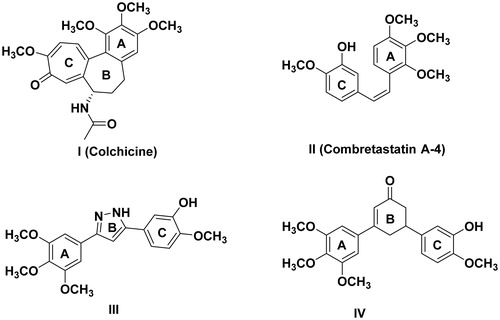
Microtubules targeting agents are known to interact with tubulin through at least three binding sites: the paclitaxel domain, vinca site and the colchicine domain. So far, tubulin binding agents can be classified into two types based on their site of action as microtubule destabilizing drugs (vinca site and the colchicine site) and microtubule-stabilizing drugs (taxane site). Out of the three binding domains, colchicine binds with high affinity to β-tubulin and forms entangled tubulin dimer, which inhibits the microtubule assemblyCitation3. Literature revealed many colchicine site inhibitors being evaluated under clinical investigation, and even more in preclinical studiesCitation4. Colchicine I is a rigid molecule whose rigidity is imparted by the B-ring which anchored rings A and C. Rings A and C are aimed to fit with hydrophobic pockets in the colchicine binding site. In addition, an H-bond acceptor (OCH3) group on ring A is a key feature of these inhibitors. Therefore, in the course of developing new more flexible colchicine analogs, many trials were made to modify the bridge between rings A and CCitation5,Citation6. More flexible derivatives involved the replacement of ring B with an olefinic bridge as in combretastatin A-4 IICitation7,Citation8, insertion of a carbonyl functionCitation9–11 or a variety of heterocyclic ringsCitation12–14 handling rings A and C, as exemplified by the pyrazole derivative IIICitation15. Alternatively, an alicyclic ring was used as demonstrated by the cyclohexenone derivative IVCitation16 ().
Figure 1. Examples of colchicine binding site inhibitors.
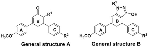
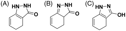
With the goal of producing new antitumor agents targeting the microtubules at the colchicine binding site, and based on the aforementioned facts, the design of the new target compounds relied on using two types of ring B models. The first involved the cyclohexenone ring (General structure A) and the other involved the indazole ring (General structure B) as linker moieties between the two hydrophobic rings A and C. Different functional groups (R1) were attached to ring B to manipulate their physicochemical properties and/or their biological activity. While retaining the H-bond acceptor methoxy group pendent on ring A, another methoxy anchor group (R2) was introduced in ring C for comparative reasons ().
Figure 2. General structures of target compounds.
The designed compounds were assessed for their antitumor activity through in vitro cytotoxicity study on selected human cancer cell lines. The mechanism of the antitumor activity was investigated through evaluating the tubulin inhibition potential of the active compounds. Finally, a molecular docking study was carried out.
Materials and methods
Chemistry
Melting points were uncorrected and were detected by open capillary tube using Electrothermal 9100 melting point apparatus (Bibby Scientific Limited, Stone, UK). Thin layer chromatography was performed using silica gel cards DC-Alufolien-Kiesel gel with fluorescent indicator UV254 using chloroform or hexane–ethyl acetate 8.5:1.5 as the eluting system and the spots were visualized using Vilber Lourmet ultraviolet lamp at λ = 254 nm. Elemental microanalyses were performed at the Regional Center for Mycology and Biotechnology, Al-Azhar University. NMR spectra were recorded at the Microanalytical unit, Faculty of pharmacy, Cairo University on Bruker Avance III spectrometer (Zurich, Switzerland) at 400 MHz for 1H and at 100 MHz for 13C. Chemical shift values (δ) were given downfield from TMS. Samples were dissolved in DMSO-d6, addition of D2O was used to confirm the exchangeable protons. Compounds 1a,b were prepared according to the previously reported procedureCitation17.
General procedure for the preparation of 2a,b
A solution of ethyl acetoacetate (1.56 ml, 12 mmol) in sodium ethoxide solution (0.3 g sodium metal in 140 ml absolute ethanol) was stirred at room temperature for 1 h. The propenone 1a,b (12 mmol) was added to the above solution with stirring. The reaction mixture was heated under reflux for 12 h and poured onto cold hydrochloric acid. The obtained solid was filtered off, washed with water, dried and crystallized from methanol.
Ethyl 6-phenyl-4-(4-methoxyphenyl)-2-oxocyclohex-3-enecarboxylate (2a)
Compound 2a was prepared from compound 1a and ethylacetoacetate. 61% yield, mp 70–73 °C. 1H NMR δ 0.96 (t, 3H, J = 7.2 Hz, CH3 ethyl), 2.98 (dd, 1H, J = 4.6 Hz, J = 17.8 Hz, H5cyclohex.ax.), 3.08 (ddd, 1H, J = 2.1 Hz, J = 11.2 Hz, J = 17.68 Hz, H5′cyclohex.eq.), 3.64 (m, 1H, H6cyclohex), 3.40 (s, 3H, OCH3), 3.93 (q, 2H, J = 7.2, CH2 ethyl), 4.13 (d, 1H, J = 3.9, H1cyclohex), 6.56 (d, 1H, J = 1.9 Hz, H3cyclohex), 7.40–7.73 (m, 9H, Ar–Hs). Anal. calcd. for C22H24O4 (350.41): C, 75.41; H, 6.33. Found: C, 75.64; H, 6.42.
Ethyl 4,6-(4-methoxyphenyl)-2-oxocyclohex-3-enecarboxylate (2b)
Compound 2b was prepared from compound 1b and ethylacetoacetate. 64% yield, mp 78–81 °C. 1H NMR δ 0.95 (t, 3H, J = 7.2 Hz, CH3 ethyl), 3.02 (m, 2H, H5cyclohex.ax., H5′cyclohex.eq.), 3.66 (m, 1H, H6cyclohex), 3.80 (s, 6H, 2OCH3), 3.94 (q, 2H, J = 7.2 Hz, CH2 ethyl), 4.11 (d, 1H, J = 3.8 Hz, H1cyclohex), 6.52 (s, 1H, H3cyclohex), 6.98–7.72 (m, 8H, Ar–Hs). Anal. Calcd. for C23H24O5 (380.43): C, 72.61; H, 6.36. Found: C, 72.88; H, 6.41.
General procedure for the preparation of 3a,b
To a solution of the ester 2a,b (10 mmol) in absolute ethanol (30 ml), 98% hydrazine hydrate (0.64 ml, 20 mmol) was added. The reaction mixture was stirred for 24 h. The precipitated solid was filtered off and recrystallized from absolute ethanol.
2-Hydroxy-4-(4-methoxyphenyl)-6-phenylcyclohexa-1,3-diene carbohydrazide (3a)
Compound 3b was prepared from compound 2b and 98% hydrazine hydrate by stirring at RT 67% yield, mp 124–127 °C. 1H NMR δ 2.69 (dd, 1H, J = 4.7, J = 17.6, H5cyclohex.ax.), 2.79 (ddd, 1H, J = 2.1 Hz, J = 11.2 Hz, J = 17.6 Hz, H5′cyclohex.eq.), 3.72 (m, 1H, H6cyclohex), 3.87 (s, 3H, OCH3), 6.30 (brs, 3H, NHNH2, D2O exchange), 6.87 (d, 1H, J = 1.8 Hz, H3cyclohex), 6.98–7.50 (m, 9H, Ar–Hs), 10.80 (brs, 1H, OH, D2O exchange). 13C NMR δ 29.8 (C6cyclohex.), 34.8 (C5cyclohex.), 55.5 (OCH3), 114.2, 114.3, 123.7, 127.6, 128.6, 131.2, 133.0, 137.1, 138.6, 142.5 (Ar–Cs), 160.4 (C=O). MS (EI): m/z (%): 336.21 (14.91). Anal. calcd. For C20H20N2O3 (336.38): C, 71.41; H, 5.99; N, 8.33. Found: C, 71.78; H, 6.07; N, 8.51.
2-Hydroxy-4,6-bis(4-methoxyphenyl) cyclohexa-1,3-diene carbohydrazide (3b)
Compound 3b was prepared from compound 2b and 98% hydrazine hydrate. 73% yield, mp 144–146 °C. 1H NMR δ 2.75 (dd, 1H, J = 4.7 Hz, J = 17.8 Hz, H5cyclohex.ax.), 2.97 (ddd, 1H, J = 2.2 Hz, J = 11.2 Hz, J = 17.7 Hz, H5′cyclohex.eq.), 3.72 (m, 1H, H6cyclohex), 3.87 (s, 6H, 2OCH3), 6.26 (brs, 3H, NHNH2, D2O exchange), 6.81 (d, 1H, J = 1.9 Hz, H3cyclohex), 6.91–7.45 (m, 9H, Ar–Hs and OH). 13C NMR δ 30.0 (C6cyclohex.), 35.1 (C5cyclohex.), 55.5, 55.6 (2OCH3), 114.2, 114.3, 123.7, 128.5, 133.1, 137.1, 137.9, 146.4 (Ar–Cs), 159.0 (C=O). MS (EI): m/z (%): 336.48 (1.10). Anal. calcd For C21H22N2O4 (336.41): C, 68.84; H, 6.05; N, 7.65. Found: C, 68.97; H, 6.13; N, 7.69.
General procedure for the preparation of 4a,b
A mixture of 2a or 2b (10 mmol) and hydrazine hydrate (0.32 ml, 10 mmol) in ethanol (20 ml) was heated under reflux for 8 h. The reaction mixture was evaporated under reduced pressure. After cooling, the reaction mixture was poured onto crushed ice and the solid thus obtained was filtered off, washed with water and crystallized from ethanol to give 4a and 4b, respectively.
4-Phenyl-6-(4-methoxyphenyl)-4,5-dihydro-1H-indazol-3-ol (4a)
Compound 4a was prepared from compound 2a and 98% hydrazine hydrate under reflux. 76% yield, mp 107–110 °C. 1H NMR δ 2.86, 2.90 (dd, 1H, J = 3.1 Hz, J = 16.7 Hz, H5indazol.eq.), 3.12 (ddd, 1H, J = 1.7 Hz, J = 8.4 Hz, J = 16.68 Hz, H5′indazol.ax.), 3.79 (s, 3H, OCH3), 4.15 (dd, 1H, J = 3.0 Hz, J = 8.2 Hz, H4indazol), 6.27 (s, 1H, NH, D2O exchange), 6.66 (d, 1H, J = 3.0 Hz, H7indazol), 7.00–7.70 (m, 9H, Ar–Hs), 10.80 (brs, 1H, OH, D2O exchange). 13C NMR δ 29.8 (C4indazol), 34.8 (C5indazol), 55.5 (OCH3), 114.4, 114.6, 126.4, 127.7, 128.5, 128.8, 129.6, 133.0 (Ar–Cs), 137.1 (C7indazol), 145.9, 146.3 (C4 of the 2 phenyl rings), 159.1 (C3indazol). MS (EI): m/z (%): 318.09 (12.02). Anal. calcd. For C20H18N2O2 (318.37): C, 75.45; H, 5.70; N, 8.80. Found: C, 75.49; H, 5.76; N, 8.94.
4,6-Bis(4-methoxyphenyl)-4,5-dihydro-1H-indazol-3-ol (4b)
Compound 4b was prepared from compound 2b and 98% hydrazine hydrate under reflux. 78% yield, mp 86–89 °C. 1H NMR δ 2.84, 2.88 (dd, 1H, J = 3.1 Hz, J = 16.8 Hz, H5indazol.eq.), 3.14 (ddd, 1H, J = 1.7 Hz, J = 8.4 Hz, J = 16.69 Hz, H5′indazol.ax.), 3.80 (s, 6H, 2OCH3), 4.16,4.18 (dd, 1H, J = 3.0 Hz, J = 8.3 Hz, H4indazol), 6.20 (s, 1H, NH, D2O exchange), 6.89 (s, 1H, H3cyclohex), 7.00–7.70 (m, 8H, Ar–Hs), 10.79 (brs, 1H, OH, D2O exchange). 13C NMR δ 33.4 (C4indazol), 35.0 (C5indazol), 55.3, 55.6 (2OCH3), 114.2, 114.4, 128.2, 128.5, 129.6, 132.7 (Ar–Cs), 137.5 (C7indazol), 157.9. 158.1 (C4 of the two phenyl rings), 160.4 (C3indazol). MS (EI): m/z (%): 348.36 (2.81). Anal. calcd. for C21H20N2O3 (348.40): C, 72.40; H, 5.79; N, 8.04. Found: C, 72.53; H, 5.84; N, 8.17.
General procedure for the preparation of 5a–f
A mixture of the corresponding hydrazide 4a,b (10 mmol) and the appropriate isothiocyanate derivative (10 mmol) in ethanol (20 ml) was heated under reflux for 3 h. The formed solid was filtered off, washed with ethanol and crystallized from ethanol.
3-Hydroxy-6-(4-methoxyphenyl)-4-phenyl-N-methyl-4,5-dihydroindazole-1-carbothioamide (5a)
Compound 5a was prepared from compound 4a and methyl isothiocyanate. 88% yield, mp 220–224 °C. 1H NMR δ 2.80, 2.84 (dd, 1H, J = 3.2 Hz, J = 16.8 Hz, H5indazol.eq), 3.00 (s, 3H, CH3), 3.11 (ddd, 1H, J = 1.8, J = 8.3 Hz, J = 16.7 Hz, H5′indazol.ax.), 3.79 (s, 3H, OCH3), 4.10, 4.14 (dd, 1H, J = 3.0 Hz, J = 8.1 Hz, H4indazol) 6.61 (d, 1H, J = 1.8 Hz, H7indazol), 7.10–8.30 (m, 9H, Ar–Hs), 10.36 (s, 1H, NH, D2O exchange), 11.05 (s, 1H, OH, D2O exchange). 13C NMR δ 31.1 (NHCH3), 34.7 (C4indazol), 36.3 (C5indazol), 55.6 (OCH3), 114.3, 114.6, 122.1, 126.9, 127.6, 128.6, 131.9, 132.1 (Ar–Cs), 137.1 (C7aindazol), 144.9 (C6indazol), 147.1, 147.7 (C4 of the 2 phenyl rings), 160.5 (C3indazol), 178.6 (C=S). MS (EI): m/z (%): 391.24 (1.80). Anal. calcd. for C22H21N3O2S (391.49): C, 67.50; H, 5.41; N, 10.73. Found: C, 67.84; H, 5.44; N, 10.49.
3-Hydroxy-4,6-bis(4-methoxyphenyl)-N-methyl-4,5-dihydroindazole-1-carbothioamide (5b)
Compound 5b was prepared from compound 4b and methyl isothiocyanate. 90% yield, mp 170–173 °C. 1H NMR δ 2.80, 2.85 (dd, 1H, J = 3.2 Hz, J = 16.8 Hz, H5indazol.eq), 2.99 (ddd, 1H, J = 1.8, J = 8.3 Hz, J = 16.7 Hz, H5′indazol.ax.), 3.08 (s, 3H, CH3), 3.79 (s, 3H, OCH3), 4.10, 4.14 (dd, 1H, J = 3.0 Hz, J = 8.1 Hz, H4indazol) 6.61 (d, 1H, J = 1.8 Hz, H7indazol), 7.10–8.30 (m, 9H, Ar–Hs), 10.30 (s, 1H, NH, D2O exchange), 11.01 (s, 1H, OH, D2O exchange). 13C NMR δ 31.2 (NHCH3), 34.9 (C4indazol), 36.0 (C5indazol), 55.4, 55.7 (2OCH3), 114.2, 114.4, 122.0, 126.9, 128.4, 131.9, 132.1 (Ar–Cs), 137.1 (C7aindazol), 144.9 (C6indazol), 158.3, 159.9 (C4 of the two phenyl rings), 160.4 (C3indazol), 178.5 (C=S). MS (EI): m/z (%): 421.48 (3.61). Anal. calcd. for C23H23N3O3S (421.51): C, 65.54; H, 5.50; N, 9.97. Found: C, 65.81; H, 5.57; N, 10.04.
3-Hydroxy-6-(4-methoxyphenyl)-4-phenyl-N-ethyl-4,5-dihydroindazole-1-carbothioamide (5c)
Compound 5c was prepared from compound 4a and ethyl isothiocyanate. 83% yield, mp 202–205 °C. 1H NMR δ 1.01 (t, 3H, J = 7.2, CH3 ethyl), 2.72, 2.74 (dd, 1H, J = 3.2 Hz, J = 16.8 Hz, H5indazol.eq), 2.83 (ddd, 1H, J = 1.8 Hz, J = 8.3 Hz, J = 16.7 Hz, H5′indazol.ax.), 3.50 (q, 2H, J = 7.2, CH2 ethyl), 3.78 (s, 3H, OCH3), 4.11,4.14 (dd, 1H, J = 3.0 Hz, J = 8.1 Hz, H4indazol), 6.60 (d, 1H, J = 1.8 Hz, H7indazol), 6.95–8.37 (m, 9H, Ar–Hs), 10.27 (s, 1H, NH, D2O exchange), 10.99 (s, 1H, OH, D2O exchange). 13C NMR δ 15.0 (CH3 ethyl), 34.6 (C4indazol), 35.8 (CH2 ethyl), 38.1 (C5indazol), 55.6 (OCH3), 114.2, 122.1, 127.0, 127.4, 128.1, 128.6, 128.9, 131.9, 132.1 (Ar–Cs), 137.2 (C7aindazol), 144.9 (C6indazol), 159.9 (C4 of the two phenyl rings), 160.5 (C3indazol), 177.6 (C=S). MS (EI): m/z (%): 405.17 (1.90). Anal. calcd. for C23H23N3O2S (405.51): C, 68.12; H, 5.72; N, 10.36. Found: C, 68.35; H, 5.81; N, 10.49.
3-Hydroxy-4,6-bis(4-methoxyphenyl)-N-ethyl-4,5-dihydroindazole-1-carbothioamide (5d)
Compound 5d was prepared from compound 4b and ethyl isothiocyanate. 86% yield, mp 210–213 °C. 1H NMR δ 1.11 (t, 3H, J = 7.2, CH3 ethyl), 2.71, 2.73 (dd, 1H, J = 3.2 Hz, J = 16.8 Hz, H5indazol.eq), 2.85 (ddd, 1H, J = 1.8 Hz, J = 8.3 Hz, J = 16.7 Hz, H5′indazol.ax.), 3.50 (q, 2H, J = 7.2, CH2 ethyl), 3.78 (s, 6H, 2OCH3), 4.12, 4.15 (dd, 1H, J = 3.0 Hz, J = 8.1 Hz, H4indazol), 6.60 (d, 1H, J = 1.8 Hz, H7indazol), 6.90–7.70 (m, 8H, Ar–Hs), 10.28 (s, 1H, NH, D2O exchange), 10.99 (s, 1H, OH, D2O exchange). 13C NMR δ 15.7 (NHCH2CH3), 31.7 (NHCH2CH3), 34.7 (C4indazol), 36.1 (C5indazol), 55.4, 55.6 (2OCH3), 114.2, 114.3, 114.5, 122.1, 126.8, 127.6, 128.6, 131.9, 132.1, 133.0 (Ar–Cs), 137.1 (C7aindazol), 145.0 (C6indazol), 157.8, 158.3 (C4 of the 2 phenyl rings), 160.5 (C3indazol), 177.6 (C=S). MS (EI): m/z (%): 435.20 (1.27). Anal. calcd. for C24H25N3O3S (435.54): C, 66.18; H, 5.79; N, 9.65. Found: C, 66.35; H, 5.82; N, 9.78.
3-Hydroxy-6-(4-methoxyphenyl)-4-phenyl-N-phenyl-4,5-dihydroindazole-1-carbothioamide (5e)
Compound 5e was prepared from compound 4a and phenyl isothiocyanate. 76% yield, mp 138–140 °C. 1H NMR δ 2.75, 2.78 (dd, 1H, J = 3.2 Hz, J = 16.8 Hz, H5indazol.eq), 2.87 (ddd, 1H, J = 1.8 Hz, J = 8.3 Hz, J = 16.7 Hz, H5′indazol.ax.), 3.80 (s, 3H, OCH3), 4.11, 4.15 (dd, 1H, J = 3.0 Hz, J = 8.1 Hz, H4indazol), 6.70 (d, 1H, J = 1.8 Hz, H7indazol), 7.00–7.80 (m, 14H, Ar–Hs), 10.06 (s, 1H, NH, D2O exchange), 10.77 (s, 1H, OH, D2O exchange). MS (EI): m/z (%): 453.11 (0.81). Anal. calcd. for C27H23N3O2S (453.56): C, 71.40; H, 5.11; N, 9.26. Found: C, 71.63; H, 5.14; N, 9.38.
3-Hydroxy-4,6-bis(4-methoxyphenyl)-N-phenyl-4,5-dihydroindazole-1-carbothioamide (5f)
Compound 5f was prepared from compound 4b and phenyl isothiocyanate. 78% yield, mp 155–159 °C. 1H NMR δ 2.78, 2.81 (dd, 1H, J = 3.2 Hz, J = 16.8 Hz, H5indazol.eq), 2.88 (ddd, 1H, J = 1.8 Hz, J = 8.3 Hz, J = 16.7 Hz, H5′indazol.ax.), 3.80 (s, 6H, 2OCH3), 4.11, 4.14 (dd, 1H, J = 3.0 Hz, J = 8.1 Hz, H4indazol), 6.70 (d, 1H, J = 1.8 Hz, H7indazol), 7.10–7.80 (m, 13H, Ar–Hs), 10.06 (s, 1H, NH, D2O exchange), 10.76 (s, 1H, OH, D2O exchange). MS (EI): m/z (%): 483.20 (2.22). Anal. calcd. for C28H25N3O3S (483.58): C, 69.54; H, 5.21; N, 8.69. Found: C, 69.78; H, 5.29; N, 8.94.
General procedure for the preparation of 6a,b
To a solution of the corresponding hydrazide 3a,b (10 mmol) and potassium hydroxide (0.56 g, 10 mmol) in absolute ethanol (5 ml), carbon disulfide (0.95 ml, 15 mmol) was added. The reaction mixture was heated under reflux for 5 h till the release of hydrogen sulfide gas ceased. After dilution with water, the reaction mixture was filtered. The filtrate was acidified with 1 N hydrochloric acid. The precipitated solid was filtered off, washed with water and crystallized from ethanol.
5-[2-Hydroxy-4-(4-methoxyphenyl)-6-phenyl-cyclohexa-1,3-dienyl]-1,3,4 oxadiazole-2–(3H)-thione (6a)
Compound 6a was prepared from compound 3a and carbon disulfide. 86% yield, mp 282–284 °C. 1H NMR δ 2.73, 2.75 (dd, 1H, J = 4.7, J = 17.6, H5cyclohex.ax.), 2.82 (ddd, 1H, J = 2.1 Hz, J = 11.2 Hz, J = 17.6 Hz, H5′cyclohex.eq.), 3.77 (m, 1H, H6cyclohex), 3.84 (s, 3H, OCH3), 6.95 (d, 1H, J = 1.8 Hz, H3cyclohex), 7.00–7.90 (m, 9H, Ar–Hs), 13.09 (s, 1H, NH, D2O exchange), 14.05 (s, 1H, OH, D2O exchange). 13C NMR δ 30.8 (C6cyclohex.), 39.4 (C5cyclohex.), 55.8 (OCH3), 114.3, 114.4, 114.9, 123.9, 127.1, 127.4, 128.1, 128.6, 129.3, 134.5, 136.0 (Ar–Cs), 159.6 (C=O), 161.1 (C2cyclohex.), 180.7 (C=S). MS (EI): m/z (%): 378.19 (2.61). Anal. calcd. for C21H18N2O3S (378.44): C, 66.65; H, 4.79; N, 7.40. Found: C, 66.82; H, 4.846; N, 7.53.
5-[2-Hydroxy-4,6-bis(4-methoxyphenyl) cyclohexa-1,3-dienyl]-1,3,4 oxadiazole-2-(3H)-thione (6b)
Compound 6b was prepared from compound 3b and carbon disulfide. 89% yield, mp 278–281 °C. 1H NMR δ 2.73, 2.75 (dd, 1H, J = 4.7, J = 17.6, H5cyclohex.ax.), 2.82 (ddd, 1H, J = 2.1 Hz, J = 11.3 Hz, J = 17.8 Hz, H5′cyclohex.eq.), 3.74 (m, 1H, H6cyclohex), 3.79 (s, 6H, 2OCH3), 6.97 (d, 1H, J = 1.8 Hz, H3cyclohex), 7.00–7.60 (m, 8H, Ar–Hs), 10.09 (s, 1H, NH, D2O exchange), 11.47 (s, 1H, OH, D2O exchange). 13C NMR δ 33.7 (C6cyclohex.), 40.6 (C5cyclohex.), 55.5, 55.7 (2OCH3), 113.9, 114.2, 114.7, 127.1, 127.4, 128.3, 128.5, 129.1, 134.5, 135.7 (Ar–Cs), 159.0 (C=O), 163.9 (C2cyclohex.), 198.7 (C=S). MS (EI): m/z (%): 408.24 (0.29). Anal. calcd. for C22H20N2O4S (408.47): C, 64.69; H, 4.94; N, 6.86. Found: C, 64.82; H, 4.98; N, 6.95.
General procedure for the preparation of 7a,b
A mixture of compound 3a,b (10 mmol) and acetylacetone (1 ml, 10 mmol) in a mixture of ethanol–acetic acid (100:10 v/v) was heated under reflux for 10 h. The reaction mixture was cooled and the precipitated solid was filtered off, washed with water, dried and crystallized from ethanol.
(3,5-Dimethyl-1H-pyrazol-1-yl) [2-hydroxy-4-(4-methoxyphenyl)-6-phenylcyclohexa-1,3-dienyl] methanone (7a)
Compound 7a was prepared from compound 3a and acetylacetone. 45% yield, mp 277–279 °C. 1H NMR δ 2.40 (s, 3H, CH3 at C3pyrazole), 2.42 (s, 3H, CH3 at C5pyrazole), δ 2.73, 2.77 (dd, 1H, J = 4.7, J = 17.6, H5cyclohex.ax.), 2.98 (ddd, 1H, J = 2.3 Hz, J = 11.3 Hz, J = 17.8 Hz, H5′cyclohex.eq.), 3.75 (m, 1H, H6cyclohex), 3.83 (s, 3H, OCH3), 6.43 (s, 1H, C4pyrazole), 6.71 (d, 1H, J = 1.8 Hz, H3cyclohex), 7.10–8.30 (m, 9H, Ar–Hs), 10.82 (s, 1H, OH, D2O exchange). 13C NMR δ 12.8, 13.7 (2 CH3), 35.8 (C6cyclohex.), 44.3 (C5cyclohex.), 55.5, 55.7 (2OCH3), 114.3, 114.8, 122.8, 128.4, 131.0, 132.1, 136.3, 139.9, 141.8, 148.4 (Ar–Cs), 159.7 (C=O), 161.3 (C2cyclohex.). MS (EI): m/z (%): 400.23 (1.00). Anal. calcd. for C25H24N2O3 (400.47): C, 74.98; H, 6.04; N, 7.00. Found: C, 75.12; H, 6.13; N, 7.24.
(3,5-Dimethyl-1H-pyrazol-1-yl) [2-hydroxy-4,6-bis(4-methoxy phenyl)cyclohexa-1,3-dienyl] methanone (7b)
Compound 7b was prepared from compound 3b and acetylacetone. 48% yield, mp 268–270 °C. 1H NMR δ 2.39 (s, 3H, CH3 at C3pyrazole), 2.45 (s, 3H, CH3 at C5pyrazole), δ 2.72, 2.75 (dd, 1H, J = 4.8, J = 17.7, H5cyclohex.ax.), 2.93 (ddd, 1H, J = 2.3 Hz, J = 11.3 Hz, J = 17.8 Hz, H5′cyclohex.eq.), 3.73 (m, 1H, H6cyclohex), 3.79 (s, 6H, 2OCH3), 6.41 (s, 1H, C4pyrazole), 6.89 (d, 1H, J = 1.6 Hz, H3cyclohex), 7.00–7.80 (m, 8H, Ar–Hs), 10.55 (s, 1H, OH, D2O exchange). 13C NMR δ 12.8,13.7 (2 CH3), 35.8 (C6cyclohex.), 44.3 (C5cyclohex.), 55.5, 55.7 (2OCH3), 114.3, 114.8, 122.8, 128.4, 131.0, 132.1, 136.3, 139.9, 141.8, 148.4 (Ar–Cs), 159.7 (C=O), 161.3 (C2cyclohex.). MS (EI): m/z (%): 430.14 (1.08). Anal. calcd. for C26H26N2O4 (430.50): C, 72.54; H, 6.09; N, 6.51. Found: C, 72.69; H, 6.08; N, 6.57.
General procedure for the preparation of 8a,b
A mixture of compound 3a,b (1 mmol), ethyl acetoacetate (0.13 ml, 1 mmol) and anhydrous potassium carbonate (0.21 g, 1.5 mmol) in ethanol (15 ml) was heated under reflux for 10 h. The reaction mixture was poured on water and the precipitated solid was filtrated off, washed with water, dried and crystallized from ethanol.
1-[2-Hydroxy-4-(4-methoxyphenyl)-6-phenyl cyclohexa-1,3-dienecarbonyl]-3-methyl-1H-pyrazole-5(4H)-one (8a)
Compound 8a was prepared from compound 3a and ethyl acetoacetate. 51% yield, mp 249–252 °C. 1H NMR δ 1.66 (s, 3H, CH3 at C3pyrazole), 1.84 (s, 2H, CH2pyrazolone), 2.81, 2.88 (dd, 1H, J = 4.6 Hz, J = 17.6 Hz, H5cyclohex.ax.), 2.92 (ddd, 1H, J = 2.1 Hz, J = 11.2 Hz, J = 17.6 Hz, H5′cyclohex.eq.), 3.72 (m, 1H, H6cyclohex), 3.81 (s, 3H, OCH3), 6.90 (s, 1H, H3cyclohex), 7.00–7.90 (m, 9H, Ar–Hs), 10.30 (s, 1H, OH, D2O exchange). 13C NMR δ 25.1 (CH3), 35.2 (C6cyclohex.), 39.3 (C5cyclohex.), 40.5 (CH2pyrazoline), 55.6 (OCH3), 69.7 (C3pyrazoline), 114.1, 114.7, 126.9, 127.4, 128.5, 128.9, 129.0, 132.1, 136.3, 139.9, 141.8, 144.5, 148.4 (Ar–Cs), 158.6 (C=O), 175.7 (C2cyclohex.). MS (EI): m/z (%): 402.17 (10.39). Anal. calcd. for C24H22N2O4 (402.44): C, 71.63; H, 5.51; N, 6.96. Found: C, 71.81; H, 5.58; N, 7.11.
1-[2-Hydroxy-4,6-bis(4-methoxyphenyl)cyclohexa-1,3-dienecarbonyl]-3-methyl-1H-pyrazole-5(4H)-one (8b)
Compound 8b was prepared from compound 3b and ethyl acetoacetate. 53% yield, mp 261–263 °C. 1H NMR δ 1.68 (s, 3H, CH3 at C3 pyrazole), 1.85 (s, 2H, CH2pyrazolone), 2.75, 2.79 (dd, 1H, J = 4.7 Hz, J = 17.7 Hz, H5cyclohex.ax.), 2.94 (ddd, 1H, J = 2.2 Hz, J = 11.3 Hz, J = 17.7 Hz, H5′cyclohex.eq.), 3.72 (m, 1H, H6cyclohex), 3.81 (s, 6H, 2OCH3), 6.82 (s, 1H, H3cyclohex), 7.00–7.69 (m, 8H, Ar–Hs), 10.50 (s, 1H, OH, D2O exchange). MS (EI): m/z (%): 432.19 (5.24). Anal. calcd. for C25H24N2O5 (432.47): C, 69.43; H, 5.59; N, 6.48. Found: C, 69.54; H, 5.63; N, 6.61.
General procedure for the preparation of 9a–f
A mixture of the corresponding hydrazide 3a,b (10 mmol) and the appropriate isothiocyanate derivative (10 mmol) in ethanol (20 ml) was heated under reflux for 5 h. The formed precipitate was filtered off, washed with ethanol and crystallized from ethanol.
1-[2-Hydroxy-4-(4-methoxyphenyl)-6-phenylcyclohexa-1,3-dienecarbonyl]-4-N- methyl thiosemicarbazide (9a)
Compound 9a was prepared from compound 3a and methyl isothiocyanate. 83% yield, mp 136–139 °C. 1H NMR δ 2.86 (dd, 1H, J = 4.7, J = 17.6, H5cyclohex.ax.), 3.10 (s, 3H, CH3), 3.18 (ddd, 1H, J = 2.1 Hz, J = 11.2 Hz, J = 17.6 Hz, H5′cyclohex.eq.), 3.60 (m, 1H, H6cyclohex), 3.79 (s, 3H, OCH3), 6.80 (d, 1H, J = 1.8 Hz, H3cyclohex), 7.10–8.3 (m, 9H, Ar–Hs), 10.80 (brs, 4H, 3NHs + OH, D2O exchange). 13C NMR δ 31.0 (CH3), 34.9 (C6cyclohex.), 38.3 (C5cyclohex.), 55.7 (OCH3), 114.3, 114.6, 127.5, 127.6, 128.6, 128.7, 131.2, 138.7 (Ar–Cs), 145.6 (C4cyclohex.), 158.9 (C4methoxyphenyl), 159.9 (C=O), 160.4 (C2cyclohex.), 175.2 (C=S). MS (EI): m/z (%): 409.32 (1.28). Anal. calcd. for C22H23N3O3S (409.50): C, 64.53; H, 5.66; N, 10.26. Found: C, 64.70; H, 5.72; N, 10.47.
1-[2-Hydroxy-4,6-bis(4-methoxyphenyl)-6-phenyl cyclohexa-1,3-dienecarbonyl]-4-N-methyl thiosemicarbazide (9b)
Compound 9b was prepared from compound 3b and methyl isothiocyanate. 80% yield, mp 147–150 °C. 1H NMR δ 2.69 (dd, 1H, J = 4.6, J = 17.5, H5cyclohex.ax.), 2.80 (ddd, 1H, J = 2.2 Hz, J = 11.3 Hz, J = 17.8 Hz, H5′cyclohex.eq.), 3.10 (s, 3H, CH3), 3.30 (m, 1H, H6cyclohex), 3.78 (s, 6H, 2OCH3), 6.60 (d, 1H, J = 1.7 Hz, H3cyclohex), 6.90–8.30 (m, 8H, Ar–Hs), 10.30 (s, 3H, 3NHs, D2O exchange), 11.01 (s, 1H, OH, D2O exchange). 13C NMR δ 31.1 (CH3), 34.9 (C6cyclohex.), 36.1 (C5cyclohex.), 55.5, 55.7 (2OCH3), 112.9, 114.2, 114.6, 122.1, 126.9, 128.2, 128.4, 128.7, 132.2, 137.1 (Ar–Cs), 144.9 (C4cyclohex.), 158.3 (C4methoxyphenyl), 159.9 (C=O), 160.5 (C2cyclohex.), 178.7 (C=S). MS (EI): m/z (%): 439.25 (3.11). Anal. calcd. for C23H25N3O4S (439.50): C, 62.85; H, 5.73; N, 9.56. Found: C, 63.04; H, 5.76; N, 9.68.
1-[2-Hydroxy-4-(4-methoxyphenyl)-6-phenylcyclohexa-1,3-dienecarbonyl]-4-N-ethyl thiosemicarbazide (9c)
Compound 9c was prepared from compound 3a and ethyl isothiocyanate. 86% yield, mp 140–143 °C. 1H NMR δ 0.91 (t, 3H, J = 7.2 Hz, CH3 ethyl), 2.62 (dd, 1H, J = 4.7 Hz, J = 17.7 Hz, H5cyclohex.ax.), 3.13 (ddd, 1H, J = 2.1 Hz, J = 11.2 Hz, J = 17.7 Hz, H5′cyclohex.eq.), 3.40 (m, 1H, H6cyclohex), 3.50 (q, 2H, J = 7.3 Hz, CH2 ethyl), 3.76 (s, 3H, OCH3), 6.60 (d, 1H, J = 1.8 Hz, H3cyclohex), 6.90–7.40 (m, 9H, Ar–Hs), 9.80 (s, 3H, 3NHs, D2O exchange), 11.96 (s, 1H, OH, D2O exchange). Anal. calcd. for C23H25N3O3S (423.53): C, 65.23; H, 5.95; N, 9.92. Found: C, 65.41; H, 6.02; N, 9.98.
1-[2-Hydroxy-4,6-bis(4-methoxyphenyl) cyclohexa-1,3-dienecarbonyl]-4-N-ethyl thiosemicarbazide (9d)
Compound 9d was prepared from compound 3b and ethyl isothiocyanate. 87% yield, mp 155–157 °C. 1H NMR δ 1.10 (t, 3H, J = 7.22, CH3 ethyl), 2.69 (dd, 1H, J = 4.7, J = 17.6, H5cyclohex.ax.), 2.83 (ddd, 1H, J = 2.1 Hz, J = 11.4 Hz, J = 17.7 Hz, H5′cyclohex.eq.), 3.40 (m, 1H, H6cyclohex), 3.60 (q, 2H, J = 7.23, CH2 ethyl), 3.79 (s, 6H, 2OCH3), 6.60 (d, 1H, J = 1.8 Hz, H3cyclohex), 6.90–7.50 (m, 8H, Ar–Hs), 10.20 (s, 3H, 3NHs, D2O exchange), 11.00 (s, 1H, OH, D2O exchange). 13C-NMR δ 15.0 (CH3 ethyl), 34.9 (C6cyclohex.), 36.0 (CH2ethyl), 38.3 (C5cyclohex.), 55.4, 56.5 (2OCH3), 112.8, 114.2,
114.5, 122.0, 127.0, 128.3, 128.5, 131.9, 132.1 (Ar–Cs), 144.9 (C4cyclohex.), 158.3 (C4methoxyphenyl), 159.9 (C=O), 160.4 (C2cyclohex.), 177.4 (C=S). MS (EI): m/z (%): 453.37 (0.45). Anal. calcd. for C24H27N3O4S (453.55): C, 63.56; H, 6.00; N, 9.26. Found: C, 63.62; H, 6.09; N, 9.43.
1-[2-Hydroxy-4-(4-methoxyphenyl)-6-phenyl cyclohexa-1,3-dienecarbonyl]-4-N-phenyl thiosemicarbazide (9e)
Compound 9d was prepared from compound 3a and phenyl isothiocyanate. 67% yield, mp 188–190 °C. 1H NMR δ 2.94 (dd, 1H, J = 4.8, J = 17.6, H5cyclohex.ax.), 3.16 (ddd, 1H, J = 2.3 Hz, J = 11.2 Hz, J = 17.8 Hz, H5′cyclohex.eq.), 3.74 (m, 1H, H6cyclohex), 3.80 (s, 3H, OCH3), 6.80 (d, 1H, J = 1.8 Hz, H3cyclohex), 7.00–7.80 (m, 14H, Ar–Hs), 10.98 (s, 3H, 3NHs, D2O exchange), 11.00 (s, 1H, OH, D2O exchange). 13C NMR δ 34.7 (C6cyclohex.), 39.3 (C5cyclohex.), 55.7 (OCH3), 114.2, 114.6, 117.3, 121.5, 123.3, 124.9, 125.1, 127.5, 127.6, 128.4, 128.8, 129.4, 131.9, 138.9 (Ar–Cs), 145.4 (C4cyclohex.), 156.2 (C4methoxyphenyl), 158.7 (C=O), 160.1 (C2cyclohex.), 180.6 (C=S). MS (EI): m/z (%): 471.18 (0.71). Anal. calcd. for C27H25N3O3S (471.57): C, 68.77; H, 5.34; N, 8.91. Found: C, 68.94; H, 5.38; N, 9.02.
1-[2-Hydroxy-4,6-bis(4-methoxyphenyl) cyclohexa-1,3-dienecarbonyl]-4-N-phenyl thiosemicarbazide (9f)
Compound 9d was prepared from compound 3b and phenyl isothiocyanate. 61% yield, mp 196–198 °C. 1H NMR δ 2.68 (dd, 1H, J = 4.7, J = 17.6, H5cyclohex.ax.), 2.73 (ddd, 1H, J = 2.2 Hz, J = 11.1 Hz, J = 17.6 Hz, H5′cyclohex.eq.), 3.71 (m, 1H, H6cyclohex), 3.84 (s, 6H, 2OCH3), 6.70 (d, 1H, J = 1.8 Hz, H3cyclohex), 6.90–7.60 (m, 13H, Ar–Hs), 9.80 (s, 3H, 3NHs, D2O exchange), 10.80 (s, 1H, OH, D2O exchange). 13C NMR δ 35.7 (C6cyclohex.), 38.6 (C5cyclohex.), 55.5, 55.8 (2OCH3), 114.2, 114.5, 117.9, 122.0, 125.0, 125.1, 126.8, 127.8, 128.5, 129.1, 129.6, 132.0, 136.8 (Ar–Cs), 142.7 (C4cyclohex.), 156.2 (C4methoxyphenyl), 159.2 (C=O), 161.7 (C2cyclohex.), 181.7 (C=S). MS (EI): m/z (%): 501.67 (1.39). Anal. calcd. for C28H27N3O4S (501.60): C, 67.05; H, 5.43; N, 8.38. Found: C, 67.13; H, 5.48; N, 8.49.
In vitro antitumor evaluation by MTT assay
Antiproliferative activity of the target compounds was determined in cells treated with the different concentrations of the tested compounds in comparison with untreated control using MTT assay as following:
Cells were grown as monolayer in media supplemented with 10% inactivated fetal bovine serum.
The monolayers of 10 000 cells were plated (104 cells/well) in a 96-well tissue culture plate and incubated for 24 h at 37 °C in a humidified incubator with 5% CO2 before treatment with the compounds to allow attachment of cell to the plate except blank wells without cells.
Different concentrations of 100, 10, 1.0, 0.1 and 0.01 μM of each tested compound and positive control drug were tested for cytotoxicity. Tetraplicate wells were prepared for each concentration in addition to cell control (cell only without compounds).
Cells were incubated with the tested compounds for 48 h into CO2 incubator at 37 °C and 5% CO2.
Culture media containing different concentration of tested compounds and dead cells were decanted leaving only viable attached cells into the tissue culture plate.
The plate was washed twice with pre-warmed phosphate buffered saline (PBS).
MTT reagent (40 μl) was added to each well including blank and negative control wells.
After addition of MTT reagent the plates were incubated in dark for 4 h for the reduction of MTT into formazan (purple needle color) by dehydrogenase activity in mitochondria of viable cells.
DMSO (150 μl) was added to each well to solubilize the purple crystals of formazan.
Absorbance was measured at 570 nm with microplate reader (ROBONIK TM P2000 Eliza plate reader; Robonik India Pvt. Ltd, Maharashtra, India).
The percentage of cell survival was calculated by the following equation:
where As is the absorbance of sample, Ab is the absorbance of blank and Ac is the absorbance of control.
The inhibitory concentration 50 (IC50) was calculated from the equation of the plot between molar concentration of the tested compounds against survival rate percent.
Tubulin polymerization assay
Standard curve construction
Seven different dilution of standard such as 2000, 1000, 500, 250, 125, 62.5, 31.2 pg/mL, and the last tubes with the blank 0 pg/mL concentration were prepared, while test drugs were taken at their IC50 concentration. The duplicate readings for each standard, control and samples were averaged and subtracted from the average zero standard optical density. A standard curve was constructed by plotting the mean OD and concentration for each standard. A best fit curve was drawn through the points on the graph, with concentration on the y-axis and absorbance on the x-axis. In order to make the calculation easier, the OD values of the standard (x-axis) were plotted against the known concentrations of the standard (y-axis), although concentration is the independent variable and OD value is the dependent variable.
Sample preparation
The cell lysates obtained after incubation of MCF-7 and HCT-116 cells with the tested compounds at their IC50 concentration were prepared according to the following:
Adherent cells should be detached with trypsin and then collected by centrifugation (suspension cells can be collected by centrifugation directly).
Cells were washed three times in cold PBS.
Cells were resuspended in PBS (1×) and the cells was subjected to ultrasonication for four times (or freeze cells at ≤ −20 °C. Thaw cells with gentle mixing. Repeat the freeze/thaw cycle for three times.)
Centrifugation was done at 1500g for 10 min at 2–8 °C to remove cellular debris.
Calculation of results
From the curve OD of each sample is converted to tubulin concentration, then percentage inhibition of tubulin polymerization can be calculated by the following equation:
Cell cycle analysis by fluorescence-activated cell sorting analysis
Fluorescence-activated cell sorting analysis following cell staining with propidium iodide (PI) was used according to the following protocol:
Approximately 106 cells (HCT-116 or MCF-7) were suspended in 0.5 ml of PBS. The suspension was gently vortexed (5 s) or gently aspirated several times with a Pasteur pipette to obtain a mono-dispersed cell suspension, with minimal cell aggregation.
Cells were fixed by transferring this suspension, with a Pasteur pipette, into centrifuge tubes containing 4.5 ml of 70% ethanol, on ice. Cells were kept in ethanol for at least 2 h at 4 °C. Cells may be stored in 70% ethanol at 4 °C for weeks.
The ethanol-suspended cells were centrifuged for 5 min at 300g. Ethanol was decanted thoroughly.
The cell pellet was suspended in 5 ml of PBS, and after about 30 s it was centrifuged at 300g for 5 min.
The cell pellet was suspended in 1 ml of PI staining solution and kept in the dark at room temperature for 30 min, or at 37 °C for 10 min.
The sample was transferred to the flow cytometer, Becton Dickinson Immunocytometry Systems and cell fluorescence was measured. Maximum excitation of PI bound to DNA is at 536 nm, and emission is at 617 nm.
Phoenix Flow Systems software (Phoenix Flow systems, Inc., San Diego, CA) was used to deconvolute the DNA content frequency histograms and to estimate the proportions of cells in the respective phases of the cycle.
The cell cycle progression was analyzed at a 10 μM concentration for 72 h.
Molecular docking procedure
X-ray crystal structure of tubulin in complex with DAMA-colchicine and the stathmin-like domain (SLD) at 3.5 Å resolution (PDB: 1SA0) was downloaded from protein data bankCitation7. All molecular modeling calculations and docking studies were carried out using Discovery Studio software v4.0.0.13259Citation18 running on a Windows7 PC.
Binding site sphere determination
The protein–ligand complex obtained from the protein data bank was prepared for docking as follows: Deletion of chains A, B and E of the protein together with co-crystallized water molecules was performed. Automatic protein preparation module was used applying CHARMm forcefield. The binding site sphere has been defined automatically by the software.
Preparation of target compounds for docking
The docked compounds were prepared for docking by applying the following protocol: 2D structures of the docked ligands were built using Marvin Sketch and copied to Discovery Studio 4. Ligands were prepared using “Prepare Ligands” protocol in Discovery Studio where hydrogen atoms were added at their standard geometry, optical isomers and 3D conformations were automatically generated.
Running docking
Docking was performed using CDOCKER protocol in Discovery Studio keeping the parameters at default. The best scoring pose of the docked compounds was recognized. Receptor–ligand interactions of the complexes were examined in 2D and 3D styles.
Results and discussion
Chemistry
The designed compounds were synthesized adopting the chemical pathways outlined in Schemes 1 and 2.
Scheme 1. Synthesis of compounds 3a,b, 4a,b and 5a–f.
Scheme 2. Synthesis of compounds 6a,b, 7a,b, 8a,b and 9a–f.
In the present work, the synthesis of the propenones 1a,b was achieved by reacting benzaldehyde or 4-methoxybenzaldheyde with 4-methoxy acetophenone in ethanol using aqueous NaOH as a catalystCitation17. The cyclohexenone intermediates 2a,b were prepared via Michael addition through a cyclo-condensation reaction between the propenones 1a,b and the β-keto ester, ethyl acetoacetate using sodium ethoxide as a catalyst. As the explored reaction was not stereo selective, two chiral centers (C1 and C6) in the structure of the cyclohexenones 2a,b were generated, which would result in a mixture of diastereomers. No attempt to separate the diastereomeric cyclohexenones was undertaken, and the cyclocondensation products were characterized in the form of the mixture originated from the synthesis. The characteristic triplet-quartet pattern confirmed the presence of the ethyl group of the ester. The characteristic signal in the 1H NMR spectrum of 2a was, however, the two protons at C-5, being magnetically nonequivalent, appeared as two different signals, the axial proton appeared as double of doublet of doublets at around δ 2.98 ppm showing geminal coupling, vicinal coupling with H-6 and long range coupling with vinyl proton H-3. The equatorial proton at C-4 appeared as doublet of doublets at around δ 3.08 ppm showing both germinal and vicinal coupling. The vinyl proton, H-3, appeared as doublet at δ 6.56 ppm. Proton at C-6 appeared as multiplet due to vicinal coupling to H-5 and H-1. H-1 proton appeared as doublet being coupled to H-6. Reaction of the cyclohexenones 2a,b with 98% hydrazine hydrate in ethanol at room temperature afforded derivatives 3a,b, respectively. Proceeding the reaction under reflux condition resulted in cyclization with formation of indazole derivatives 4a,b. The appearance of the OH stretching band confirmed the presence of the enol tautomer, which resulted in the loss of one of the two chiral centers. According to a previous report on the tautomeric forms of indazole, the obtained compounds 4a,b could be present in three tautomeric forms A, B and C (). The absence of carbonyl bands in the IR spectra of the products ruled out lactam structures A and BCitation19. The 1H NMR spectra of the indazole derivatives exhibited three protons in the sp3 shift range (H-5eq, H-5ax and H-4). H-5eq and H-4 appeared as doublet of doublets at around δ 2.90 and 4.18 ppm, respectively. While the signal for H-5ax appeared as doublet of doublet of doublets at δ 3.12 ppm showing geminal coupling with H-5eq, vicinal coupling with H-4 and long range proton coupling with H-7. The vinylic proton at H-7 appeared as doublet at δ 6.89 ppm with J= 1.7 Hz.
Figure 3. Tautomeric forms of 1H-indazol-3-ol.
Target compounds 5a–f were prepared by reacting compounds 4a,b with the appropriate substituted alkyl/aryl isothiocyanate in absolute ethanol under reflux condition.
Substituted 1,3,4-oxadiazole-2(3H)-thione derivatives 6a,b were synthesized by reacting the hydrazide derivatives 3a,b with carbon disulfide in absolute ethanol in the presence of potassium hydroxide. Pyrazole derivatives 7a,b were synthesized via cyclo-condensation of acetyl acetone with the hydrazide derivatives 3a,b in a mixture of ethanol and glacial acetic acid. The yield was found to be solvent-dependent as cyclocondensation in a 10:1 (v/v) mixture of ethanol–acetic acid afforded the corresponding pyrazole derivatives in high yield, while the yield decreased upon using a mixture of ethanol and triethyl amine. Using ethyl acetoacetate as a β-diketone, cyclocondensation reaction with the appropriate hydrazides 3a,b afforded the 3-methyl-1H-pyrazole-5-(4H)-ones 8a,b, respectively. Finally, reaction of isothiocyanates with hydrazides 3a,b in absolute ethanol under reflux furnished the corresponding thiosemicarbazides 9a–f.
The structures of all the synthesized compounds were confirmed using the EI MS, FT-IR, 1H and 13C NMR spectral analyses.
Biological screening
In vitro antitumor evaluation by MTT assay
The antiproliferative activity of the target compounds against colon cancer HCT-116 and breast cancer MCF-7 cell lines was measured at Vacsera, Egypt. The MTT method of assay was adopted and the IC50 values are listed in .
Table 1. Antiproliferative activity against HCT-116 cell line and MCF-7.
An overview of the results of MTT assay revealed that few compounds exhibited IC50 values lower than or slightly higher than colchicine. Concerning the antitumor activity against colon HCT-116 tumor cell line, the obtained results showed that compounds 4b, 5e and 5f exhibited higher potency than colchicine. Furthermore, compound 5f revealed comparable activity to doxorubicin, while compounds 3b, 8b and 9d exerted moderate activity. Regarding antitumor activity against MCF-7 breast tumor cell line, it can be revealed that compound 5e demonstrated higher potency than colchicine. Meanwhile, compounds 4b and 5f were less active than colchicine. The cyclohexenols 3a,b displayed IC50 of 55.35–63.39 μM, respectively. Regarding the effect of substitution on the phenyl ring (R1) the 4-methoxy derivative 3b showed higher activity than the unsubstituted derivative 3a. Interestingly, structure rigidification of 3a,b into the indazole derivatives 4a,b resulted in increase in the antitumor activity especially for the 4-methoxyphenyl derivative 4b (IC50= 6.78 and 11.40 μM against HCT-116 and MCF-7 cells, respectively). Structure extension of the indazole derivatives 4a,b with N-substituted carbothioamide moiety was successful only with the N-phenyl derivatives 5e,f (IC50 ranging from 5.50 to 11.55 μM). The N-methyl (5a,b) and N-ethyl derivatives (5c,d) were inactive. Unfortunately, introduction of heterocylic rings at position 1 of the cyclohexanol nucleus as in compounds 6a,b, 7a,b, 8a,b did not reveal any advantage toward the activity of the compounds, compared to their precursor less bulky hydrazides 3a,b. In conclusion, it could be revealed that the bicylic indazole derivatives 4a,b and 5e,f were the most potent of all derivatives. The hydrazides 3a,b and the azacyclic related derivatives 6a,b, 7a,b, 8a,b showed only moderate activity. The thiosemicarbazides 9a–f and N-methyl 5a,b and N-ethyl 5c,d indazole-1-carbothioamide derivatives were the least potent.
Tubulin polymerization inhibition assay
Further investigation to assess the mechanism of action of the most active compounds in the MTT assay as potential tubulin polymerization inhibitors was carried out using tubulin polymerization assay. The percentage inhibition of tubulin polymerization following sandwich enzyme immunoassay by ELISA method using Enzyme-linked Immunosorbent Assay Kit was performed. Results are summarized in . Percentage inhibition of tubulin polymerization was performed on the compounds with the highest activity profile in the MTT assay, namely, 4b, 5e and 5f. The tested compounds showed percentage inhibition of tubulin in both cell line homogenates ranging from 79.72% to 89.31%. Compound 5e was the most active on HCT-116 and 5f was the most active on MCF-7 cells. It is noteworthy that activities of the tested compounds were comparable to that of colchicine or even higher especially on HCT-116 cells homogenate.
Table 2. Percentage inhibition of tubulin polymerization.
Cell cycle analysis
It was hypothesized that the mechanism of action of compounds 4b, 5e and 5f involved arresting the process of mitosis. Accordingly, cell cycle analysis was performed on HCT-116 and MCF-7 cells after treatment with these compounds. Upon exposure of the cells to the tested compounds, the percentages of cells in the G0/G1 phase of the cell cycle in both cell lines, were markedly decreased, especially with compound 5e, while the percentages in the G2/M phase of the cell cycle increased. Compound 5e had the highest effect on G2/M phase in both cell lines (, and ). Compared with the untreated control, tested compounds disturbed the cell cycle strongly at G2/M phase, which was in agreement with the proposed mechanism of action.
Figure 4. Cell cycle analysis histograms for HCT-116 cells. (A) Control, (B) 4b, (C) 5e and (D) 5f.
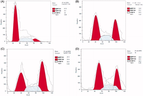
Figure 5. Cell cycle analysis histograms for MCF-7 cells. (A) Control, (B) 4b, (C) 5e and (D) 5f.
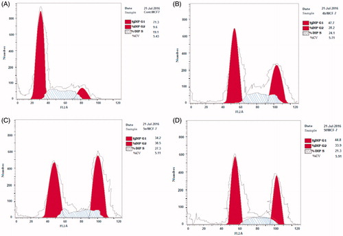
Table 3. Results of cell cycle analysis in HCT-116 and MCF-7 for compounds 4b, 5e and 5f.
Molecular modeling
Based on the results of the tubulin polymerization assay, docking of the most active compounds 4b, 5e and 5f was performed at X-ray crystal structure of tubulin in complex with (N-deacetyl-N-(2-mercaptoacetyl)colchicine) (DAMA-colchicine) and the SLD at 3.5 Å resolution (PDB: 1SA0)Citation7 using Discovery Studio 4 software packageCitation18 to shed light on their potential binding modes and investigate their similarity to the native ligand. Since the synthesized compounds are chiral, both isomers were enrolled in the docking study. illustrates the bonding and the nonbonding interactions of the docked compounds with amino acids of the active site. To ensure the validity of the docking protocol, re-docking of the co-crystallized DAMA-colchicine into the active site of tublin was performed. The coordinates of the best scoring docking pose of DAMA-colchicine was compared with its coordinates in the co-crystallized PDB file based on the binding mode and root mean square deviation (rmsd). The docking validation showed a near perfect alignment with the original ligand as obtained from the X-ray resolved pdb file. The re-docked ligand showed an rmsd of 0.6995 Å with CDOCKER interaction energy of −55.6986 and the same binding interactions. The binding site of DAMA-colchicine is composed of two hydrophobic cavities accommodated by the phenyl ring and tropone ring of DAMA-colchicine. Essential hydrogen bond of CYS241 with a methoxy group in DAMA-colchicine was reported. Thiol and the carbonyl of tropone ring were engaged in two hydrogen bonds with THR179 and VAL181, respectively.
Table 4. Results of the molecular docking study.
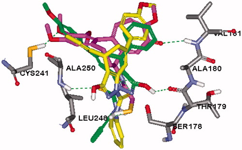
Analysis of the docking results revealed that the docked compounds showed comparable CDOCKER energy to the reference ligand and they interacted with variable amino acids previously reported in molecular modeling studies of CSIsCitation20. A properly positioned OH group at pyrazole ring of both isomers of compound 4b ( and ) and R isomers of compounds 5e and 5f was engaged in hydrogen bond interaction with SER178 or THR179, respectively ( and ), this hydrogen bond was reported to increase the activityCitation21. The S isomers of compounds 5e and 5f were flipped in such a way that their OH group was forming a hydrogen bond with Ala250 ( and ). Additional hydrogen bond interaction was observed at the methoxy group of compound 5f with CYS241 which was reported to be crucial for CBSIsCitation20. It is worth mentioning that although the hydrogen bonding to CYS241 was not reported in the docking poses of compounds 4b and 5e, a methoxy group in these compounds was in the vicinity of this amino acid ( and ). Moreover, pyrazole ring shows Pi interaction with LEU 248 in S isomer of 5e and 5f or with LYS254 or LYS352 in their R isomer, in addition to other valuable hydrophobic interactions. These results suggested that the new compounds had the potential to exhibit antitumor activity through inhibition of tubulin polymerization.
Figure 6. 2D interaction diagram of the top docking pose of the R isomer of compound 4b.
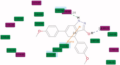
Figure 7. Overlay of the top docking poses of R (green) and S (yellow) isomers of 4b in the active site of tubulin (PDB: 1SA0).
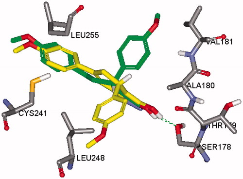
Figure 8. 2D interaction diagram of the top docking pose of the R isomer of compound 5e.
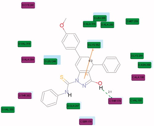
Figure 9. 2D interaction diagram of the top docking pose of the R isomer of compound 5f.
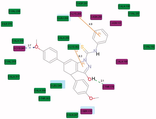
Figure 10. 2D interaction diagram of the top docking pose of the S isomer of compound 5e.
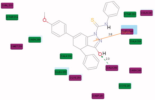
Figure 11. Overlay of the top docking poses of R (green), S (yellow) isomers of 5e and DAMA-colchicine (magenta) in the active site of tubulin (PDB: 1SA0).
Conclusion
Twenty two new target compounds were designed as inhibitors of tubulin polymerization relying on using two types of ring B models (cyclohexenone and indazole) to replace the central ring in colchicine. The designed compounds were assessed for their antitumor activity through in vitro cytotoxicity study on HCT-116 and MCF-7 cancer cell lines. Few compounds exhibited IC50 values lower than or slightly higher than colchicine. The bicylic indazole derivatives 4a,b and 5e,f were the most potent of all derivatives. Derivatives 4b and 5e exhibited higher potency than colchicine against colon HCT-116 tumor cell. Compound 5f revealed comparable activity to colchicine. Compound 5e demonstrated higher potency than colchicine against MCF-7 breast tumor cell line. The mechanism of the antitumor activity of the most active compounds 4b, 5e and 5f was investigated through evaluating the tubulin inhibition potential of the active compounds. These indazole derivatives 4b, 5e and 5f showed percentage inhibition of tubulin in both cell line homogenates ranging from 79.72% to 89.31%. The effects of 4b, 5e and 5f on cell cycle in HCT-116 and MCF-7 cell lines were analyzed revealing an increase of cell percentage at G2/M phase. Molecular docking was performed to reveal the interaction of the active compounds into the colchicine binding site of tubulin. Thereby, it could be claimed that the indazole derivatives represented a promising starting point for further study.
Disclosure statement
The authors report that that they have no conflicts of interest.
References
- Kandakatla N, Ramakrishnan G, Karthikeyan J, Chekkara R. Pharmacophore modeling, atom based 3D-QSAR and docking studies of chalcone derivatives as tubulin inhibitors. Orient J Chem 2014;30:1083–98.
- Sutkovic J. In silico prediction of three-dimensional structure and interactome analysis of Tubulin Alpha subfamily of Arabidopsis thaliana. Netw Biol 2014;4:47.
- Lu Y, Chen J, Xiao M, et al. An overview of tubulin inhibitors that interact with the colchicine binding site. Pharm Res 2012;29:2943–71.
- Li DD, Qin YJ, Zhang X, et al. Combined molecular docking, 3D‐QSAR, and pharmacophore model: design of novel tubulin polymerization inhibitors by binding to colchicine‐binding site. Chem Biol Drug Des 2015;86:731–45.
- Choudhury GG, Banerjee A, Bhattacharyya B, Biswas BB. Interaction of colchicine analogues with purified tubulin. FEBS Lett 1983;161:55–9.
- Jin Y, Qi P, Wang Z, et al. 3D-QSAR study of combretastatin A-4 analogs based on molecular docking. Molecules 2011;16:6684–700.
- Ravelli RB, Gigant B, Curmi PA, et al. Insight into tubulin regulation from a complex with colchicine and a stathmin-like domain. Nature 2004;428:198–202.
- Zardiny Z, Gholami S, Ramazani MA. Structure activity relationship (SAR) investigation on the binding properties of ligands similar to combretastatin to colchicine binding site of tubulin protein as anti-cancer drug. Res Pharm Sci 2012;7:S568.
- Attia SM. Molecular cytogenetic evaluation of the mechanism of genotoxic potential of amsacrine and nocodazole in mouse bone marrow cells. J Appl Toxicol 2013;33:426–33.
- Lu Y, Li CM, Wang Z, et al. Discovery of 4-substituted methoxybenzoyl-aryl-thiazole as novel anticancer agents: synthesis, biological evaluation, and structure–activity relationships. J Med Chem 2009;52:1701–11.
- Rischin D, Bibby DC, Chong G, et al. Clinical, pharmacodynamic, and pharmacokinetic evaluation of BNC105P: a phase I trial of a novel vascular disrupting agent and inhibitor of cancer cell proliferation. Clin Cancer Res 2011;17:5152–60.
- Bailly C, Bal C, Barbier P, et al. Synthesis and biological evaluation of 4-arylcoumarin analogs of combretastatins. J Med Chem 2003;46:5437–44.
- Flynn BL, Flynn GP, Hamel E, Jung MK. The synthesis and tubulin binding activity of thiophene-based analogues of combretastatin A-4. Bioorg Med Chem Lett 2001;11:2341–3.
- Wang L, Woods KW, Li Q, et al. Potent, orally active heterocycle-based combretastatin A-4 analogs: synthesis, structure–activity relationship, pharmacokinetics, and in vivo antitumor activity evaluation. J Med Chem 2002;45:1697–711.
- Johnson M, Younglove B, Lee L, et al. Design, synthesis, and biological testing of pyrazoline derivatives of combretastatin-A4. Bioorg Med Chem Lett 2007;17:5897–901.
- Ruprich J, Prout A, Dickson J, et al. Design, synthesis and biological testing of cyclohexenone derivatives of combretastatin-A4. Lett Drug Des Discov 2007;4:144–8.
- Montes-Avila J, Díaz-Camacho SP, Sicairos-Félix J, et al. Solution-phase parallel synthesis of substituted chalcones and their antiparasitary activity against Giardia lamblia. Bioorg Med Chem 2009;17:6780–5.
- Dassault Systèmes BIOVIA. Discovery studio modeling environment, Release 4. San Diego, CA: Dassault Systèmes; 2015.
- Shakil N, Singh MK, Sathiyendiran M, et al. Microwave synthesis, characterization and bio-efficacy evaluation of novel chalcone based 6-carbethoxy-2-cyclohexen-1-one and 2H-indazol-3-ol derivatives. Eur J Med Chem 2013;59:120–31.
- Botta M, Forli S, Magnani M, Manetti F. Molecular modeling approaches to study the binding mode on tubulin of microtubule destabilizing and stabilizing agents. In: Carlomagno T, ed. Tubulin-binding agents: synthetic, structural and mechanistic insights. Berlin (Heidelberg): Springer Berlin Heidelberg; 2009:279–328.
- O’Boyle NM, Greene LM, Bergin O, et al. Synthesis, evaluation and structural studies of antiproliferative tubulin-targeting azetidin-2-ones. Bioorg Med Chem 2011;19:2306–25.