
Abstract
To identify the metabolite distribution in ascidian, we have applied an integrated liquid chromatography– tandem mass spectrometry (LC–MS) metabolomics approach to explore and identify patterns in chemical diversity of invasive ascidian Styela plicata. A total of 71 metabolites were reported among these alkaloids, fatty acids and lipids are the most dominant chemical group. Multivariate statistical analysis, principal component analysis (PCA) showed a clear separation according to chemical diversity and taxonomic groups. PCA and partial least square discriminant analysis were applied to discriminate the chemical group of S. plicata crude compounds and classify the compounds with unknown biological activities. In this study, we reported for the first time that a partially purified methanol extract prepared from the ascidian S. plicata and Ascidia mentula possess antitumor activity against four tumor cell lines with different tumor histotype, such as HeLa (cervical carcinoma), HT29 (colon carcinoma), MCF-7 (breast carcinoma) and M14 (melanoma). S. plicata fraction SP-50 showed strong inhibition of cell proliferation and induced apoptosis in HeLa and HT29 cells, thus indicating S. plicata fraction SP-50 a potential lead compound for anticancer therapy. The molecular mechanism of action and chemotherapeutic potential of these ascidian unknown biomolecules need further research.
Introduction
Ascidian species possess a broad range of biodiversity of marine natural products (MNPs) because of their great ability to synthesize bioactive substances and serve as a vital source of new therapeutics. Due to their richness in biomolecules or secondary metabolite production, ascidians represent an important beginning for the discovery of new marine drugs and could be used as novel chemical scaffoldsCitation1. Interestingly, in Japan and Korea, ascidians are used as commercial seafood (e.g. Styela plicata, Microcosmus exasperates, Halocynthia rortezi) because of their high content of proteins, amino acids and micronutrientsCitation2,Citation3. Hence, it is important to study their chemical diversity, pharmacological properties when considering their food applications. The marine ascidians metabolome possesses valuable resource of MNPs and another organism such as sponge, corals and algae are likely produced much more metabolites than have been discovered so farCitation1.
In analytical chemistry, metabolomics is an emerging field of systematic study of the unique chemical fingerprints of flora and faunal living cells and biofluidsCitation4,Citation5. Metabolomics using mass spectrometry (MS) techniques allows qualitative and quantitative profiling of small biomolecules in marine organisms. This novel approach can be applied to reveal metabolic differences between ascidian species, to find differences in their profiles and to identify novel potential chemical lead for clinical biomarker discovery. Mass spectrometry-based metabolomic profiling provides a potent approach to quantifying and identifying the chemical diversity of living organisms and developing metabolic maps. It has been wildly applied in food and nutritional science, pharmacology and toxicology research, functional genomics, drug discovery to clinical development of new drugs and quality control particularly of chemotaxonomic identification of flora and faunaCitation6. Liquid chromatography–mass spectrometry (LC–MS) coupled with UV detection has traditionally lead biodiscovery efforts, but generally only detects highly abundant compounds and yields little structural information for dereplication of known compoundsCitation7. MS-based metabolomic platforms in MNPs research have increased the dynamic range of compound detection and dramatically decreased the quantity of material required. In spite of these advantages, the biggest challenge of MS-based metabolic profiling is processing and prioritizing the metabolomics data sets before the determination of the chemical structure of these metabolitesCitation6. A typical metabolomics profiling requires an enormous number of samples to generate the results that are statistically rigorous. Besides, highly sensitive and accurate instrumentation and powerful software tools (e.g. XCMS-METLIN) are essential to address the vast amount of data generated by these experiments. The recent development in the field of natural products chemistry and LC–MS/NMR-based metabolomics research on marine origin secondary metabolites exhibits diverse range of biological properties for developing new therapies to improve the health of individuals across the universe suffering from various deadly diseases such as infectious disease malaria, HIV, neurological and immunological diseases and cancer. The application of LC–MS-based metabolic profiling of biological systems has gained more extensive use in identifying drug metabolite, developing metabolite maps and lending clues mechanism of bioactivationCitation6. However, the knowledge of the metabolite accumulates in different Mediterranean ascidians chemical diversity is scarce.
In this study, we investigated the metabolite distribution of invasive ascidian S. plicata and Mediterranean ascidian A. mentula using LC–MS-based metabolomics methodCitation8 and multivariate statistical analysis with the aim of revealing unknown biomolecules from the ascidian S. plicata. In an earlier study, Halouska et al.Citation9 predicted the in vivo mechanism of action for drug leads against antitubercular activity using NMR metabolomics and orthogonal partial least square-discriminant analysis (OPLS-DA). In his study, he reported that compound separated from known drugs in the OPLS-DA scores plot, could be inferred new mechanism of action and has potentially valuable a new antibiotic agent. In this study, we have applied LC–MS metabolic profiling and partial least square-discriminant analysis (PLS-DA) to profile in vitro mechanism of action of known pharmacological potencies of metabolites identified in ascidian S. plicata. Mainly, we are processed to apply this information to classify the ascidian metabolites with known and unknown pharmacological potencies demonstrated antibacterial and antitumor action.
Recently, the European Network of Cancer Registries has reported more than 24 million cases of cancer diseases that have spread in 27 European nations. In the year 2012, around 3.45 million new cancer cases and 1.75 million deaths from cancer have been estimated in EuropeCitation10. Biomolecules derived from ascidian has aid potential candidate as anticancer drugs and for the development of novel therapeutic targets. For instance, the most well-known compound, ecteinascidin 743 (Yondelis®), isolated from the ascidian Ecteinascidia turbinate and marketed by PharmaMar was approved by the European Commission and Food and Drug Administration (FDA) to treat soft-tissue sarcomas and ovarian cancer disease. Another important antitumor drug plitidepsin (Aplidin®), isolated from Aplidium albicans, is in clinical usage for the treatment of acute lymphoblastic leukemia and several other ascidian MNPs are at various stages of clinical trials, biotechnological and industrial applicationsCitation11.
The objectives of this study were to assess metabolic profiling of Mediterranean ascidian using LC–MS combined with multivariate statistical analysis (PCA, PLS-DA) according to their mass spectral variables and classify the metabolites of known and unknown pharmacological application. In addition, we aimed to identify active fractions using bioassay-guided fractionation, structure elucidates active biomolecules and screen their antitumor efficacy against cervical, colon cancer and other tumor cell lines using MTT bioassay to find the antitumor activity of ascidian species.
Materials and methods
Chemicals
MeOH – methanol, CHCl3 – chloroform, CH2Cl2 – dichloromethane, Na2SO4 – sodium sulfate, formic acid, TFA – trifluroacetic acid (Sigma-Aldrich, St. Louis, MO) were used as solvents for the extraction of crude extracts preparation and successive partition of the aqueous phase. DMSO – dimethyl sulfoxide, isopropanol, PBS – phosphate-buffered saline and PI – propidium iodide were used for screening biological activity of ascidians.
Sample collection and extraction
Ascidians were collected by snorkeling in Lake Faro, Messina coast, Italy at a depth 3–5 m and identified to the lowest taxonomic levelCitation12. A voucher specimen is deposited at the Department of Biological and Environmental Science, Messina, Italy. Crude extracts from the nonindigenous ascidian S. plicata (weight 42.3 g) and Mediterranean-native species A. mentula (24 g) have been extracted by Soxhlet extraction and ultra-sonication method using MeOH/CHCl3. The chemical characterization of both crude compounds was determined using Fourier transform infra-red spectroscopy (FTIR) and 1H-NMR techniques. The IR spectral data were examined byCitation13, to differentiate the both ascidian based on the functional group of chemical components (Table S1 is given in Supplementary Information). However, nontargeted FTIR fails to monitor the species-specific metabolites and variation of chemical structure because of the limited specificity and sensitivity.
LC–MS-based metabolomics
LC–MS spectrum analysis of ascidian crude extracts (CE) was performed on a Waters ZQ-LC–MS electrospray ionization (ESI) single quadrupole mass spectrometer using a C18 column 3.3 μm, 15 cm ×2.1 mm with following mobile phases: (A) H2O 0.1% formic acid and (B) MeOH 0.1% formic acid (sample input and acquisition; 0.1 ml/min flow rate and 5–20 μl injection volume). For our integrated metabolite profiling of ascidian species, we utilized LC–MS mass data acquisition and an open source platform MS software XCMS-METLINCitation14. The raw data file was analyzed using XCMS software combined with METLIN, and for MS data, the following libraries were used HMDB, KEGG and LMIDCitation14,Citation15. LC–MS spectral data of each single sample were collected, the data will be preprocessed before the principal component analysis (PCA). The data normalization was identified using descriptive analysis followed by log transformation and Pareto scaling method. Partial least square discriminant analysis (PLS-DA) was performed to explore and identify patterns in chemical diversity of ascidian S. plicata. All the statistical analyses were performed using STATISTICA (ver. 10.0; Tulsa, OK) and PAST software (ver. 2.17C; Norway) for Windows.
HPLC separation of fractions and compound identification
A deep brownish gum (2.5 g) obtained from S. plicata was dissolved in 2 ml of DMSO and the extracts were equally divided. All solvents used for high-performance liquid chromatography (HPLC) and MS were HPLC grade (Sigma-Aldrich), and the H2O was Millipore Milli-Q PF filtered. The fractions were further diluted with 5 ml of MeOH, 1 ml of extract was transferred onto cotton and kept 30 min to dry and independently subjected to HPLC separation on a betasil C18 column (5 μm, 150 × 21.2 mm). Two of these columns were connected in HPLC and the combined columns eluted with a linear gradient of H2O containing 1% TFA at 9 ml/min flow rate over 60 minCitation16. The fractions were collected by autosampler and further acquired 1H-NMR of all the 60 fractions (Figure S1); combined all the fractions with a similar chemical shift. From these, the following fractions SP-8, SP-28, SP-50, SP-53, SP-55 was selected and further purified by HPLC (Figure S2) and screened against four tumor cell lines. The pyrimidine nucleoside (1) and long-chain fatty acid (2) were isolated from SP-8, SP-53 and their tentative structure was elucidated using 2D-NMR spectroscopy. 1H-NMR data of ascidian CE were recorded by Agilent 500 MHz NMR using 5-mm NMR tube and 1H-NMR, 13C and other 2D-NMR spectroscopy of individual fractions were acquired by Agilent 600 MHz NMR using 3-mm tube and the data were analyzed by MestreNova software, version 10.01 (Mestrelab Research).
Antitumor activity of ascidian compounds
Cell culture
BJ-EHLT (human fibroblasts), HeLa (cervical carcinoma), MCF7 (breast carcinoma) cells were cultured in Dulbecco's modified Eagle medium (DMEM), while HT29 (colon carcinoma), M14 (melanoma), HT1080 (fibrosarcoma) cells were cultured in Roswell Park Memorial Institute medium (RPMI medium). Cells were maintained at 37 °C, 5% CO2, 95% air and 100% humidity. 20 mM stock solution was prepared from crude and purified fractions dissolved in DMSO and stored at −20 °C. 1000–3000 cells in 200 μl were seeded in 96-well plates. Cells were treated for 48–72 h with concentrations ranging from 1 to 50 μM.
Cell viability
The effect of ascidian compounds on tumor cell proliferation was analyzed by measuring 3-(4,5-dimethyl-2-thiazolyl)-2,5-diphenyl-2H tetrazolium bromide dye absorbance of cells following manufacturer’s protocol. Cell viability in treated versus untreated control cells was calculated for each concentration of drugs used as “optical density (OD) of treated cells/OD of control cells” ×100. Data were analyzed by the median-effect method to determine the concentration that causes 50% of cell viability inhibition (IC50). Significant differences between the mean of control versus various doses were tested by one-way analysis of variance (ANOVA), followed by Dunnett’s post hoc test for multiple comparisons using GraphPad Prism 6, Windows 10, CA. Statistically, significant differences were assumed at p< 0.05 (*p< 0.05, **p< 0.01).
Cell cycle and apoptosis analysis
Cell-cycle distribution by PI staining was performed as previously describedCitation17. Briefly, 50,000 cells were cultured in 6-well plates, treated for 72 h with concentrations ranging from 1 to 50 μM, collected by centrifugation, fixed in cold 70% ethanol and stained with a PBS solution containing PI (62.5 mg/ml; Sigma-Aldrich) and RNase A (1.125 mg/ml; Sigma-Aldrich). Samples were acquired with C6 Accuri Flow cytometry (BD Bioscience) and the percentage of cells in the different phases of cell cycle and in the sub-G1 compartment was calculated. Apoptosis was quantified by cytofluorimetric analysis staining cells simultaneously with FITC-annexin V and the nonvital dye PI (Immunological Sciences, Rome, Italy), following the manufacturer’s instruction. At the end of incubation with the respective reagents, samples were analyzed with C6 Accuri Flow cytometry (BD Bioscience). About 20,000 events were acquired and gated using forward scatter and side scatter to exclude cell debris. Bivariate analysis allows the discrimination of viable cells (FITC−/PI−), early apoptotic (FITC+), and late apoptotic or necrotic cells (FITC+/PI+).
Results and discussion
Comparative analysis of ascidian metabolome
The challenge of untargeted LC–MS-based metabolic profiling is to find a technological approach that allows the reproducible identification, combined with adequate statistical analysis and quantification of the highest possible number of metabolites. LC–MS metabolomic analysis was performed for the rapid identification of novel biomolecules from both ascidians S. plicata and A. mentula MeOH crude compounds. The analysis of the LC–MS chromatogram of ascidian samples with XCMS-METLIN software revealed a total of 71 metabolite peaks with containing between 105 and 1365 m/z in S. plicata and 21 metabolites peaks containing between 117 and 519 m/z in A. mentula extracts, the complete characterization of all detected metabolites would be impossible. In this study, the maximum numbers of metabolites were produced by nonindigenous ascidian S. plicata compared to Mediterranean-native species A. mentula. Fatty acids, cembranoid, pyrimidine and alkaloid compounds are the most dominant compounds in S. plicata. In A. mentula, alkanes and flavonoids are prepotent. Significant metabolic differences were observed between both ascidian species. XCMS analysis provided statistical information about the differences between two ascidian species, including p values, fold change and LC retention time comparisons. Features considered to be present in the samples met the following criteria: p > 0.01, low fold change (<1.5) and <0.3 min deviation in LC retention time. We defined a unique feature as one with a p values of 0.01 and fold change of 1.5 or higher. Mass spectrum of all the metabolite peaks was analyzed by manually to confirm the comparative analysis of XCMS study. As suggested by Sidebottom et al.Citation18, we eliminated those features not meeting the specifications due to their higher possibility that could be false positives from the peak picking algorithmCitation15,Citation18. Besides, the ionization state and the presence of adducts (e.g. [M + Na]2+;[M + H + Na]2+) was evaluated to verify that the correct parent ion species was determinedCitation18. Relative peak metabolites identified in both ascidian species were given in Supplementary Tables S2 and S3.
From the identified metabolites, antibacterial and antiprotozoal macrolides, acetylapramycin (m/z 291.65) and antimalarial kabiramide B (m/z 483.4) were previously reported in sponge Pachastrissa nux collected in ThailandCitation19. Another neuroactive benzomorphan small molecule, pentazocine glucuronide (m/z 253.61) is a kappa opioid receptor agonist was reported in S. plicataCitation20. Another, antibiotic metabolite β-lactam dicloxacillin sodiumCitation21 was reported in MeOH extracts of A. mentula. However, this is the first report for the presence of these compounds in tunicates. Similar to this study using XCMS platform, 150 metabolite features were identified from actinomycete Streptomyces coelicolor M145 and discovered 16 siderophores not previously reported in this strainCitation18. Besides fatty acids and polyunsaturated aldehydes are the main metabolites in the interaction of the marine organism with their environmentCitation22,Citation23. In aquatic ecosystem, a highly unsaturated fatty acids are essential to transfer carbon between primary producers and consumersCitation24. More abundant of fatty acid metabolites present in S. plicata may help to compete with other native species in the Mediterranean Sea. Interestingly, the presence of metabolite decarbamoylgonyautoxin 1 (m/z 138.35) and lyngbyatoxin (m/z 216.65) molecule were first reported in S. plicata which is causing paralytic shellfish poisoning (PSP)Citation25 and could be utilized to biotoxin monitoring for shellfish harvesting in Lake Faro, which is completely absent in A. mentula. The first report of PSP syndrome in human after consumption of ascidian Microcosmus vulgaris was reported in CroatiaCitation25. Results present in this study suggest that ascidian S. plicata collected in Messina coast is not recommended for human consumption and could be cause serious economic loss of shellfish aquaculture. However, further studies of these biotoxins in ascidian species and continuous environmental monitoring is urgently required.
Chemotaxonomic classification
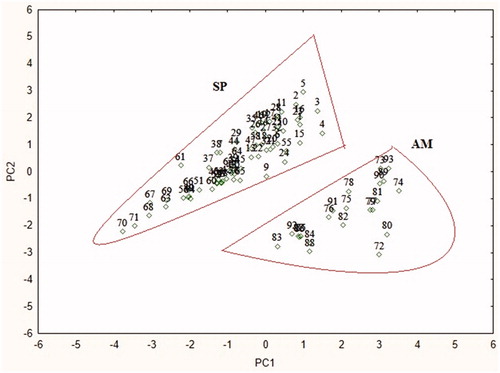
A total of 92 metabolites mass spectral variables, chemical group composition and taxonomic group of ascidian species were subjected to PCA. A data matrix was constructed using the taxonomic group and chemical composition as given in Tables S2 and S3. In PCA, the loading scatter plot clearly discriminates the two ascidian species separately based on the metabolites highly contribute to discriminate the samples (Figure S3). In , the discrimination of two ascidian species can be observed clearly, the native ascidian species A. mentula distinctly discriminated from the invasive ascidian S. plicata and plotted separately. The variables of fatty acids, carotenoids were highly contributed to clear discrimination of both ascidian species. Similar results of this present study, the chemotaxonomy of marine plants and organism have been reported using GC-MS, NMR based metabolomics approach using multivariate statistical analysisCitation6,Citation15,Citation26. Based on the results of this study, it can be concluded that PCA analysis of LC–MS mass spectral variables could be utilized as a reliable tool for species discrimination and taxonomic classification of marine ascidians.
Figure 1. Chemotaxonomic classification of ascidian using mass spectral variables. Species SP: Styela plicata; AM: Ascidia mentula.
Multivariate statistical analysis
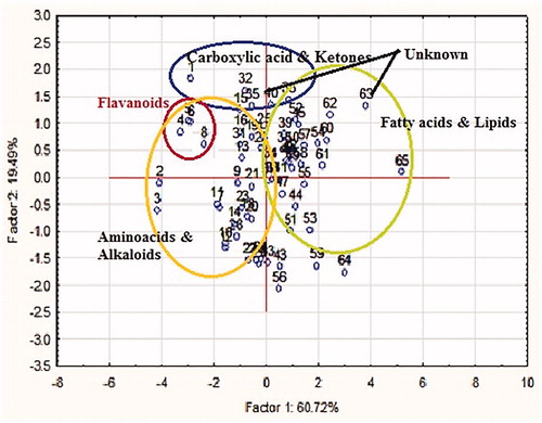
One of the prospective application of our untargeted metabolomic analysis in marine ascidian aims to identify potential species with novel biomolecules and classify compounds with an unknown mechanism of actions. We have chosen ascidian S. plicata on the basis of maximum chemical diversity and pharmacological potentials to classify their chemical compounds using multivariate statistical analysis PCA and PLS-DACitation9,Citation15. Using relative quantification and PCA clustering of all annotated metabolites in positive ionization mode, clear differences were noted in chemical compounds of S. plicata. PCA scores plot demonstrated the clustering pattern of seven different classes of known metabolites (). Loadings of group 1 (principal component (60.72%) and group 2 (19.49%) is complex as samples overlapped. In this study PC1, corresponding to the higher eigen value, is mostly correlated with the variable retention time, peak maximum intention and adduct molecular weight (a strong positive correlation that these axes are highly related to differences between the groups). In the first discrimination function, fatty acids and lipids discriminate compared to other metabolites reported in ascidian S. plicata. Prodigious, fatty acids and lipids metabolites were found more abundant in this species (Table S2). In second group PC2, amino acids, flavonoids and carboxylic acid showed very different distribution pattern in the S. plicata. Marshall et al.Citation26 have reported metabolites, fatty acids and steroids are highly contributed to the discrimination of algae Chattonella sp. Similar to this present study, the metabolites of Egyptian soft corals were identified using MS/NMR-based metabolomics approach and coral metabolites were discriminated using OPLS-DA methodCitation27.
Figure 2. PCA demonstrating the clustering of chemical groups of known metabolites.
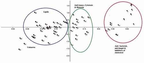
In this study, we utilized LC–MS m/z features of S. plicata and PLS-DA to profile biological potentials of known metabolites used as antibacterial, antifungal and antitumor. Herein, we aim to use this information to classify the metabolites with unknown pharmacological potentials and mechanism of action. A representative of PLS-DA scores plot demonstrates clustering pattern of chemical groups with known mechanisms and in vitro biological potencies of S. plicata (). Each point in PLS-DA scores plot represents electron ionization of LC–MS spectrum of specific metabolites. A quality of assessment score (R2) of 0.545 and (Q2) of 0.48 was obtained, indicating an excellent and highly reliable modelCitation9,Citation27. Correspondingly; PLS-DA is preferred as long as cross validation verifies a reliable model. The PLS-DA scores plot consists of three separate clustering patterns, which revealed that each group has considerably different potency on the metabolome of S. plicata. Importantly, fatty acids and lipids group metabolites were distinct and separate clusters from other biologically active metabolites inhibiting antibacterial, antifungal and antitumor growth (biological activity of these metabolites were retrieved from databases HMDB, KEGG, LIMD). The biomolecules with similar biological activity or therapeutic targets will have a similar impact on the metabolome of S. plicata and will cluster together in PLS-DA scores plotCitation10. Hence, the action of a novel chemical lead can be inferred from its clustering in PLS-DA scores plot relative to drugs with defined biological targets. Similar statistical method was applied to predict in vivo mechanism of action to find therapeutic drug leads against M. smegmatis using NMR metabolomicsCitation9. Generally, if the chemical lead is separated from known compounds in the PLS-DA scores plotCitation9, then this result would suggest a new mechanism of action and could be serve as potentially valuable new clinical biomarkersCitation15. The method, using LC–MS and multivariate analysis, could be a useful tool for quality control issues and effectively utilized in drug discovery process.
Figure 3. PLS-DA scores plot demonstrating the clustering pattern of Styela plicata metabolites with known biological potencies.
Structure elucidation of compounds
HPLC analysis of S. plicata CE is given in (Figure S1). Different retention times (RT) with various combinations of water and MeOH were used for the present HPLC method. Fraction SP-8 was chosen from the 60 HPLC fractions and purified further (Figure S4), which were analyzed by LC–MS to investigate the m/z of this purified fraction m/z 128.06. Using an extensive 1D and 2D NMR spectral data (Table S4), dihydro-5-methylpyrimidine – 2, 4 (1H, 3H)-dione (1) was tentatively identified from the fraction SP-8 with a molecular formula of C5H6N2O2 (NMR spectral was enclosed in Supplementary Information).
Further, the purified fractions (SP-28, SP-50, SP-53, SP-55) molecular weight, 1H-chemical shifts and 2D NMR spectroscopy was recorded on LC–MS and NMR spectrometry. Remarkably, all the fractions were identified as long-chain fatty acids and molecular weight of all the fractions was acquired using mass spectroscopy and given in Supplementary information (Figure S5–S6). Fatty acid, nonanoic acid was tentatively identified from the fraction SP-53 (NMR spectral data were enclosed in Supplementary information). Similarly, a modified octapeptide, plicatamide and their analogs have been isolated from the same ascidian S. plicata collected at San Diego BayCitation28. In this study, pyrimidine derivatives, nonanoic acid along with three long-chain fatty acid fractions were isolated in S. plicata. Initially, Wasylyk and AlamCitation29 isolated diterpene styelidae from the same ascidian species collected in California. Anti-inflammatory heparin-like analogs dermatasulfate was isolated in S. plicata collected in Brazil coastCitation30. Recently, six MNPs (e.g. tetillapyrone, nortetillapyrone, two nucleosides thymidine) were reported in S. plicata collected in China coastCitation31.
Antitumor activity of ascidian extracts
The crude extracts of ascidians have shown a lot of interest due to their higher antibacterial, antifungal and cytotoxicity activitiesCitation16,Citation32. The MeOH extracts of ascidians S. plicata and A. mentula exhibited significant cytotoxicity and strongly inhibited nitric oxide (NO) production against human embryonic kidney cells (HEK-293) at concentration 1 mg/ml as reported in the previous studyCitation33.
The effect of ascidian CE of S. plicata and A. mentula on proliferation of four tumor cell lines from different origin was tested using MTT assay. As reported in , IC50 values for S. plicata CE range from 41 to 125 μmol/l. In particular, the S. plicata showed the highest activity against HeLa cervical cancer (IC50 41.15 μmol/l), HT29 colon cancer (IC50 42 μmol/l) and HT1080 fibrosarcoma cells (IC50 52 μmol/l), while MCF7 breast cancer cells were more resistant (IC50 125 μmol/l). On the contrary, the response of the four cell lines to A. mentula CE was less heterogeneous IC50 values ranging from 39 to 58 (HeLa, IC50 53 μmol/l; HT29, IC50 39 μmol/l; MCF7, IC50 58.3 μmol/l; and HT1080, IC50 50 μmol/l). Similarly, Nurhayati et al.Citation34 have reported modest anticancer activity of sponge Cinachyrella sp crude extracts against HeLa cells (IC50 82.744 μg/ml).
Table 1. Antitumor activity of methanol extracts of ascidians in four tumor cell lines determined by MTT assay.
Then, the response of HeLa cells to different time exposure (24–96 h) and concentrations (10–100 μmol/l) of S. plicata extracts was analyzed. As reported in a significant reduction of cell viability was observed at the longest time exposure (96 h) and at the highest concentrations used (20 and 50 μmol/l). Similarly, a significant reduction of cell viability was observed when HeLa cells were exposed to 10–50 μM
Figure 4. Cytotoxicity of ascidian crude extracts against HeLa cells. Analysis of cell viability by MTT assay using CE of Styela plicata () and Ascidia mentula () at concentrations ranging from 1 to 100 μM for 24–96 h. The results represents the mean ± SEM of replicates. One-way ANOVA were calculated between contol and treated cells. *p < 0.05, **p < 0.01.
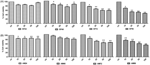
A. mentula extracts for 24–96 h (). Similar to our results Aiello et al.Citation35 have reported two new sulfated metabolites, 3,7,11,15-tetramethyl-hexadecan-l,19-sodium disulfate and heneicosane-l,21-sodium disulfate from the same ascidian species with modest antiproliferative activity against human melanoma IGR-1 (IC50>110, 100 μg/ml) and murine monocyte/macrophage J774 (IC50>180, >170 μg/ml). The extract of Didemnum ligulum exhibited modest cytotoxicity against HCT-8 cells (IC50 35.3 μg/ml) and Didemnum psammatodes fraction 1 showed strongest cytotoxicity against Molt-4 cells (IC50 35.3 μg/ml)Citation36,Citation37.
Antitumor activity of S. plicata fractions
shows the effect of five S. plicata fractions (SP-8, SP-28, SP-50, SP-53, SP-55) on HeLa cells determined by MTT assay. SP-8, SP-28, SP-53, SP-55 fractions showed modest antitumor activity against HeLa cells with IC50 values ranging from (37–46 μmol/l); on the other hand, the fraction SP-50 showed strongest growth inhibition (IC50 33 μmol/l). Remarkably, SP-50 did not induce significant cell cytotoxicity in BJ-EHLT human fibroblasts (). To study in depth the cytotoxicity effect of SP-50 the most effective fraction on tumor cells, we extended our analysis on a panel of tumor cell lines, including HT29, MCF7 breast cancer and M14 melanoma cells. As reported in , IC50 values ranging from 32 to 41 μmol/l were observed when HT29 (IC50 31.66 μmol/l), MCF7 (37.29 μmol/l) and M14 (IC50 41.05 μmol/l) cells were exposed to the fraction. A dose-dependent reduction of cell viability by fraction SP-50 was observed in all cell lines tested . Accordingly with MTT assay results, our data showed a significant decrease in the number of viable cells in two cancer cells lines (HT-29 and HeLa) after 72 h of treatment with higher doses of SP-50 fraction (Figure S14).
Figure 5. Dose-dependent cytotoxicity of fraction SP-50 of ascidian S. plicata against tumor and normal cells. Analysis of cell viability by MTT assay against normal BJ-EHLT (A) and four tumor (B–E) cell lines exposed to SP-50 at concentrations ranging from 1 to 50 μM for 24–96 h. The results represents the mean ± SEM of replicates. One-way ANOVA were calculated between contol and treated cells. *p < 0.05, **p < 0.01.
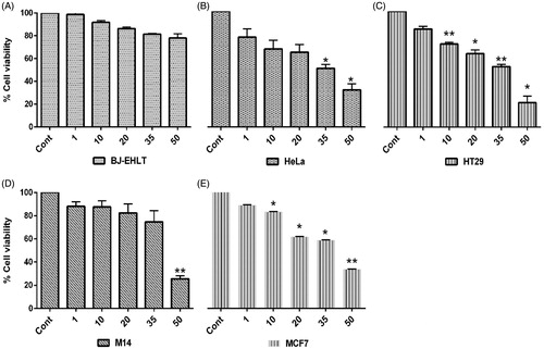
Table 2. Antitumor activity of S. plicata fractions by MTT assay.
Similar to this study, Eudistoma vannamei fraction 17 showed antiproliferative activity (IC50 value 1.85 μg/ml) and induced apoptosis and necrosis of human promyelocytic leukemia HL-60 cellsCitation36. Also, compounds, norteillapyrone and nucleosides thymidine were reported from the same ascidian with poor efficacy against Artemia salina (18.3%) at concentration 50 μg/mlCitation31. Fatty acids metabolites could play an essential role in various biological functions, including cell proliferation and apoptosis induction. Moreover, inhibition of fatty acid oxidations blocks tumor growth of breast cancerCitation38,Citation39. The biological activity of fatty acids on cell proliferation is highly influenced by saturation of the moleculeCitation39.
Apoptosis induction
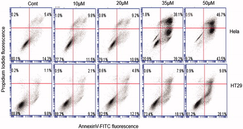
Programed cell death or apoptosis is an important phenomenon in cancer cell death. Cell shrinkage, membrane blebbing, nuclear condensation and DNA fragmentation are the characteristic features of apoptosisCitation40. To further determine whether the antiproliferative effects of S. plicata fractions were related to the induction of apoptosis and/or necrosis, SP-50-treated cells were analyzed with flow cytometry by using annexin V/propidium iodide (PI) staining that allows the discrimination of viable cells (annexin V+/PI+), early apoptotic (annexin V+/PI−), and late apoptotic or necrotic cells (annexin V+/PI+). As depicted in , SP-50 induced apoptosis in a concentration-dependent manner in both the cell lines analyzed. In particular, 24 h of SP-50 with 50 μM treatment increased the percentage of both early (about 43% compared with untreated) and late apoptotic or necrotic events (about 47% compared with untreated) in HeLa cells.
Figure 6. Flow cytometric analysis of apoptotic cells by annexin V/propidium iodide staining in HeLa and HT29 cells untreated or treated for 24 h with at the indicated concentrations. A representative experiment is shown. Annexin V+/PI − and annexin V+/PI+-stained cells were considered early apoptotic and late apoptotic or necrotic cells, respectively. Percentage of the different population of stained cells is shown.
In agreement with these results, a concentration-dependent increase in the Sub-G1 peak was observed after 72 h of treatment in both HeLa and HT29 treated cells when compared with untreated cells (Figures S12 and S13). Taken together, these data indicate that fraction SP-50 inhibited cell proliferation in both cell lines tested with higher effect when compared to other fractions of S. plicata and that the reduction in the cell viability was associated with the induction of apoptosis.
Similarly, induction of apoptosis has been reported with ecteinascidin-743 (ET-743) from E. turbinataCitation11, aplidine from A. albicansCitation11, CiEx, a partially purified compound from Ciona intestinalisCitation41 and PI-8 fraction isolated from Polyclinium indicumCitation42, and methyl ester mixtures of D. psammatodes exhibited strong apoptotic induction against HL-60 cellsCitation37. It is possible that S. plicata fraction SP-50 due to specificity of cancer cells, binds to DNA, inhibits cell division and triggers apoptosis similarly to ET-743Citation43 and (dehydrodidemnin B) from A. albicansCitation44. The marked arrest of cells in Sub-G1 phase and DNA fragmentation by the fraction SP-50 suggesting its possible use as anticancer drug. We can hypothesize a similar mechanism of action to the ascidian metabolites didemnin B and aplidine, which interfered with the synthesis of DNA and proteins and induced cell-cycle arrestCitation45,Citation46. The meridianin E from A. meridianumCitation47 and lissoclinolide from Lissoclinum patellaCitation48 showed similar arrest of G2/M phase in the cell cycle of target cells.
Conclusions
In conclusion, from an ecological perspective, the metabolome data suggest that secondary metabolites could apparently account chemical defense in ascidian S. plicata and rich resource of compounds with cytotoxic properties. LC–MS metabolomics of ascidian and PLS-DA resulted in distinct clustering patterns correlating with in vitro or in vivo biological activity of metabolites. The clustering of ascidian biomolecules relative to known biological activity could be used to discriminate the compounds based on pharmacological properties and classify the compounds with unknown biological activities. This study indicates that the presence of several metabolites in ascidian S. plicata crude extracts can be used effectively in pharmaceutical industries during drug discovery program. In this study, two biotoxin metabolite were identified in ascidian S. plicata; therefore, ascidian caught in Lake Faro not safe for human consumption. The results of cell-cycle analysis indicate that fraction SP-50-inhibited cell proliferation in both cell lines tested and that the reduction in the cell viability was associated with induction of apoptosis and/or necrosis, being SP-50 the strongest apoptosis inducer with highest growth inhibition against HeLa cells compared to other fractions of S. plicata. The outcomes from this present study suggest that S. plicata fraction SP-50 is a useful anticancer compound, which enhances a therapeutic efficacy; further studies are necessary to clarify this point. However, further studies of isolation pure compounds and structural identification of these active compounds present in S. plicata and molecular mechanism of pure compounds induced apoptosis are need to be discovered. The results of this study open new hope for discovering novel marine drugs from Mediterranean ascidian with potential therapeutic in the treatment of cervical and colon cancer disease.
IENZ_1266344_Supplementary_Material.pdf
Download PDF (1.1 MB)Acknowledgements
Author PSK thanks University of Messina for the award of Non-EU doctoral research fellowship 2013–2015. Author PSK gratefully thanks Prof. R. Quinn, Eskitis Institute of Drug Discovery, Griffith University, Brisbane – Australia for Visiting Researcher Position – 2014. Author PSK thanks Dr. Donatella Del Bufalo, Italy for Visiting Researcher position -2015 and financial support for cell culture and flow cytometry experiments. Authors thank Prof. Giacobbe’s group, UNIME for Ascidian collection and taxonomy and research advice for this work. Author PSK thanks to Prof. Emilio De Domenico, UNIME for his helps in this project and encouragement.
Disclosure statement
The authors report no conflicts of interest. The authors alone are responsible for the content and writing of this article.
Additional information
Funding
References
- Davidson BS. Ascidians: producers of amino acid-derived metabolites. Chem Rev 1993;93:1771–91.
- Yamaguchi K, Konosu S. Extractive nitrogenous constituents of two species of edible ascidians Styela clava and S. plicata. Nippon Suisan Gakkaishi 1991;57:169–74.
- Oh KS, Kim JS, Heu MS. Food constituents of edible ascidians Halocynthia roretzi and Pyura michaelseni. Korean J Food Sci Tech 1997;29:955–62.
- Kuhlisch C, Pohnert G. Metabolomics in chemical ecology. Nat Prod Rep 2015;32:937–55.
- Powers R. NMR metabolomics and drug discovery. Magn Reson Chem 2009;47:S2–11.
- Goulitquer S, Potin P, Tonon T. Mass spectrometry-based metabolomics to elucidate functions in marine organisms and ecosystems. Mar Drugs 2012;10:849–80.
- Lang G, Mayhudin NA, Mitova MI, et al. Evolving trends in the dereplication of natural product extracts: new methodology for rapid, small-scale investigation of natural product extracts. J Nat Products 2008;71:1595–9.
- Neumann S, Böcker S. Computational mass spectrometry for metabolomics: identification of metabolites and small molecules. Anal Bioanal Chem 2010;398:2779–88.
- Ferlay JE, Steliarova-Foucher J, Lortet-Tieulent Sonia Rosso JWW, et al. Cancer incidence and mortality patterns in Europe: estimates for 40 countries in 2012. Eur J Cancer 2013;49:1374–403.
- Halouska S, Fenton RJ, Barletta RG, Powers R. Predicting the in vivo mechanism of action for drug leads using NMR metabolomics. ACS Chem Biol 2011;7:166–71.
- Molinski TF, Doralyn S, Dalisay Sarah L, et al. Drug development from marine natural products. Nat Rev Drug Discov 2009;8:69–85.
- Kott P. Tunicata. Zoo Cata Australia 1998;34:51–252.
- Griffiths P, de Hasseth JA, Fourier Transform Infrared Spectrometry (2nd ed.). New York: Wiley-Interscience; 2007.
- Smith CA, O'Maille G, Want EJ, et al. METLIN: a metabolite mass spectral database. Ther Drug Monit 2005;27:747–51.
- Mahieu NG, Genenbacher JL, Patti GJ. A roadmap for the XCMS family of software solutions in metabolomics. Curr Opin Chem Biol 2016;30:87–93.
- Tran TD, Pham NB, Ekins M, et al. Isolation and total synthesis of stolonines A–C, unique taurine amides from the Australian marine tunicate Cnemidocarpa stolonifera. Mar Drugs 2015;13:4556–75.
- Di Martile M, Desideri M, De Luca T, et al. Histone acetyl transferase inhibitor CPTH6 preferentially targets lung cancer stem-like cells. Oncotarget 2016;7:11332–48.
- Sidebottom AM, Johnson AR, Karty JA, et al. Integrated metabolomics approach facilitates discovery of an unpredicted natural product suite from Streptomyces coelicolor M145. ACS Chem Bio 2013;8:2009–16.
- Sirirak T, Kittiwisut S, Janma C, et al. Kabiramides J and K, trisoxazole macrolides from the sponge Pachastrissa nux. J Nat Prod 2011;74:1288–92.
- Anzenbacher P, Zanger UM, (Eds.), Metabolism of drugs and other xenobiotics. Weinheim, Germany: Wiley-VCH; 2012.
- Lacey CS. Interaction of dicloxacillin with warfarin. Ann Pharmacother 2004;38:898.
- Stoecker D. Relationship between chemical defense and ecology of benthic ascidians. Mar Eco Prog Ser 1980;3:257–65.
- Pohnert G. Diatom/copepod interactions in plankton: the indirect chemical defense of unicellular algae. ChemBioChem 2005;6:946–59.
- Müller-Navarra DC, Brett MT, Liston AM, Goldman CR. A highly unsaturated fatty acid predicts carbon transfer between primary producers and consumers. Nature 2000;403:74–7.
- Roje-Busatto R, Ujević I. PSP toxins profile in ascidian Microcosmus vulgaris (Heller, 1877) after human poisoning in Croatia (Adriatic Sea). Toxicon 2014;79:28–36.
- Marshall JA, Nichols PD, Hallegraeff GM. Chemotaxonomic survey of sterols and fatty acids in six marine raphidophyte algae. J Appl Phycol 2002;14:255–65.
- Farag MA, Porzel A, Al-Hammady MA, et al. Soft corals biodiversity in the Egyptian Red Sea: a comparative MS and NMR metabolomics approach of wild and aquarium grown species. J Proteome Res 2016;15:1274–87.
- Tincu JA, Menzel LP, Azimov R, et al. Plicatamide, an antimicrobial octapeptide from Styela plicata hemocytes. J Biol Chem 2003;278:13546–53.
- Wasylyk JM, Alam M. Isolation and identification of a new cembranoidditerpene from the tunicate Styelaplicata. J Nat Pro 1989;52:1360–2.
- Belmiro CL, Castelo-Branco MT, Melim LM, et al. Unfractionated heparin and new heparin analogues from ascidians (chordate-tunicate) ameliorate colitis in rats. J Biol Chem 2009;284:11267–78.
- Ping SU, Yan XU, Jialin Y, Chang-Yun W. Study on the chemical constituents and bioactivity of the ascidian Styelaplicata, China. Chin J Mar Drug 2014;33:1–6.
- Jimenez PC, Fortier SC, Lotufo TM, et al. Biological activity in extracts of ascidians (Tunicata, Ascidiacea) from the northeastern Brazilian coast. J Exp Mar Boil Ecol 2003;287:93–101.
- Satheesh Kumar P, Morabito R, Remigante A, et al. Biological activity of extract from Styela plicata and Ascidia mentula (Ascidiacea). J Biol Res 2016;89:5812–18.
- Nurhayati APD, Pratiwi R, Wahyuono S, Fadlan A. Isolation and identification of alkaloid compound of marine sponge Cinachyrella sp. (Family Tetillidae). J Adv Botany Zool 2014;2:1–4.
- Aiello A, Fattorusso E, Menna M, et al. Novel anti-proliferative alkylsulfates from the Mediterranean tunicate Ascidia mentula. Tetrahedron 1997;53:5877–82.
- Jimenez PC, Wilke DV, Takeara R, Lotufo TM, et al. Cytotoxic activity of a dichloromethane extract and fractions obtained from Eudistoma vannamei (Tunicata: Ascidiacea). Comp Biochem Physiol A Mol Integr Physiol 2008;151:391–8.
- Takeara R, Jimenez PC, Wilke DV, et al. Antileukemic effects of Didemnum psammatodes (Tunicata: Ascidiacea) constituents. Comp Biochem Physiol A Mol Integr Physiol 2008;151:363–9.
- Crunkhorn S. Breast cancer: inhibiting fatty acid oxidation blocks tumour growth. Nat Rev Drug Discov 2016;15:310.
- Hardy S, El-Assad W, Przybytkowski E, et al. Saturated fatty acid-induced apoptosis in MDA-MB-231 breast cancer cells. A role for cardiolipin. J Biol Chem 2003;278:31861–70.
- Faivre S, Chièze S, Delbaldo C, et al. Phase I and pharmacokinetic study of aplidine, a new marine cyclodepsipeptide in patients with advanced malignancies. J Clin Oncol 2005;23:7871–80.
- Russo GL, Ciarcia G, Presidente E, et al. Cytotoxic and apoptogenic activity of a methanolic extract from the marine invertebrate Ciona intestinalis on malignant cell lines. Med Chem 2008;4:106–9.
- Rajesh RP, Ramasamy SM, Murugan A. Anticancer activity of the ascidian Polyclinum indicum against cervical cancer cells (HeLa) mediated through apoptosis induction. Med Chem 2010;6:396–405.
- Takebayashi Y, Goldwasser F, Urasaki Y, et al. Ecteinascidin 743 induces protein-linked DNA breaks in human colon carcinoma HCT116 cells and is cytotoxic independently of topoisomerase I expression. Clin Cancer Res 2001;7:185–91.
- Sakai R, Kishore V, Kundu B, et al. Structure-activity relationships of the didemnins. J Med Chem 1996;39:2819–34.
- Erba E, Serafini M, Gaipa G, et al. Effect of aplidin in acute lymphoblastic leukaemia cells. Br J Cancer 2003;89:763–73.
- Ko-Hsiu L, Ko-Huang L, Hsien-Hua L, et al. Induction of caspase-3-dependent apoptosis in human leukemia HL-60 cells by paclitaxel. Clin Chim Acta 2005;357:65–73.
- Gompel M, Leost M, Joffe EBD, et al. Meridianins, a new family of protein kinase inhibitors isolated from the ascidian Aplidium meridianum. Bioorg Med Chem Lett 2004;14:1703–7.
- Richardson AD, Ireland CA. A profile of the in vitro antitumor activity of lissoclinolide. Toxicol Appl Pharmacol 2004;195:55–61.