
Abstract
Twenty-three novel resveratrol-based cinnamic ester hybrids were designed and synthesized. All the compounds were evaluated for their anti-inflammatory activity using RAW264.7 cells. Among them, compound D15 was found to be the most potent one in inhibiting NO production in LPS-stimulated RAW264.7 cells. The further study indicated that compound D15 could suppress expression of proteins iNOS, COX-2, p-p65, and p-IκB LPS-induced. Immunofluorescence further revealed compound D15 could reduce activation p65 in nuclei. All the results indicated that the anti-inflammatory activity of title compound may partly due to its inhibitory effect on the NF-κB signaling pathway.
Introduction
Inflammation is a common and essential pathological process that the immune system uses as response to a large variety of stimuli such as injury or infectionCitation1. The ubiquitously expressed nuclear transcription factor-κB (NF-κB), plays a core role in the inflammatory response by regulating the expression of various genes encoding pro-inflammatory cytokines, adhesion molecules, chemokines, growth factors, and inducible enzymes such as cyclooxygenase-2 (COX-2) and inducible nitric oxide synthase (iNOS)Citation2,Citation3. A growing body of evidences indicate that the inflammatory process may cause tissue damage and a host of diseases such as cancerCitation4,Citation5. Nonsteroidal anti-inflammatory drugs (NSAIDs) have been considered as one of the most widely used medicines for alleviation of pain, fever as well as inflammations. However, the traditional NSAIDs drug is often accompanied by severe adverse effects, the main of which include gastrointestinal adverse effectsCitation6. Therefore, the development of novel therapeutic agentswith improved pharmaceutical profiles is now in urgent need.
Natural products have long been deemed as lead templates for the design and discovery of novel anti-inflammatory agentsCitation7,Citation8. Resveratrol (trans-3,5,4’-trihydroxylstilbene, 1, ), a natural product with a stilbene structure, is without a doubt the most famous dietary polyphenol presents in medicinal plantsCitation9. In recent years, resveratrol has been extensively investigated as cardioprotective, anti-oxidative, anticancer, anti-aging agentsCitation10–15, and treatment of type II diabetesCitation16–18. Resveratrol also plays an important anti-inflammatory role in human umbilical vascular endothelial cellCitation19. More and more evidences have indicated that resveratrol is expected to be a new moiety for treatment of inflammation by reducing proinflammatory cytokines. It also can alleviate inflammation LPS-induced in Caco-2 and SW480 human colon cancer cell through inhibiting NF-κB pathway, protect LPS-induced extracellular lipoperoxidationCitation20–22. Besides, resveratrol shows anti-inflammatory activity by inhibiting the TLR4/NF-κB/STAT signaling cascadeCitation23, modulating the cytokines-stimulated activation of SAPK/JNK pathwayCitation24, affecting MAPKs signaling cascadesCitation25. Furthermore, resveratrol has been reported to be endowed with the capacity of protecting the gastric mucosa against the side effect NSAID-inducedCitation26.
Figure 1. Structure of resveratrol (1) and cinnamic acid (2).
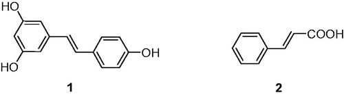
On the other hand, cinnamic acid (2, ) is a natural organic acid in plants with high safety and a variety of pharmacological activities, such as antioxidant, antimicrobial, anticancer, and anti-inflammatory activitiesCitation27,Citation28. Due to their common occurrence in plants and low toxicity, cinnamic acid derivatives have been evaluated as pharmacologically active compoundsCitation29. Among them, cinnamamide derivatives were identified as neuroprotective, anti-microbial, anti-nociceptive, and anti-inflammatoryCitation28,Citation30–32. In our previous research, we have reported a series of novel resveratrol-cinnamamide hybrids with significant antitumor activityCitation33. As a part of our continuous interest in search of active natural analogs with anti-inflammatory activity, herein, some new resveratrol-based cinnamic esters were synthesized and evaluated as anti-inflammatory. Molecular docking studies were consequently performed to identify the possible binding mode.
Experimental part
Chemistry
General
The 1H NMR and 13 C NMR spectra were recorded on a VNMRS600 model spectrometer in DMSO solutions at room temperature with TMS as an internal standard. Chemical shifts (d) for 1H NMR and 13 C NMR spectra were reported in parts per million to residual solvent protons. Melting points were measured on a Boetius micro melting point apparatus. EI-MS were obtained on a Mariner System 5304 mass spectrometer.
Synthesis of compounds D1–23
To a solution of compound 3 (1 mmol, 0.340 g), N, N'-dicyclohexylcarbodiimide (DCC) (1.2 mmol, 0.206 mg), 4-dimethylaminopyridine (DMAP) (0.1 mmol, 0.012 mg) in dichloromethane (10 ml) was added the proper aromatic substituted phenolic compound (1.1 mmol). The mixture was stirred for 2 h at room temperature. After completion of the reaction (TLC analysis), the mixture was diluted with water and extracted with dichloromethane. The combined organic layers were dried over anhydrous sodium sulfate, concentrated in vacuo, and purified by column chromatography on silica gel (PE:EtOAc = 12:1), to afford the corresponding pure products D1–23.
Biological test
Cell culture
Mouse macrophage cell line RAW 264.7 was purchased from the Type Culture Collection of Chinese Academy of Sciences (Shanghai, China). Cells were cultured in DMEM medium supplemented with 10% FBS (Tianhang Biotechnology, Zhejiang, China), 100 U/mL penicillin-G and 100 g/ml streptomycin (Beyotime, Shanghai, China) at 37 °C in an atmosphere of 5% CO2.
Assay for NO production
NO production was quantified by nitrite accumulation in the culture medium using the Griess reaction. Briefly, RAW264.7 cells were pretreated with compounds for 4 h, and then stimulated with or without LPS (1 µg/mL) for 24 h. The isolated supernatants were mixed with an equal volume of Griess reagent (Beyotime Biotechnology, China). NaNO2 was used to generate a standard curve, and nitrite production was determined by measuring the optical density at 540 nm by a microplate reader (M1000, TECAN, Austria GmbH, Austria)Citation34.
Cell viability assay
Cell cytotoxicity was evaluated by MTT assay as reportedCitation35. The medium was changed before the assay. Mouse macrophage cell line RAW 264.7 were seeded in 96-well plates, after serum starvation overnight, and cells were exposed to D15 (6.25–50 µM) for 24 h. MTT dissolved in phosphate buffered saline (PBS) and was added to the culture medium to reach a final concentration of 0.5 mg/mL. After incubation at 37 °C for 4 h, the culture media containing MTT were removed, and then DMSO was added into each well and the absorbance at 570 nm was measured by a microplate reader (TECAN M1000, Austria GmbH, Austria).
Western blot
The RAW 264.7 cells were plated at a density of 5 × 10⁵ cells/well, which were treated with compound D15 (10, 20, 40 µM) and Bay 11–7082 (0.3 µg/mL) for 12 h, and then stimulated by LPS (1 ng/mL) for 3 h (p65, IκB). Subsequently, cells were lysed with RIPA lysis buffer (Beyotime, Shanghai, China). Whole extracts were prepared, and the protein concentrations were determined using a BCA protein assay kit (Boster, Wuhan, China). Equal amounts of protein lysates (30 μg) were separated by SDS-PAGE (10%, 80 V for 30 min and then 120 V for 60 min). The proteins were transferred onto a PVDF membrane (Millipore Corp, Billerica, MA). Then the PVDF membranes will be incubated in TBS/Tween-20 containing 5% nonfat dry milk at 37 °C for 3 h. After blocking, the PVDF membranes were incubated with specific primary antibodies overnight at 4 °C. Rabbit monoclonal antibodies against NF-κB p65, NF-κB phospho-p65, NF-κB IκB, NF-κB phospho-IκB (Cell Signaling Technology) and mouse monoclonal anti-β-actin (ZSGB-BIO, Beijing, China) were used at 1:1000. Following incubation with primary antibodies, blots were washed three times in TBS/Tween-20 before incubation at 37 °C for about 1 h in goat anti-mouse or goat anti-rabbit horseradish peroxidase (Santa Cruz Biotechnology, Santa Cruz) conjugate antibody at 1:10 000 dilution in TBS/Tween-20 containing 5% nonfat dry milk. After extensive washing in TBS/Tween-20 for another three times, the membranes were detected by the enhanced chemiluminescence system. Proteins were visualized with ECL chemiluminescent kit (ECL-plus, Thermo Fisher Scientific, Waltham, MA). Autoradiographs were scanned using an Image-Pro Plus Imaging analysis software (Media Cybernetics, Rockville, MD)Citation35.
Immunofluorescence assay
Cells were pretreated with compound D15 and Bay 11–7082 (0.3 µg/mL) for 12 h before stimulation with 1 ng/ml LPS for 3 h. The cells were fixed with ice-acetone for 15 min, permeabilized with 0.3% TritonX-100 in PBS for 15 min, and then blocked with PBS (Boster, Wuhan, China) containing 5% bovine serum albumin (BSA, Sigma, St. Louis, MO) for 1 h. The cells were then incubated with the primary antibody against NF-κB p65 (1:500) overnight at 4 °C, followed by detection with a FITC-conjugated anti-rat IgG (Molecular Probes, Beijing, China) in the dark for 40 min at 37 °C. Nuclear staining was incubated with 4',6-diamidino-2-phenylindole, dilactate (DAPI; Invitrogen, Carlsbad, CA). Cells were washed and imaged using an inverted fluorescence microscope (Olympus, Tokyo, Japan)Citation35.
X-ray crystallography
The crystallographic data for compound D2 were collected on a Bruker Smart 1000 CCD area detector diffractometer. Equipped with Mo Kα (λ = 0.71073 Å) radiation using ω-scan mode. Empirical absorption correction was applied to the data. The structures were solved by direct methods and refined by full-matrix least-squares methods on F2. All non-hydrogen atoms were located from the trial structure and then refined anisotropically. All hydrogen atoms were generated in idealized positions and were assigned fixed isotropic thermal parameters at 1.2 times the equivalent isotropic U of the atoms to which they are attached and allowed to ride on their respective parent atoms. The contributions of these hydrogen atoms were included in the structure-factors calculations.
Molecular docking
Molecular docking of compound D15 into the three-dimensional COX-2 complex structure (PDB code: 1cx2) was carried out by using the Discovery Studio 2017 (D S 2017, Accelrys, Inc., San Diego, CA) software. The three-dimensional structures of the compounds were constructed by using Chem. 3 D ultra 12.0 software (Chemical Structure Drawing Standard, Cambridge Soft Corporation, Cambridge, MA), then they were energetically minimized by using MMFF94 with 5000 iterations and minimum RMS gradient of 0.10. The crystal structures of protein complex complexity were retrieved from the RCSB Protein Data Bank (http://www.rcsb.org/pdb/home/home.do) and prepared by DS 2017 with all bound waters and ligands eliminated from the protein and the polar hydrogen added to the protein. The molecular docking procedure was performed by using CDOCKER protocol for receptor-ligand interactions section of DS 2017.
Results and discussion
Chemistry
The synthetic route was depicted in Scheme 1. Compound 3, which was prepared as previously describedCitation33, was reacted with substituted aromatic phenolic compounds, in presence of N, N’-dicyclohexylcarbodiimide (DCC) and 4-dimethylaminopyridine (DMAP) to give the title compounds, see .
Scheme 1. Reagents and conditions: (i) DCC, DMAP, DCM, substituted aromatic phenolic compounds, room temperature, 2 h.
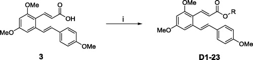
Table 1. Chemical structures of the title compounds.
The purity and spectra of all compounds were detailed in Supporting Information. All the obtained compounds gave satisfactory elementary analysis and spectroscopic data. 1H NMR,13 C NMR and ESI MS spectra were consistent with the structures. Furthermore, the structure of compound D2 was also confirmed by single crystal X-ray diffraction analysis.
Compound D2 was crystallized in the monoclinic space group P1c1. The crystal data and refinement data are listed in . Selected bond lengths and angles are given in . All bond lengths are within normal ranges. As shown in , C7-C8 and C15-C16 are both in trans form and the bond lengths of 1.327(7) and 1.321(7) Å conform to the value for double C-C bond, respectively. Similarly, the bond length of C17-O4 (1.179(7) Å) conforms to the value for C=O bond, which is a little shorter than the length of C=O (1.225(5) Å) that we observed previously in a resveratrol amide derivative (E)-3–(2,4-dimethoxy-6-((E)-4-methoxystyryl)phenyl)-1–(3,5-dimethylpiperidin-1-yl)prop-2-en-1-oneCitation33. The dihedral angle between the phenyl rings C9-C10-C11-C12-C13-C14 and C18-C19-C20-C21-C22-C23-C24 is 87.8 (2)° while the dihedral angle between the phenyl rings C1-C2-C3-C4-C5-C6 and C18-C19-C20-C21-C22-C23-C24 is 41.4 (2)°.
Figure 2. Crystal structure of compound D2.
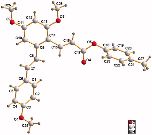
Table 2. Crystallographic data and structure refinements for compound D2.
Table 3. Selected bond lengths (Å) and angles (°) for compound D2.
Biological evaluation
Inhibition of NO production LPS-stimulated RAW264.7 cells
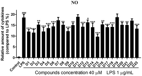
NO is a key pro-inflammatory mediator and excessive production of NO was proved to be associated with the pathogenesis of inflammation diseasesCitation34. It is generally accepted that NO inhibitors may offer potential opportunity to identify new therapeutic method for the inflammatory diseasesCitation36. To determine the potential anti-inflammation activity of these hybrids, LPS-induced RAW 264.7 cells were used as inflammatory cell model to evaluate the effects of compounds D1–23 against release of NO. As shown in , it could be seen that most compounds reflected certain effects at 40 µM. Compound D1 exhibited relatively good inhibitory activity. In general, the introduction of methyl group to the phenyl ring (D1) could lead to an obvious decline in potency (D3–D8). However, compound D2 is an exception. The introduction ofpara-CH3 group to D1 exhibited better inhibitory activity. Moreover, there was no positive influence on inhibitory activity appending electron-with drawing group like chlorine and iodine at the benzene ring of compound D1. The 4-chloro substituted analog compound D9 exhibited almost the same inhibitory activity as compound D1. However, he replacement of the chlorine atom with an iodine one caused significant decrease of activity. What is more, there was a great decrease in inhibitory activity of compound D11 when introduced 3′,5′-dimethyl groups onto compound D9. Compound D15 showed the most potent anti-inflammatory activity. Modifications of D15 with naphthalene ring and biphenyl group generated D16–23 leaded to obviously weaker inhibitory activity than compound D15. Comparison of the activity of compounds D19, D20 and D21, it could be seen that the position of the substituent grouphad an obvious effect on activity. These preliminary SAR results could be very helpful for our further SAR study. To get a better insight into the mechanism of LPS-induced NO release inhibition, the most potent compound D15 was subjected to the further study.
Figure 3. Effects of compounds D1–D23 on production of NO by RAW264.7 cell RAW264.7 cells were pretreated with D1–D23 (40 μM) for 4 h, and then stimulated with or without LPS (1 μg/mL) for 24 h. NO production was measured using nitrite and nitrate assay. ###p < .001 compared with unstimulated cells, *p < .05, **p < .01 and ***p < .001 compared with LPS-stimulated cells; Data were from at least three independent experiments, each performed in duplicate.
Compound D15 inhibited LPS-induced inflammatory mediators
To investigate the safety of the selected compounds, the potential cytotoxicity of compound D15 against the RAW264.7 cells was evaluated using MTT assay. As depicted in , compound D15 did not affect the cell viability from 6.25 to 50 μM, indicating that compound D15 was non-cytotoxic at the above concentrations. Therefore, compound D15 was used for the subsequent study from 6.25 to 50 μM.
Figure 4. Compound D15 inhibited LPS-induced inflammatory response in RAW 264.7 cells. Cells were treated with compound D15 (10, 20, 40 µM) for 12 h, and then stimulated by LPS (1 µg/ml) for 3 h. Cell viability was evaluated using the MTT assay. NO production was measured using nitrite and nitrate assay. iNOS and COX-2 expression were detected by Western blot analysis. (A) Cell viability assay; (B) Quantitative analysis of NO production. (C) Quantitative analysis of iNOS and COX-2 expression, β-actin was used as loading control. ###p < .001 compared with unstimulated cells, **p < .01 and ***p < .001 compared with LPS-stimulated cells. Data were from at least three independent experiments, each performed in duplicate.
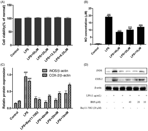
Inflammation-related diseases are closely related to the expressions of iNOS and COX-2. Thus, the inhibitory effects of compound D15 on LPS-mediated expression of iNOS and COX-2 were analyzed by Western blot. As shown in , it could be seen that the LPS (1 µg/mL) stimulation significantly induced generation of NO production as well as the expressions of iNOS and COX-2. The results preliminary demonstrated that title compound could significantly inhibited LPS-induced expressions of iNOS and COX-2 in RAW264.7 cells.
Compound D15 inhibited LPS-induced NF-κB activation
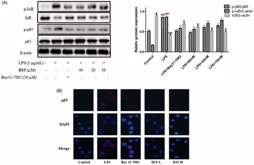
The NF-κB transcription factor family is a pleiotropic regulator of many cellular signaling pathways, providing a mechanism for the cells in response to a wide variety of stimuli to inflammation, which can activate the NF-κB signaling pathwayCitation37. Subsequently, NF-κB will be phosphorylated and the activated NF-κB will translocate from cytoplasm to nucleus promoting transcription of various inflammatory marker genes, including cytokines, chemokines, iNOS, and COX-2Citation35. In order to understand the effect of title compound D15 on NF-κB signaling LPS-induced, the relative levels of proteins p-IκB, IκB, p-p65 and p65 were examined by western blot. As shown in , LPS markedly upregulated the expressions of p-p65 and p-IκB compared to normal group. However, compound D15 concentration-dependently inhibited the expressions of above mentioned proteins LPS-induced, preliminary indicating that compound D15couldinhibit the activation of NF-κB. Consistently with the inhibitory effects of compound D15 on the expressions of proteins p-p65 and p-IκB LPS-induced, immunofluorescence staining further confirmed that LPS stimulated cells showed a clear and positive labeling for the activation p65 in nuclei. Compared to bay 11–7082, compound D15 could reduce this effect ().
Figure 5. Compound D15 suppressed LPS-induced activation of NF-κB signaling pathway in RAW 264.7 cells. After pretreatment with D15 (10 ∼ 40 µM), RAW 264.7 cells were stimulated with LPS (1 µg/mL) for 30 min. The total and phosphorylation levels of NF-κB were detected by Western blot. (A) Quantitative analysis of p-IκB and p-p65, total IκB and p65 were used as loading control, respectively. (B) Immunofluorescence analysis of compound D15. ###p < .001 compared with unstimulated cells, *p < .05 and ***p < .001 compared with LPS-stimulated cells. Data were from at least three independent experiments, each performed in duplicate.
Molecular docking
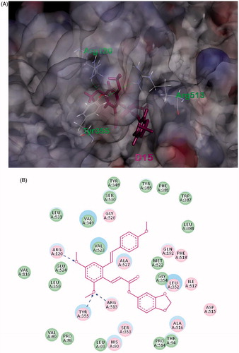
Molecular docking is an application wherein molecular modeling techniques are used to predict how a protein (enzyme) interacts with small molecules (ligands). In order to get more insight into anti-inflammatory mechanism of compound D15 and obtain more SAR clues, molecular docking studies were performed using the reported COX-2 inhibitor complex structure (PDB code: 1cx2) which obtained from the RCSB protein data bank (http://www.pdb.org)Citation38. The molecular docking procedure was performed by DS 2017 as described previously (Discovery Studio 2017, Accelrys, Inc., San Diego, CA)Citation39,Citation40.
The binding mode of compound D15 within COX-2 was depicted in . Visual inspection of the pose of D15 into the COX-2-binding site revealed that it has suitable shape complementarity with the binding pocket (), thus showed favorable binding affinity (-CDOCKER_INTERACTION_ENERGY = -43.35 kcal/mol) to the receptor via variety of interactions. The model suggests that extensive hydrophobic interactions are formed between D15 and the binding pocket of COX-2. Furthermore, its twomethoxyl groups in A-ring of resveratrol moiety formed three H-bond interactions with ARG120 (angle N-H···O = 144.0°, distance = 1.86 Å), TYR355 (angle N-H···O = 131.8°, distance = 2.05 Å), ARG513 (angle N-H···O = 143.8°, length = 2.15 Å), respectively, which were simultaneously contributed to the combination. Combining with the results of bioassays above, it was found that compound D15 might be a potential inhibitor of the COX-2 through above synergic effect, which provided more insight into its anti-inflammatory mechanism and SAR clues for further optimization of resveratrol-based anti-inflammatory agents.
Figure 6. (A) Binding model of D15 (purple) in the active site of COX-2. The H-bond is displayed as blue dashed line. (B) 2Dprojection drawing of D15 docked into COX-2 active site.
Conclusions
In summary, a novel series of resveratrol-based cinnamic ester hybrids were designed, synthesized and characterized. Their anti-inflammatory activities were evaluated in a LPS-induced RAW264.7 cell model. Among the synthesized compounds, compound D15 was found to be the most potent one to suppress NO production in LPS-induced RAW264.7 cells. Western blot experiments indicated that compound D15 inhibited LPS-induced protein expression. Furthermore, immunofluorescence revealed that compound could D15 lightly reduce activation p65 in nuclei. All the results indicate that the anti-inflammatory role of compound D15 may partly due to its inhibitory effect on the NF-κB signaling pathway LPS-induced RAW 264.7 cells.
IENZ_1381090_Supplementary_Material.pdf
Download PDF (1.6 MB)Additional information
Funding
References
- Ferrero-Miliani L, Nielsen OH, Andersen PS, et al. Chronic inflammation: importance of NOD2 and NALP3 in interleukin-1beta generation. Clin Exp Immunol 2007;147:227–35.
- Zhang Z, Rigas B. NF-kappaB, inflammation and pancreatic carcinogenesis: NF-kappaB as a chemoprevention target (review). Int J Oncol 2006;29:185–92.
- Perkins ND. Integrating cell-signalling pathways with NF-kappaB and IKK function. Nat Rev Mol Cell Biol 2007;8:49–62.
- Libby P, Ridker PM, Maseri A. Inflammation and atherosclerosis. Circulation 2002;105:1135.
- Azizi G, Navabi SS, Alshukaili A, et al. The role of inflammatory mediators in the pathogenesis of Alzheimer’s disease. Sultan Qaboos Univ Med J 2015;15:305–16.
- Wu J, Li J, Cai Y, et al. Evaluation and discovery of novel synthetic chalcone derivatives as anti-inflammatory agents. J Med Chem 2011;54:8110–23.
- Chen LZ, Sun WW, Bo L, et al. New arylpyrazoline-coumarins: Synthesis and anti-inflammatory activity. Eur J Med Chem 2017;138:170–81.
- Lv XH, Ren ZL, Liu P, et al. Design, synthesis and biological evaluation of novel nicotinamide derivatives bearing a substituted pyrazole moiety as potential SDH inhibitors. Pest Manag Sci 2017; 73:1585–92.
- Gakh AA, Yu AN, Kiselevsky MV, et al. ChemInform abstract: dihydro-resveratrol—a potent dietary polyphenol. Cheminform 2011;42.
- Pervaiz S. Chemotherapeutic potential of the chemopreventive phytoalexin resveratrol. Drug Resist Updat 2004;7:333–44.
- Baur JA, Sinclair DA. Therapeutic potential of resveratrol: the in vivo evidence. Nat Rev Drug Discov 2006;5:493–506.
- Csuk R, Albert S, Siewert B, Schwarz S. Synthesis and biological evaluation of novel (E) stilbene-based antitumor agents. Eur J Med Chem 2012;54:669–78.
- Androutsopoulos VP, Ruparelia KC, Papakyriakou A, et al. Anticancer effects of the metabolic products of the resveratrol analogue, DMU-212: structural requirements for potency. Eur J Med Chem 2011;46:2586–95.
- İlhami G. Antioxidant properties of resveratrol: a structure-activity insight. Innovative Food Sci Emerg Technol 2010;11:210–18.
- Innocenti A, Gülçin I, Scozzafava A, Supuran CT. Carbonic anhydrase inhibitors. Antioxidant polyphenols effectively inhibit mammalian isoforms I-XV. Bioorg Med Chem Lett 2010;20:5050–3.
- Lolicato F, Raudino A, Milardi D, La RC. Resveratrol interferes with the aggregation of membrane-bound human-IAPP: A molecular dynamics study. Eur J Med Chem 2015;92:876–81.
- Evers F, Jeworrek C, Tiemeyer S, et al. Elucidating the mechanism of lipid membrane-induced IAPP fibrillogenesis and its inhibition by the red wine compound resveratrol: a synchrotron X-ray reflectivity study. J Am Chem Soc 2009;131:9516–21.
- Sciacca MF, Chillemi R, Sciuto S, et al. Interactions of two O-phosphorylresveratrol derivatives with model membranes. Arch Biochem Biophys 2012;521:111–16.
- Liu CW, Sung HC, Lin SR, et al. Resveratrol attenuates ICAM-1 expression and monocyte adhesiveness to TNF-α-treated endothelial cells: evidence for an anti-inflammatory cascade mediated by the miR-221/222/AMPK/p38/NF-κB pathway. Sci Rep 2017;7:44689.
- Poulsen MM, Fjeldborg K, Ornstrup MJ, et al. Resveratrol and inflammation: Challenges in translating pre-clinical findings to improved patient outcomes. Biochim Biophys Acta 2015;1852:1124–36.
- Das S, Das DK. Anti-inflammatory responses of resveratrol. Inflamm Allergy Drug Targets 2007;6:168–73.
- Wang G, Hu Z, Fu Q, et al. Resveratrol mitigates lipopolysaccharide-mediated acute inflammation in rats by inhibiting the TLR4/NF-κBp65/MAPKs signaling cascade. Sci. Rep 2017;7:45006.
- Capiralla H, Vingtdeux V, Zhao H, et al. Resveratrol mitigates lipopolysaccharide- and Aβ-mediated microglial inflammation by inhibiting the TLR4/NF-κB/STAT signaling cascade. J Neurochem 2012;120:461–72.
- Serra D, Rufino AT, Mendes AF, et al. Resveratrol modulates cytokine-induced JAK/STAT activation more efficiently than 5-aminosalicylic acid: an in vitro approach. PLoS One 2014;9:109048.
- Wang Y, Cao J, Fan Y, et al. Artemisinin inhibits monocyte adhesion to HUVECs through the NF-κB and MAPK pathways in vitro. Int J Mol Med 2016;37:1567–75.
- Zhu YD, Fu JS, Shurlknight KL, et al. Novel resveratrol-based aspirin prodrugs: synthesis, metabolism, and anticancer activity. J Med Chem 2015;58:6494–506.
- De P, Baltas M, Bedos-Belval F. Cinnamic acid derivatives as anticancer agents-a review. Curr Med Chem 2011;18:1672–703.
- Sova M. Antioxidant and antimicrobial activities of cinnamic acid derivatives. Mini-Rev Med Chem 2012;12:749.
- Giles F, Fischer T, Cortes J, et al. A phase I study of intravenous LBH589, a novel cinnamic hydroxamic acid analogue histone deacetylase inhibitor, in patients with refractory hematologic malignancies. Clin Cancer Res 2006;12:4628–35.
- Gunia-Krzyżak A, Pańczyk K, Waszkielewicz AM, Marona H. Cinnamamide derivatives for central and peripheral nervous system disorders–a review of structure-activity relationships. Chemmedchem 2015;10:1302–25.
- Musso DL, Cochran FR, Kelley JL, et al. Indanylidenes. 1. Design and synthesis of (E)-2-(4,6-difluoro-1-indanylidene)acetamide, a potent, centrally acting muscle relaxant with antiinflammatory and analgesic activity. J Med Chem 2003;46:399–408.
- Chen GZ, Zhang YL, Liu X, et al. Discovery of a New Inhibitor of Myeloid Differentiation 2 from cinnamamide derivatives with anti-inflammatory activity in sepsis and acute lung injury. J Med Chem 2016;59:2436–51.
- Yao RS, Lu XQ, Guan QX, et al. Synthesis and biological evaluation of some novel resveratrol amide derivatives as potential anti-tumor agents. Eur J Med Chem 2013;62:222–31.
- Tang ML, Zhong C, Liu ZY, et al. Discovery of novel sesquistilbene indanone analogues as potent anti-inflammatory agents. Eur J Med Chem 2016;113:63–74.
- Wang ZS, Chen LZ, Zhou HP, et al. Diarylpentadienone derivatives (curcumin analogues): Synthesis and anti-inflammatory activity. Bioorg Med Chem Lett 2017;27:1803–7.
- Anning PB, Coles B, Morton J, et al. Nitric oxide deficiency promotes vascular side effects of cyclooxygenase inhibitors. Blood 2006;108:4059–62.
- Shih RH, Wang CY, Yang CM. NF-kappaB signaling pathways in neurological inflammation: a mini review. Front Mol Neurosci 2015;8:77.
- Kurumbail RG, Stevens AM, Gierse JK, et al. Structural basis for selective inhibition of cyclooxyenase-2 by anti-inflammatory agents. Nature 1996; 384:644–8.
- Wu GS, Robertson DH, Brooks CL, III, Vieth M. Detailed analysis of grid-based molecular docking: A case study of CDOCKER-A CHARMm-based MD docking algorithm). J Comput Chem 2003;24:1549–62.
- Discovery studio. San Diego (CA): Accelrys Software Inc; 2017. Available from: http://accelrys.com/products/collaborative-science/biovia-discovery-studio [last accessed 24 Sept 2017].