
Abstract
α-Fluorinated chalcones were prepared and evaluated for their cell growth inhibitory properties against six human cancer cell lines. The most potent chalcone 4c demonstrated excellent selective toxicity against cancer cells versus normal human cells, with IC50 values at nanomolar concentration ranges against 5 cancer cell lines. A further study revealed that 4c could bind to the colchicine site of tubulin, disrupt the cell microtubule networks, and effectively inhibit tubulin polymerisation. Cellular-based mechanism studies elucidated that 4c arrested MGC-803 cell cycle at G2/M phase. In addition, 4c dose-dependently caused Caspase-induced apoptosis of MGC-803 cells through mitochondrial dysfunction. Notably, compound 4c was found to inhibit the HUVECs tube formation, migration, and invasion in vitro. Furthermore, our data suggested that treatment with 4c significantly reduced MGC-803 cells metastasis and proliferation in vitro. Overall, this work showed that chalcone hybrid 4c is a potent inhibitor of tubulin assembly with prominent anti-angiogenesis and anti-cancer properties.
1. Introduction
Cancer is one of the most formidable afflictions, which represents a leading cause of death worldwideCitation1. Drug resistance, side effects and lack of efficacy associated with current cancer chemotherapy demand the design of new and safer anticancer drugsCitation2. Vascular Disrupting Agents (VDAs) constitute an innovative and complementary approach to other standard anticancer therapies, due to the low toxicity to the normal cell, the broad-spectrum of activity against tumours, and tolerance to the development of resistanceCitation3. These compounds have been found to cause a rapid and selective shutdown in tumour blood flow to solid tumours, while the blood flow in normal tissues was leaved to be relatively intact. As a result, the endothelial cells of immature vasculature shutdown and blood flow within the tumour rapidly and dramatically reduced. Finally, extensive tumour necrosis will occur due to starved of oxygen and nutrientsCitation4.
The most studied group of VDAs is microtubule-destabilizing agents that interact at the colchicine-binding site of the αβ-tubulin dimerCitation5. Compared with other site binders, colchicine-site binders have several advantages including simpler structure, low toxicity, improved aqueous solubility and fewer issues of multidrug resistanceCitation6. The normal function of endothelial cells, including movement, invasion, attachment, arrangement, and proliferation, is highly dependent on the tubulin cytoskeleton. It has been shown that endothelial cells of immature vasculature have a less developed actin cytoskeleton and are therefore more sensitive to the effects of VDAs. Consequently, the vascular targeting properties of VDAs were invariably linked to the microtubule-binding properties of the compound. In general, colchicine-site binders have anti-angiogenesis or disrupting established tumour vasculature effects or bothCitation7. Combretastatin A4(CA-4), a representative colchicine-site binder, exhibit prominent anti-angiogenic property within solid tumour, suggesting that this class of compounds could target the tumour vasculature and ultimately lead to tumour necrosis ()Citation8. The water-soluble phosphate prodrug CA-4P (Zybrestat TM) has been approved as an orphan drug for anaplastic thyroid cancer treatment by FDACitation9. However, Z-natural stilbene compound CA-4 is prone to isomerise to the more stable E-isomer during storage and administration, which displayed a dramatically reduced activity.
Figure 1. Representive tubulin inhibitors with anti-angiogenesis potency.
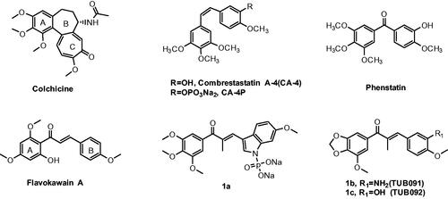
CA-4 has provided a structural model for the design of new analogs with anti-vascular and anticancer profiles. Chalcones, bearing an α, β-unsaturated carbonyl moiety, can be viewed simplistically as keto-stilbenes and mimics a portion of CA-4Citation10. Accordingly, chalcones have been used as a source of microtubule-destabilizing agents binding at the colchicine-site with the purpose to improve the chemical stability of CA-4. Flavokawain A was a promising apoptosis inducer and arrested cell cycle at G2/M against HER2-overexpressing breast cancer cells ()Citation10. Incorporated the aryl substitution pattern of CA-4 into chalcones afford compound 1a, which displayed low cytotoxicity towards normal cells and greater metabolic stability than CA-4, along with potent antiproliferative activity against drug resistant cellsCitation11. Chalcone 1b (TUB091) exhibit vascular disrupting effects and anti-metastatic activity similar to that of CA-4PCitation12.
The introduction of fluorine atoms into a bioactive molecule might affect the binding affinity with the target protein, pKa value, lipid solubility, metabolic pathways, molecular conformation and pharmacokinetic propertiesCitation13. For a specific molecule, the biological effect brought by fluorine substitution is difficult to predict. Therefore, in the process of drug optimisation, "fluorine scan" has become a fundamental routine work. Furthermore, the benzoheterocycle has assumed special significance in pharmaceutical chemistry and serve as unique versatile scaffolds for drug designCitation14. In continuation of our efforts to discover and develop novel bioactive molecules that target the tubulin-microtubule systemCitation15, we have incorporated a fluorine atom in the α-position of chalcone and substituted one of the two phenyl rings with benzoheterocycle nucleus. In all cases, as a feature structure of tubulin-binding agents, the typical 3,4,5-trimethoxyphenyl of CA-4 was retained in order to achieve the outstanding compounds. Herein, we would like to report full details of the synthesis, characterisation, and evaluation of their tumour growth inhibition against cancer cells. The lead compound 4c was also elucidated for its fundamental cytotoxic mechanisms, anti-angiogenesis, and anti-metastatic properties in vitro and in vivo.
2. Results and discussion
2.1. Chemistry
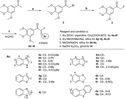
A novel series of chalcone derivatives containing α-fluoro moiety was synthesised using the procedure described in Scheme 1. The intermediate 2 was obtained by bromination at the α-position of starting material 3,4,5-trimethoxyacetophenone with tetrabutylammonium tribromide. The resulting bromide 2 was subjected to nucleophilic substitution reaction, affording fluoride 3 in the presence of potassium fluoride in dry acetonitrile. A Knoevenagel condensation between intermediate 3 and a wide range of heterocyclic aromatic aldehyde was promoted by appropriate base to provide the target chalcone analogues 4a–4t. It should be noted the amount of base may be range from 0.01 to 100 equivalents.
Scheme 1. Reagents and conditions: (a) TBABr3, DCM: MeOH = (5:2), rt, overnight; (b) KF,18-crown-6, MeCN; (c) MeOH or ethanol or THF or Dioxane, appropriate base, 0–75 °C.
2.2. Anti-cancer activity
2.2.1. Antiproliferative activity and SARs
To discover novel tubulin polymerisation inhibitors, we initially determined the anti-proliferative activity of all synthesised compounds 4a–4t against MCF-7 (human breast cancer cells), MGC-803 (human gastric cancer cells), HepG2 (human liver cancer cells), Hela (human cervical cancer cells), A549 (human non-small cell lung cancer) and U937 (human leukaemia cells) cell lines using the SRB and CCK-8 assay, while the colchicine site tubulin polymerisation inhibitor CA-4 was used as a positive control. The results listed in indicated that this series of chalcones analogues exhibited decent growth-inhibitory activity.
Table 1. Antiproliferative activities of all target compounds against different human cell lines
We synthesised six indole ring-B chalcone derivatives, of which 4b (2-phenyl) and 4e (2-methyl) showed comparable antiproliferative activity, while 4c (6-bromide) exhibited the most promising and broad-spectrum anti-tumour cell proliferation activity among all tested compounds, with IC50 values of 0.025 µM against Hela and U937, respectively. However, the introduction of t -CO2Me group at the 6-, 5- and 4-positions of indoles (4a, 4d, and 4f) resulted in a significant decrease in the anti-proliferative activity (IC50 > 5 µM), suggesting that the donating/withdrawing properties and steric hinderance of the substituents on ring-B might play a comprehensive role on the inhibitory activity. Comparing three benzothiophene ring-B chalcones (4g, 4h, and 4i), it was found that the activity was significantly affected by the position of benzothiophene attached to α, β-unsaturated ketone and the tendency of the inhibition activity is 3-position >2-position. Compared with the parent compound 4h, chalcone 4i containing methyl substation at 3-position showed diminished cytotoxic activity towards all tested cell lines. Interestingly, this SARs were also valid in benzofuran series, namely, chalcone with an CH3 group at the 3-position (4p) caused a remarkable decrease in antiproliferative activity, and the relationships between the location of heterocycles and the antiproliferative activities were still 3-position(comparable to 5-position) >2-position. Among all benzofuran chalcone derivatives, compound 4n (2,3-dihydro-benzofuran ring-B) had a more potent inhibitory effect (IC50=0.031–0.364 µM) towards several tested cancer cell lines. From the comparison of 4k (C4) with the inactive analogues 4l (C5, 6-Br) and 4r (C5, 6-NO2), we could further surmise that the linkage position of ring-B on α, β-unsaturated ketone may play a more critical role in anti-proliferation effect than properties of substituent on ring-B, such as electronegativity and atom size. Based on these findings, compound 4c was selected as the optimised compound for further studies, and to prove its mode of action.
2.2.2. Analysis of tubulin polymerisation in vitro
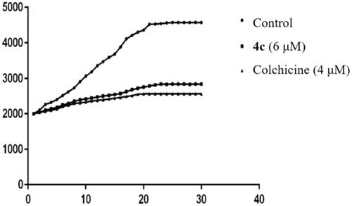
Among chalcones containing α- fluoro moiety mentioned above, 4c showed the best activity against six cell lines with an IC50 value of 0.025–0.254 µM. Accordingly, 4c was chosen to undergo the microtubule polymerisation assay in vitro and to demonstrate whether these α-fluoro chalcone derivatives are tubulin targeting agents. Colchicine, a microtubule-destabilizing agent (MDA), is known to significantly inhibit self-polymerisation of tubulin. The microtubule kinetic experiments were performed with 4 µM of colchicine as a positive control and 6 µM of 4c as the experimental group according to the method originally described by J. M. Andreu et al. with some modificationsCitation16. Purified and unpolymerised tubulin was incubated with the tested compounds and the increased tendency of fluorescence intensity shown in control samples was obviously slowed down. As shown in , compound 4c exhibited a similar action to that of colchicine, which suggest that 4c could be used as a potential MDA for cancer therapy ().
Figure 2. Effect of 4c on tubulin polymerisation in vitro. Data are presented as the mean ± SD from three independent experiments.
2.2.3. The selectivity of 4c towards normal cells
The effect of compound 4c on three types of normal human cell lines including LO2 (human hepatocyte cells), 293 T (human embryonic kidney cell) and THP-1-derived macrophages was then determined. In contrast with the positive control CA-4, 4c showed low toxicity to non-malignant cell lines especially macrophages with an IC50 value of 1210 nΜ and the selectivity ratio towards MGC-803 cells and macrophage cells was up to 21.2-folds ().
Table 2. Growth inhibitory effects of 4c on macrophage.
2.2.4. Docking profiles of compound 4c with tubulin
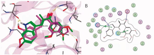
In order to explore the interaction mode of compound 4c with the colchicine binding sites of tubulin, molecular docking study was performed by using MOE (PDB code:5JVD). It should be noted that chalcones with a fluorine atom in the α-position have been shown by X-ray analysis to adopt the s-trans conformation, typical of α-methyl chalconesCitation12. As shown in , the superimposition of the best poses of compound 4c with the original ligand TUB092(α-methyl chalcone) in the colchicine binding pocket clearly demonstrated that 4c adopt almost identical binding position to that of TUB092. The 3,4,5-trimethoxyphenyl moiety of 4c was well buried into the hydrophobic pocket shaped by the side chains of β-tubulin residues Leu252, Ala250, Cys241, Leu242 and Ile318. The N atom of indole ring and carbonyl of 4c formed a hydrogen bond with residue αThr179 and βLys352 respectively, which was resembled with that of 4′-methoxy oxygen at ring-B and carbonyl of TUB902. Meanwhile, 2 D analysis of the interactions revealed that a CH-π interaction could be formed between the ring-A of 4c and βLeu255, which is highly consistent with that of TUB092. In terms of 4c, further stabilisation is provided by a cation-π interaction between the indole ring and the side chain of βLys 352. This non-polar force together with hydrogen bonds strongly help compound 4c to anchor in a dominant conformation in the binding site of tubulin and these results can describe the inhibitory effect of compound 4c.
Figure 3. Proposed binding models for 4c (green) with tubulin (PDB code:5JVD). (A) The superimposed conformation with TUB092(carmine). The hydrogen bonds were shown in yellow dashed lines. (B) The 2 D interactions between 4c and tubulin.
2.2.5. Anti-microtubule effects in MGC-803 cancer cells
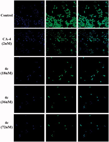
Since tubulin-microtubule system plays a crucial role in mechanical solidity, cell motility and mitosis, an immunofluorescent staining assay was further carried out in MGC-803 cells to evaluate the inhibitory effects of 4c on microtubule cytoskeleton. The morphological changes of microtubules were observed by laser confocal microscopy, as shown in . In the solvent-treated control, the intracellular network of microtubules assembled regularly and formed normal intact network with fine filaments, whereas, cells treated with 4c at different concentrations (0, 18, 36, and 72 nΜ) led to remarkable disruption of microtubule reticular network. Even at concentration as low as 18 nm, the microtubule spindle shrunk around the centre of the cells and the cellular structure was destroyed distinctly, which is very similar to positive control microtubule-destabilizing agent CA-4. Upon treatment with higher concentrations of 4c, the dispersed tubulin dimers were even more obvious and there is no tubular structure left. Such morphological microtubules changes indicated that compound 4c destroyed the microtubule organisation, thus preventing the mitosis of MGC-803 at much lower effective concentration, which might eventually induce cell apoptosis.
Figure 4. Effects of 4c on the cellular microtubule network visualised by immunofluorescence.
2.2.6. Cell cycle analysis
Most inhibitors of microtubule polymerisation are able to block mitosis in G2/M phase, which ultimately leads to apoptosis by disrupting tubulin-microtubule balanceCitation17. To elucidate whether the cytotoxicity of 4c was due to cell cycle arrest, flow cytometry analysis and PI marking method were used to evaluate the effects of 4c on the MGC-803 cell cycle distribution. As presented in , treatment of 4c at concentrations of 18, 36, 72nΜ caused a rise of cells in G2/M population from 32.15% to 50.47%, 83.65% and 90.21% respectively, indicating that 4c arrested MGC-803 cell cycle at the G2/M phase. A significant difference was also observed when we further evaluated its effects using different time points (). MGC-803 cells treated with 4c (36 nM) can be arrested in the G2/M phase from 30.37% at 6 h to 45.37% at 24 h.
Figure 5. Compound 4c induced G2/M arrest in MGC-803 cancer cells. (A) The percentages of cells at different phase of the cell cycle after treatment with various concentrations of 4c. (B) Histograms display the percentage of cell cycle distribution. (C) The percentage of different phase of the cell cycle after treatment with the same concentration for different times. (D) Histograms display the percentage of cell cycle distribution for different times. (E) Western blotting analysis on the effect of 4c on the G2/M regulatory proteins. (F) Histograms display the density rations of p-Cdc2, CyclinB1 and P21.
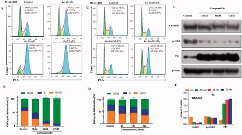
Cdc2 kinase, a member of Ser/Thr protein family, is considered as a driving force of G2/M transition. Activation of Cdc2 catalysed the phosphorylation of a variety of substrate proteins, thus promoting cell transition from G2 phase to M phaseCitation18. Cyclin B1 plays a vital role to control cell transition from G2 to M phaseCitation19. P21 also effectively regulates cell cycle progress by increasing the intracellular G2/M phase ratio. Therefore, the expression of cell cycle checkpoint protein was investigated. showed that chalcone 4c decrease the phosphorylation levels of Cdc2 in a concentration-dependent manner. We further observed that 4c caused a decrease in Cyclin B1 and increased the expression of tumour suppressor protein P21, which confirmed a G/M phase arrest by 4c. These results were consistent with flow cytometry analysis and microtubule immunofluorescence assay, which confirmed the mechanism of the cell cycle blocking effect at the cellular biochemical level.
2.2.7. Cell apoptosis assay
According to above-mentioned cell cycle analysis, we suppose that 4c might induce cells apoptosis, like other microtubule polymerisation inhibitors. Thus, the effects of 4c on MGC-803 cell apoptosis were examined by flow cytometry. As shown in , after treating MGC-803 cells with 4c (18, 36, 72nΜ) for 48 h, flow analysis showed that 4c induced apoptosis in a dose-dependent manner, with the percentage of apoptotic cells ranging from 19.12% to 57.99%, compared with 1.99% in the control group.
Figure 6. Compound 4c induced apoptosis in MGC-803 cancer cells. (A) MGC-803cells stained with Annexin V/PI, followed by flow cytometric analysis. (B) Western blot analysis of apoptosis-related proteins. (C) Histograms display the percentage of apoptosis cells. (D) Histograms display the density ratios of Caspase -3/-7/-9, cleaved Caspase -3/-7/-9, Parp and cleaved Parp.

The Caspase family proteins are considered to be critical regulators of cell apoptosis, in which Caspase-3 is one of the most important "executioner" of apoptosis induction, capable of cleaving many important cellular matrices and represent the hallmark of apoptosisCitation20. To explore the apoptosis-associated protein alterations, the expression of Caspase -3/-7/-9 and PARP was analysed. As presented in , MGC-803 cells was treated with 4c at different concentrations (18, 36 and 72 nM) for 48 h, an up-regulation of cleaved Caspase-3/-7/-9, the activated form of Caspase family apoptotic proteins, was observed. 4c treatment resulted in a cleavage of Caspase-9, indicating that it activated the exogenous apoptotic pathway and further processed other Caspase members such as Caspase-3 and -7, thereby initiating the Caspase cascade. As a result of these alteration, its substrate protein PARP, a marker of nuclear fragmentation and chromatin condensation, was cleaved and activated. These results demonstrated that compound 4c could trigger the Caspase cascade and the cleavage of PARP, eventually inducing apoptosis of MGC-803 cells.
2.2.8. In vitro evaluation of mitochondrial membrane potential (MMP) and reactive oxygen species (ROS) generation
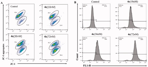
There is a close relationship between mitochondria and apoptosis, and a decrease in mitochondrial membrane potential is one of the irreversible events that occur early in apoptosis cells. Therefore, effect of compound 4c on MMP of MGC-803 cells was examined by cationic JC-1 staining method and flow cytometry. As illustrated in , with the concentration of 4c ranging from 0 to 72 nΜ, the red fluorescence intensity decreased from 92.4 to 41.3%, and the green fluorescence intensity increased from 7.13 to 55.6%, indicating that 4c produced the collapse of MMP and eventually induced MGC-803 cell apoptosis.
Figure 7. (A) Effects of 4c on the MMP of MGC-803 cells; (B) The generation of ROS was measured using DCFH-DA.
It is now generally accepted that mitochondrial dysfunction is the main source of elevated ROS levels, accompanied by changes in mitochondrial membrane potential and activation of downstream Caspases. To further evaluate the alterations of intracellular ROS levels after cells exposed to 4c, flow cytometry was analysed with 2′,7′-Dichlorodihydrofluorescein diacetate (DCFH-DA) as fluorescent probe. As shown in , 4c induced intracellular ROS generation in a dose-dependent manner. The ratio of DCF positive cells was increased from 0.17% (control) to 93.6% in cells incubated with 72 nM of 4c. According to above-mentioned results, we can deduce that 4c induced cell apoptosis through a variety of ways including the disruption of the mitochondrial membrane potential, accumulation of intracellular ROS level and regulation of apoptosis-related proteins expression in cancer cells.
2.2.9. Migration and invasion assay of MGC-803 cells
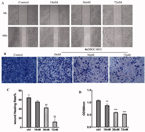
Metastasis is one of the leading causes of morbidity and mortality of cancer patients. Microtubules-targeting inhibitors have also been reported to interfere with cell migration and invasion. Accordingly, the in vitro would healing assay and Transwell assays were performed to investigate the inhibitory effect of compound 4c on the migration and invasion of MGC-803 cells. As shown , the scratches healing rate in the control group was approximately 64%. By contrast, the cells treated with 4c have much smaller scratched areas recovery at all concentrations. In particular, the migration rates were only 10% at 72 nM of 4c treatment after 48 h, which indicated that 4c could significantly impaired the migration of MGC-803 cells, and it was positively correlated with the concentration of 4c. Cell invasion is an important malignant behaviour during cancer metastasis. In Tanswell assay, MGC-803 cells were seeded into upper chambers with pre-coated Matrigel stimulating base membrane in order to assess the cellular invasion. The data shown in demonstrated that after 4c treatment, MGC-803 cells exhibited significant reduced invasion ability, mainly characterised by the decrease in absorbance value of crystal violet and the amount of MGC-3 cell invasion from the upper layer to lower layer compared with control group. These results suggested that compound 4c could inhibit tumour cell migration and invasion in a dose-dependent manner in vitro.
Figure 8. Effects of compound 4c on the migration and invasion of MGC-803 cells. (A) Images of MGC-803 cells migration inhibited by 4c; (B) Suppressing effects of 4c on the invasion of MGC-803 cells; (C) Histograms display the percentage of healing cells; (D) Histograms display the percentage of invasion cells.
2.3. Anti-angiogenesis activity
Quite a few classic tubulin polymerisation inhibitor, e.g. Taxol, 2-methoxyestradiolCitation21, or CA-4Citation22, show anti-angiogenic activity in vitro and in vivo. Due to its disorganised structure and permeability, the tumour vascular system has long been regarded as a unique target for chemotherapeutic agents that can inhibit the formation of new tumour vessels (anti-angiogenic effect) or destroy existing vessels (vascular disruption effect). For this reason, we further evaluated compound 4c for inhibitory effects on the formation of vessel-like structure on Matrigel, as well as on the angiogenesis in vitro and in vivo.
2.3.1. Migration and invasion assay of HUVEC
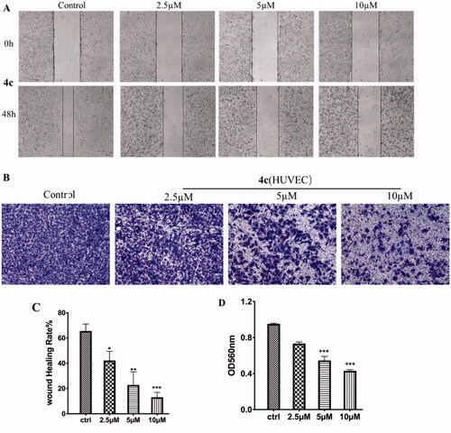
HUVECs (human umbilical vein endothelial cells) grown on Matrigel were commonly used as a tube formation model for studying the physiological and pathological processes of the vascular system in vitro, and a widely studied human endothelial cell type in angiogenesis. The effect of 4c on endothelial cell migration was assessed by using HUVECs. Standardised wounds were made in confluent monolayers of HUVECs and then exposed to compound 4c at 2.5, 5 and 10 µM for 48 h. As shown in , the wound closure rates after 48 h of 4c treatment was only 11% at the dose of 10 µM, which is much slower than 65% of the control group. We further evaluated the capacity of 4 s on endothelial cell invasion using a Transwell assay. As shown in , the control HUVECs that was not treated displayed a complete invasion from the upper chamber to lower one. However, after exposed to 4c at 2.5, 5 and 10 µM for 24 h, the number of cells invaded into lower chamber was gradually decreased in a dose-dependent manner.
Figure 9. Effects of compound 4c on the migration and invasion of HUVEC. (A) Images of HUVEC migration inhibited by 4c; (B) Suppressing effects of 4c on the invasion of HUVEC; (C) Histograms display the percentage of healing cells; (D) Histograms display the percentage of invasion cells.
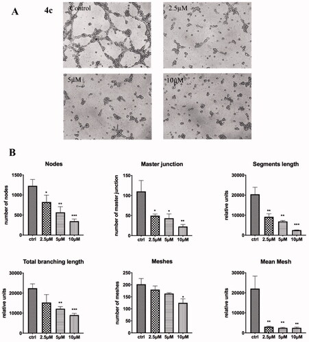
2.3.2. Tube formation of HUVECs
The formation of HUVEC tube represents the key steps in angiogenesis. Thus, we further tested the anti-vascular activity of 4c by performing a tube formation assay. As shown in HUVECs in the control group formed integrated tubules with multicentric junctions on the Matrigel matrix, while the tubes were fragmentary after exposure to 4c. HUVEC cord formation, tube area and length were dramatically suppressed even at relatively lower dose of 2.5 µM, with almost no tube structure observed. Statistical analysis showed that, compared to the control group, 4c severely impaired the tube formation ability of HUVEC. Collectively, these results highlighted that compound 4c has potent anti-vascular activity in vitro, which is evidenced by the disruption of tubule-like HUVECs, interference with HUVECs cell migration and invasion.
Figure 10. Effects of compounds 4c on the tube formation of HUVECs in vitro; (A) Images for the tube-forming inhibitory activities for 4c (0, 2.5, 5, 10 µM) in HUVECs; (C) Statistical analysis for the tube-forming capability. *p < 0.05, **p < 0.01,vs. control; ***p < 0.001 vs. control.
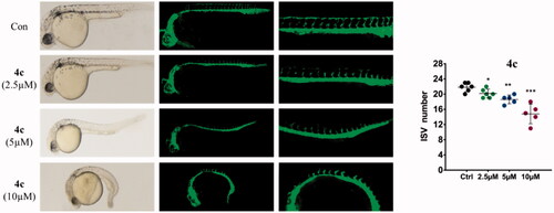
2.3.3. Anti-angiogenesis in zebrafish embryos
Since zebrafish embryos emerged as a powerful model for studying human diseases, the transgenic zebrafish with green fluorescent protein (GFP) labelled endothelial cells have been used extensively for testing angiogenesis in vivo. In this work, an anti-angiogenetic activity of 4c was confirmed by Tg(flk1: EGFP) zebrafish embryos in vivo assay. Fluorescently labelled 3hpf period embryos were treated with different doses of 4c and incubated at 28.5 °C. Until 30hpf, zebrafish was imaged under a fluorescent microscopy to observe the affection of applied agents on intersegmental vessels (ISVs) development. As shown in the , ISVs were clearly observed in newly born zebrafish embryos of solvent-treated control group, which were growing at regular longitudinal and equidistant intervals. Upon treatment with 4c, the number and length of ISVs were all dose-dependently reduced. The embryos exhibited the anti-angiogenic phenotype already at 2.5 µM dose. At the dose ≥ 5 µM, embryos exhibited marked delay in development and only scattered or fragmentary blood vessels were observed, indicating that 4c could effectively blocked the growth of normal angiogenesis.
Figure 11. Anti-angiogenic effect of compounds 4c in zebrafish embryos assay. Zebrafish embryos were incubated with 4c at 0.5, 1, and 2 µM for 24 h.
3. Conclusions
In conclusion, a series of α-fluoro chalcone derivatives was designed, synthesised, and evaluated for their antiproliferative activity, where 4c exhibited significant inhibition with IC50 ranging from 0.025 µM to 0.202 µM against five of the tested cell lines. Compound 4c, which has a bromide substituted on indole moiety ring-B, remarkably suppressed tubulin polymerisation and disrupted microtubule networks of MGC-803 cells. Molecular docking results show that 4c can bind to colchicine sites. Moreover, 4c induced MGC-803 cell cycle arrest at the G2/M phase by regulation of G2/M-related protein expression of p-Cdc2, Cyclin B1 and p21. Besides, 4c induced MGC-803 cell apoptosis, which might be the resulting effects of the cleavage of apoptotic related protein, including Caspase-3/-7/-9 and cleaved PARP. Additionally, 4c depolarised the mitochondria membrane potentials and induced ROS generation in MGC-803 cells.
The evaluation of capillary-like tube formation in vitro indicated that 4c has an inhibitory potency on HUVECs cells proliferation, which was consistent with its anti-angiogenesis activity of zebrafish embryos in vivo. Moreover, compound 4c dose-dependently inhibited the migration and invasion of HUVECs in vitro. Finally, the anti-metastasis and anti-invasion potential of 4c were confirmed in MGC-803 cells by wound healing and Transwell assay on Matrigel. Therefore, 4c deserved further investigation as a novel, structurally simple and efficacious tubule polymerisation inhibitor, and anti-angiogenesis agents for the treatment of cancer.
4. Experimental
4.1. Chemistry
All chemicals were obtained from commercial sources without further purification unless otherwise stated. Solvents and reagents were dried and purified through routine protocols. Column chromatography (CC) was performed on silica gel (200–300 mesh, Qingdao Ocean Chemical Company, China). Thin-layer chromatography (TLC) was used to monitor reactions on GF254 silica gel glass plates (Qingdao Ocean Chemical Co., China). 1HNMR and 13CNMR were recorded on a Bruker AV-400 (400 MHz) NMR instrument at 400 and 100 MHz for deuterated chloroform (CDCl3) or deuterated dimethylsulphoxide (DMSO-d6) solutions, respectively. Chemical shifts were reported in parts per million (ppm) with tetramethylsilane as an internal standard. High resolution mass spectra (HRMS) were recorded with a Q-TOF micron spectrometer.
4.1.1. 2-Bromo-1–(3,4,5-trimethoxyphenyl)ethan-1-one(2)
To a solution of 3,4,5-trimethoxyacetophenone (1.68 g, 8 mmol) in 100 ml DCM and 40 ml MeOH, was added TBABr3 and the reaction mixture was stirred at r.t. for 8 h. The solvent was removed under reduced pressure, and the residue was purified by column chromatography (PET: EA = 5:1) to obtain compound 4 as a white solid (2.0 g, 87%) . 1H NMR (400 MHz, CDCl3) δ 7.25 (s, 2H), 4.41 (s, 2H), 3.94 (s, 3H), 3.93 (s, 6H).
4.1.2. 2-Fluoro-1–(3,4,5-trimethoxyphenyl)ethan-1-one (3)
Potassium fluoride (4.8 g, 84 mmol) was dried in an infra-red oven for 8 h in advance. Compound 2 (2.0 g, 7 mmol) and 18-Crown-6 (453 mg, 1.26 mmol) were dissolved in anhydrous acetonitrile and added dropwise through a dropping funnel to a flask containing potassium fluoride, and the reaction mixture was stirred at reflux for 16 h. The solvent was removed and the residue was extracted with DCM, dried over MgSO4, concentrated, and purified by column chromatography (PET: EA = 6:1) to give compound 3 as a light yellow solid(1.3g, 85%). 1H NMR (400 MHz, CDCl3) δ 7.16 (s, 2H), 5.55 (s, 1H), 5.43 (s, 1H), 3.94 (s, 3H), 3.92 (s, 6H).
4.1.3. General procedure for synthesis of compound 4a∼4f
The intermediate 3 (50 mg, 0.22 mmol) and corresponding 1H-indole-3-carbaldehyde(1.25 equivalent) was dissolved in anhydrous ethanol, a small amount of piperidine and glacial acetic acid was added, and then the reaction mixture wasstirred at 65 °C for 4 h. The crude mixture was allowed to cooled down to 0 °C and stirred further for 30 min, then filtrated through Celite, washed with anhydrous ethanol, to obtain the target product 4a∼4f.
4.1.3.1. Methyl(Z)-3–(2-fluoro-3-oxo-3–(3,4,5-trimethoxyphenyl)prop-1-en-1-yl)-1H-indole-6-carboxylate (4a)
M. p. 207.3–209.1 °C; 1H NMR (400 MHz, CDCl3) δ 9.33 (s, 1H), 8.26 − 8.19 (m, 1H), 8.09 (s, 1H), 7.93 (dd, J = 8.4, 1.3 Hz, 1H), 7.76 (d, J = 8.4 Hz, 1H), 7.35 (d, J = 37.5 Hz, 1H), 7.21 (s, 2H), 3.97 (s, 3H), 3.96 (s, 3H), 3.94 (s, 6H); 13 C NMR (101 MHz, DMSO-d6) δ 185.30(d, J = 30.3 Hz), 166.78(s), 153.11(d, J = 258.6 Hz), 152.62(s), 139.94 (d, J = 12.1 Hz),141.18(s), 135.51(s), 133.94(d, J = 3.0 Hz), 133.82(s), 131.79(s), 129.74(s), 123.57(s), 121.14(s), 119.03(s), 115.00(d, J = 10.1 Hz), 114.03(s), 107.25(s), 106.59(s), 60.15(s), 56.06(s), 51.92(s); HRMS(ESI-TOF) m/z [M + H]+ calcd for C22H20FNO6, 414.1353, found 414.1339.
4.1.3.2. (Z)-2-fluoro-3–(2-phenyl-1H-indol-3-yl)-1–(3,4,5-trimethoxyphenyl)prop-2-en-1-one (4 b)
M. p.150.7–152.4 °C; 1H NMR (400 MHz, CDCl3) δ 8.79 (d, J = 14.1 Hz, 1H), 8.10 (d, J = 7.3 Hz, 1H), 7.55 − 7.52 (m, 2H), 7.49 − 7.42 (m, 4H), 7.33 − 7.26 (m, 2H), 7.305 (d, J = 40.2 Hz, 1H) 7.16 (s, 2H), 3.92 (s, 3H), 3.85 (s, 6H); 13 C NMR (101 MHz, CDCl3) δ 186.79(s), 186.52(d, J = 28.3 Hz), 153.58(d, J = 265.6 Hz), 152.83(s),141.94(s), 136.34(s), 132.03(s), 131.39(s), 129.23(s), 129.08(s), 128.93(s), 126.46(s), 123.68(s), 122.65, 122.48(s), 121.62 (d, J = 4.0 Hz), 115.56(d, J = 10.1 Hz), 111.22(s), 106.96(d, J = 5.1 Hz),106.66(d, J = 2.0 Hz), 60.97(s), 56.27(s); HRMS(ESI-TOF) m/z [M + H]+ calcd for C26H22FNO4, 432.1611, found 432.1620.
4.1.3.3. (Z)-3–(6-bromo-1H-indol-3-yl)-2-fluoro-1–(3,4,5-trimethoxyphenyl)prop-2-en-1-one (4c)
M. p. 229.0–229.6 °C; 1H NMR (400 MHz, CDCl3) δ 8.73 (s, 1H), 7.90 (d, J = 2.2 Hz, 1H), 7.64 − 7.57 (m, 2H), 7.35 (dd, J = 8.6, 1.6 Hz, 1H), 7.20 (d, J = 0.8 Hz, 2H), 3.96 (s, 3H), 3.93 (s, 6H); 13 C NMR (101 MHz, DMSO-d6) δ 185.27(d, J = 25.3 Hz), 153.02(d, J = 258.6 Hz), 152.61(s), 141.14(s), 137.04(s), 131.83(s), 131.67(s), 131.54(s), 125.23(s), 123.49(s), 120.97(d, J = 3.0 Hz), 115.19(s), 115.07(s), 114.769 s), 107.15(d, J = 1.0 Hz), 106.59(d, J = 2.0 Hz), 60.15(s), 56.05(s); HRMS(ESI-TOF) m/z [M + H]+ calcd for C20H17BrFNO4, 434.0398, found 434.0397.
4.1.3.4. Methyl(Z)-3–(2-fluoro-3-oxo-3–(3,4,5-trimethoxyphenyl)prop-1-en-1-yl)-1H-indole-5-carboxylate (4d)
M. p. 80.4–81.8 °C; 1H NMR (400 MHz, CDCl3) δ 9.16 (s, 1H), 8.50 (d, J = 4.0 Hz, 1H), 8.03 − 7.96 (m, 2H), 7.48 (d, J = 8.6 Hz, 1H), 7.38 (d, J = 37.3 Hz, 1H), 7.21 (s, 2H), 3.98 (s, 3H), 3.96 (s, 3H), 3.94 (s, 6H); 13 C NMR (101 MHz, DMSO-d6) δ 185.38(d, J = 25.3 Hz), 166.92(s), 152.98(d, J = 258.6 Hz), 152.61(s), 141.21(s),138.89(s), 132.51(d, J = 11.1 Hz), 131.84(s), 125.69(s), 123.36(s), 122.04(s), 121.65(d, J = 4.0 Hz), 115.16(d, J = 11.1 Hz), 112.30(s), 108.17(s), 106.72(d, J = 3.0 Hz), 60.15(s), 56.05(s), 51.82(s); HRMS (ESI-TOF) m/z [M + H]+ calcd for C22H20FNO6, 414.1339, found 414.1350.
4.1.3.5. (Z)-2-fluoro-3–(2-methyl-1H-indol-3-yl)-1–(3,4,5-trimethoxyphenyl)prop-2-en-1-one (4e)
M. p. 204.0–206.4 °C;1H NMR (400 MHz, CDCl3) δ 8.51 (s, 1H), 7.95 (d, J = 8.0 Hz, 1H), 7.34 − 7.30 (m, 1H), 7.25 − 7.19 (m, 4H), 7.23 (d, J = 40.4 Hz,1H), 3.95 (s, 3H), 3.93 (s, 6H), 2.54 (s, 3H); 13 C NMR (101 MHz, Chloroform-d) δ 186.72(d, J = 27.3 Hz), 152.88(s), 152.57(d, J = 261.6 Hz),141.87(s), 139.88(s), 135.76(s), 126.42(s), 122.68(s), 121.28(d, J = 4.0 Hz), 121.13(s), 115.05(d, J = 10.1 Hz), 110.78(s), 106.95(d, J = 4.0 Hz), 106.42(d, J = 1.0 Hz), 61.01(d, J = 2.0 Hz), 56.33(d, J = 2.0 Hz), 12.95; HRMS (ESI-TOF) m/z [M + H]+ calcd for C21H20FNO4, 370.1455, found 370.1444.
4.1.3.6. Methyl(Z)-3–(2-fluoro-3-oxo-3–(3,4,5-trimethoxyphenyl)prop-1-en-1-yl)-1H-indole-4-carboxylate (4f)
M. p. 118.0–119.0 °C; 1H NMR (400 MHz, CDCl3) δ 9.45 (s, 1H), 8.23 (d, J = 2.9 Hz, 1H), 8.12 (d, J = 39.9 Hz, 1H), 7.80 (dd, J = 7.5, 0.9 Hz, 1H), 7.65 (dd, J = 8.1, 0.9 Hz, 1H), 7.29 (d, J = 7.8 Hz, 1H), 7.18 (s, 2H), 3.98 (s, 6H), 3.94 (s, 3H), 3.80 (s, 3H); 13 C NMR (101 MHz, DMSO-d6) δ 185.77(d, J = 23.23 Hz), 167.81(s), 152.51(d, J = 26.26 Hz), 140.66(d, J = 74.74 Hz), 137.30(s), 124.15(s), 123.59(s), 122.97(s), 121.68(s), 118.83 (d, J = 6.06 Hz), 106.34(s), 55.89(d, J = 2.02 Hz), 55.47(s), 51.66(s); HRMS (ESI-TOF) m/z [M + H]+ calcd for C22H20FNO6, 414.1353, found 414.1338.
4.1.4. General procedure for synthesis of compound 4 g∼4j,4s,4t
38 mg of sodium (1.65 mmol) was dispersed into anhydrous methanol under argon atmosphere. After the disappearance of sodium, the intermediate 3 (50 mg, 0.22 mmol) and corresponding aromatic aldehyde (1.25 equivalents) was added and stirred at room temperature for 10 min and then stirred at 65 °C for additional 5 h. After completion of the reaction, the mixture was precipitated after 30 min of stirring under ice bath, filtered, and washed with cold methanol, to afford the target compounds.
4.1.4.1. (Z)-3-(benzo[b]thiophen-3-yl)-2-fluoro-1–(3,4,5-trimethoxyphenyl)prop-2-en-1-one (4 g)
M. p. 118.0–119.0 °C; 1H NMR (400 MHz, CDCl3) δ 8.24 (s, 1H), 7.90 (dd, J = 19.6, 7.5 Hz, 2H), 7.46 (dtd, J = 17.8, 7.2, 1.1 Hz, 2H), 7.33 (d, J = 35.7 Hz, 1H), 7.25 (d, J = 1.0 Hz, 2H), 3.97 (s, 3H), 3.94 (s, 6H); 13 C NMR (101 MHz, DMSO-d6) δ 152.70(d, J = 28.28 Hz), 142.18(d, J = 35.35 Hz), 138.96(d, J = 33.33 Hz), 137.57(s), 132.14(s), 130.93(s), 125.17(d, J = 12.12 Hz), 124.84(s), 124.57(s), 123.02(d, J = 12.12 Hz), 122.90(s), 122.12 (s), 121.70(s), 111.66(s), 106.97(d, J = 50.5 Hz), 59.99(d, J = 23.23 Hz), 56.13(s), 55.65(s) ; HRMS (ESI-TOF) m/z [M + H]+ calcd for C20H17FO4S, 373.0910, found 373.0989.
4.1.4.2. (Z)-3-(benzo[b]thiophen-2-yl)-2-fluoro-1–(3,4,5-trimethoxyphenyl)prop-2-en-1-one (4 h)
M. p. 122.2–123.6 °C;1H NMR (400 MHz, CDCl3) δ 7.85 (m, 2H), 7.64 (s, 1H), 7.40 (m, 2H), 7.32 (d, J = 35.0 Hz, 1H), 7.24 (d, J = 1.1 Hz, 2H), 3.96 (s, 3H), 3.94 (s, 6H); 13 C NMR (101 MHz, CDCl3) δ 185.23(d, J = 27.3 Hz),155.53(d, J = 276.7 Hz), 153.02(s), 142.81(s), 142.11(s), 142.03(s), 138.81(s), 133.97(d, J = 5.1 Hz), 130.80(s), 129.17 (d, J = 5.1 Hz), 126.09(s), 124.89(s), 124.42(d, J = 9.1 Hz), 122.31(s), 114.42(d, J = 9.1 Hz), 107.15(d, J = 6.1 Hz), 61.04(d, J = 2.0 Hz), 56.39(d, J = 1.0 Hz); HRMS (ESI-TOF) m/z [M + H]+ calcd for C20H17FO4S, 373.0910, found 373.0968.
4.1.4.3. (Z)-2-fluoro-3–(3-methylbenzo[b]thiophen-2-yl)-1–(3,4,5-trimethoxyphenyl)prop-2-en-1-one (4i)
M. p. 140.5–143.4 °C; 1H NMR (400 MHz, CDCl3) δ 7.89 − 7.82 (m, 1H), 7.78 (dd, J = 6.1, 2.9 Hz, 1H), 7.51 (d, J = 35.5 Hz, 1H), 7.45 − 7.40 (m, 2H), 7.27 − 7.24 (m, 2H), 3.96 (s, 3H), 3.94 (s, 6H), 2.55 (s, 3H); 13 C NMR (101 MHz, CDCl3) δ 185.27(d, J = 27.3 Hz), 155.28(d, J = 275.7 Hz), 153.00(s), 142.75(s), 141.36(d, J = 10.1 Hz), 139.05(s), 136.60(d, J = 3.0 Hz), 130.97(d, J = 2.0 Hz), 128.65(d, J = 7.1 Hz), 126.39(s), 124.50(s), 122.81(d, J = 3.0 Hz), 122.33(s), 112.43(d, J = 9.1 Hz), 107.18(d, J = 6.1 Hz), 106.07(s), 61.02(s), 56.37(s), 12.55(s); HRMS (ESI-TOF) m/z [M + Na]+ calcd for C21H19FO4S, 387.1066, found 387.1054.
4.1.4.4. (Z)-3-(1H-benzo[d]imidazol-2-yl)-2-fluoro-1–(3,4,5-trimethoxyphenyl)prop-2-en-1-one (4j)
M. p. 178.9–180.9 °C; 1H NMR (400 MHz, CDCl3) δ 10.38 (s, 1H), 7.74 (s, 1H), 7.62(s,1H),7.30 (d, J = 3.6 Hz, 2H), 7.20 (s, 2H), 6.62 (s, 1H), 3.96 (s, 3H), 3.92 (s, 6H); 13 C NMR (101 MHz, CDCl3) δ 185.06(d, J = 27.27 Hz), 157.43(d, J = 274.72 Hz), 153.14(s), 144.24(d, J = 3.03 Hz), 143.23(s), 130.13(s), 128.83(s), 110.61(d, J = 6.06 Hz), 107.10(d, J = 4.04 Hz), 61.09(s), 56.40(s), 30.57(s), 13.74(s); HRMS (ESI-TOF) m/z [M + H]+ calcd for C19H17FN2O4, 357.1251, found 357.1270.
4.1.4.5. (Z)-3-(dibenzo[b, d]thiophen-2-yl)-2-fluoro-1–(3,4,5-trimethoxyphenyl)prop-2-en-1-one (4 s)
M. p. 142.6–143.8 °C; 1H NMR (400 MHz, CDCl3) δ 8.50 (s, 1H), 8.21 (dd, J = 5.8, 3.1 Hz, 1H), 7.94 − 7.86 (m, 2H), 7.81 (dd, J = 8.4, 1.6 Hz, 1H), 7.53 − 7.47 (m, 2H), 7.24 (d, J = 1.1 Hz, 2H), 7.10 (d, J = 36.5 Hz, 1H), 3.97 (s, 3H), 3.95 (s, 6H); 13 C NMR (101 MHz, CDCl3) δ 186.48(d, J = 29.3 Hz), 155.97(d, J = 273.7 Hz), 153.01(s), 142.59(s), 141.39(s), 139.72(s), 136.12(s), 135.05(s), 131.16(s), 127.79(d, J = 4.0 Hz), 127.34(s), 124.82 (s), 123.76(d, J = 9.1 Hz), 123.22(s), 122.96(s), 121.76(s), 119.96(d, J = 5.1 Hz), 107.13(d, J = 5.1 Hz), 61.05(s), 56.41(s); HRMS (ESI-TOF) m/z [M + H]+ calcd for C24H19FO4S, 423.1066, found 423.1050.
4.1.4.6. (Z)-3-(benzofuran-3-yl)-2-fluoro-1–(3,4,5-trimethoxyphenyl)prop-2-en-1-one (4t)
M. p.103.7–104.8 °C;1H NMR (400 MHz, CDCl3) δ 8.23 (s, 1H), 7.74 (s, 1H), 7.57 (s, 1H), 7.41 − 7.34 (m, 2H), 7.24 (d, J = 1.2 Hz, 2H), 7.16 (d, J = 37.8 Hz, 1H), 3.97 (s, 3H), 3.94 (s, 6H). 13 C NMR (101 MHz, Chloroform-d) δ 155.02(s), 153.03(s), 148.46(s), 147.77(d, J = 15.15 Hz), 125.76(s), 125.41(s), 123.63(s), 119.63(d, J = 3.03 Hz), 111.87(s), 109.48(d, J = 12.12 Hz), 107.35(s), 107.10(d, J = 5.05 Hz), 106.76(s), 61.05(s), 56.38(s); HRMS (ESI-TOF) m/z [M + H]+ calcd for C20H17FO5, 357.1138, found 357.1143.
4.1.5. General procedure for synthesis of compound 4k∼4q
To a solution of corresponding aromatic aldehyde (1 equivalent) in 5 ml anhydrous methanol, was added NaOH (4 equivalents) and compound 3 (1 equivalent), and then stirred at room temperature for 5 min and at 65 °C for additional 2 h. Reaction completion was monitored by TLC. The reaction mixture was precipitated after cooling and filtered to obtain the target compounds 4k∼4q.
4.1.5.1. (Z)-3-(benzo[d][1,3]dioxol-4-yl)-2-fluoro-1–(3,4,5-trimethoxyphenyl)prop-2-en-1-one (4k)
M. p. 148.1–149.1 °C; 1H NMR (400 MHz, CDCl3) δ 7.45 (d, J = 8.0 Hz, 1H), 7.19 (s, 2H), 7.05 (d, J = 36.9 Hz, 1H), 6.93 − 6.82 (m, 2H), 6.02 (s, 2H), 3.95 (s, 3H), 3.92 (s, 6H); 13 C NMR (101 MHz, CDCl3) δ 186.27(d, J = 29.3 Hz), 156.18(d, J = 275.7 Hz), 152.96(s), 147.43(s),146.92(s), 142.60(s), 130.96(s), 122.32(d, J = 13.1 Hz), 122.02(s), 113.86(s), 112.44(d, J = 7.1 Hz), 109.80(d, J = 2.0 Hz), 107.14(d, J = 5.1 Hz), 101.28(s), 61.01(d, J = 1.0 Hz), 56.34(d, J = 2.0 Hz); HRMS (ESI-TOF) m/z [M + H]+ calcd for C19H17FO6, 361.1087, found 361.1084.
4.1.5.2. (Z)-3–(6-bromobenzo[d][1,3]dioxol-5-yl)-2-fluoro-1–(3,4,5-trimethoxyphenyl)prop-2-en-1-one (4l)
M. p.156.3–157.8 °C; 1H NMR (400 MHz, CDCl3) δ 7.53 (s, 1H), 7.26 (d, J = 36.0 Hz, 1H), 7.18 (s, 2H), 7.11 (s, 1H), 6.06 (s, 2H), 3.95 (s, 3H), 3.94 (s, 6H); 13 C NMR (101 MHz, CDCl3) δ 186.49(d, J = 27.3 Hz), 155.06(d, J = 273.7 Hz), 152.98(s), 149.61(d, J = 2.0 Hz), 147.72(s), 142.55(s), 131.01(s), 124.38(d, J = 4.0 Hz), 118.79(d, J = 3.0 Hz), 117.93(d, J = 2.0 Hz), 113.18(s), 110.33(d, J = 15.2 Hz), 107.08(d, J = 4.0 Hz), 102.36(s), 61.01(d, J = 1.0 Hz), 56.32(d, J = 1.0 Hz).HRMS (ESI-TOF) m/z [M + H]+ calcd for C19H16BrFO6, 439.0193, found 439.0175.
4.1.5.3. (Z)-3-(benzofuran-5-yl)-2-fluoro-1–(3,4,5-trimethoxyphenyl)prop-2-en-1-one (4 m)
M. p. 94.1–94.2 °C;1H NMR (400 MHz, CDCl3) δ 8.03 (s, 1H), 7.68 (d, J = 2.3 Hz, 1H), 7.55 (d, J = 8.8 Hz, 1H), 7.21 (d, J = 1.2 Hz, 2H), 7.02 (d, J = 36.6 Hz, 1H), 6.83 (s, 1H), 3.96 (s, 3H), 3.93 (s, 6H); 13 C NMR (101 MHz, CDCl3) δ 186.65(d, J = 28.3 Hz), 155.34(d, J = 272.3 Hz),152.97(s), 146.11(s), 142.51(s), 131.25(s), 128.15(s), 127.30(d, J = 8.1 Hz), 124.04(d, J = 9.1 Hz), 120.40(d, J = 6.1 Hz), 111.96(s), 111.42(s), 107.11(d, J = 5.1 Hz), 106.85(s),65.57(s), 61.01(d, J = 1.0 Hz), 56.37(d, J = 1.0 Hz); HRMS (ESI-TOF) m/z [M + H]+ calcd for C20H17FO5, 357.1138, found 357.1142.
4.1.5.4. (Z)-3–(2,3-dihydrobenzofuran-5-yl)-2-fluoro-1–(3,4,5-trimethoxyphenyl)prop-2-en-1-one (4n)
M. p. 100.8–103.7 °C;1 H NMR (400 MHz, CDCl3) δ 7.65 (s, 1H), 7.46 (d, J = 8.4 Hz, 1H), 7.17 (s, 2H), 6.88 (d, J = 28.6 Hz, 1H), 6.82 (s, 1H), 4.65 (t, J = 8.7 Hz, 2H), 3.94 (s, 3H), 3.92 (s, 6H), 3.27 (t, J = 8.6 Hz, 2H); 13 C NMR (101 MHz, CDCl3) δ 186.40(d, J = 28.28 Hz), 161.94(s), 152.93(s), 142.26(s), 132.12(d, J = 7.07 Hz), 131.53(s), 128.19(s), 127.46(d, J = 10.1 Hz), 124.12(d, J = 5.05 Hz), 120.72(d, J = 12.12 Hz), 109.86(s), 106.99(d, J = 5.05 Hz), 71.93(s), 61.00(s), 56.34(s), 29.33(s); HRMS (ESI-TOF) m/z [M + H]+ calcd for C20H19FO5, 359.1289, found 381.1286.
4.1.5.5. (Z)-3-(benzofuran-2-yl)-2-fluoro-1–(3,4,5-trimethoxyphenyl)prop-2-en-1-one (4o)
M. p. 115.5–116.6 °C; 1H NMR (400 MHz, CDCl3) δ 7.65 (d, J = 7.8 Hz, 1H), 7.52 (d, J = 8.2 Hz, 1H), 7.39 (d, J = 7.2 Hz, 1H), 7.32 (s, 1H), 7.28 (s, 1H), 7.23 (s, 2H), 7.08 (d, J = 34.4 Hz,1H), 3.96 (s, 3H), 3.94 (s, 6H); 13 C NMR (101 MHz, CDCl3) δ 185.09(d, J = 27.3 Hz), 155.29(d, J = 203.0 Hz), 153.04(s), 148.75(s), 142.89(s), 130.63(s), 128.48(s), 126.37(s), 123.53(s), 121.85(s), 112.13(d, J = 12.1 Hz), 112.01(s), 111.48(s), 109.30(d, J = 9.1 Hz), 107.16(d, J = 5.1 Hz), 106.16(s), 61.04(d, J = 2.0 Hz), 56.39(d, J = 1.0 Hz); HRMS (ESI-TOF) m/z [M + H]+ calcd for C20H17FO5, 357.1138, found 357.1127.
4.1.5.6. (Z)-2-fluoro-3–(3-methylbenzofuran-2-yl)-1–(3,4,5-trimethoxyphenyl)prop-2-en-1-one (4p)
M. p.117.4–118.8 °C; 1H NMR (400 MHz, CDCl3) δ 7.55 (dd, J = 12.7, 8.0 Hz, 2H), 7.39 (d, J = 7.2 Hz, 1H), 7.30 (d, J = 1.4 Hz, 2H), 7.28 (s, 1H), 7.09 (d, J = 34.5 Hz, 1H), 3.96 (s, 3H), 3.95 (s, 6H), 2.40 (d, J = 1.4 Hz, 3H); 13 C NMR (101 MHz, CDCl3) δ 185.45(d, J = 27.3 Hz), 155.40(d, J = 3.0 Hz), 154.96(d, J = 281.8 Hz), 152.96(s), 145.72(d, J = 5.1 Hz), 142.79(s), 130.80(d, J = 2.0 Hz), 129.07(s), 126.78(s), 122.99(s), 122.01(d, J = 5.1 Hz), 111.52(s), 107.28(d, J = 6.1 Hz), 106.07(s), 105.76(d, J = 8.1 Hz), 61.01, 56.35, 8.83; HRMS (ESI-TOF) m/z [M + H]+ calcd for C21H19FO5, 371.1295, found 371.1284.
4.1.5.7. (Z)-3-(dibenzo[b,d]furan-2-yl)-2-fluoro-1–(3,4,5-trimethoxyphenyl)prop-2-en-1-one (4q)
M. p. 114.0–116.4 °C; 1H NMR (400 MHz, CDCl3) δ 8.36 (s, 1H), 7.99 (s, 1H), 7.81 (d, J = 10.2 Hz, 1H), 7.61 (dd, J = 8.3, 6.5 Hz, 2H), 7.51 (d, J = 7.7 Hz, 1H), 7.39 (s, 1H), 7.24 (d, J = 0.9 Hz, 2H), 7.16 (d, J = 22.4 Hz, 1H), 3.97 (s, 3H), 3.95 (s, 6H); 13CNMR (101 MHz, CDCl3) δ 186.55(d, J = 28.3 Hz), 156.84(d, J = 3.0 Hz), 156.9(s), 155.52(d, J = 254.5 Hz), 142.53(s), 131.21(s), 130.17(d, J = 8.1 Hz), 127.87(s), 126.34(d, J = 8.1 Hz), 125.08(s), 123.63(s), 123.23(s), 123.17(d, J = 9.1 Hz), 120.92(s), 120.04(d, J = 6.1 Hz), 112.22(d, J = 32.3 Hz), 107.10(d, J = 5.1 Hz), 61.05(s), 56.39(s); HRMS (ESI-TOF) m/z [M + H]+ calcd for C24H19FO5, 407.1295, found 407.1281.
4.1.6. General procedure for synthesis of compound 4r
Compound 3 (50 mg, 0.22 mmol), 2-nitro piperonal (45 mg, 0.23 mmol), NaOH (20 mg, 0.5 mmol), K2CO3 350 mg (2.5 mmol) were added to the mortar, mixed with an appropriate amount of silica gel, stirred and then ground. Reaction completion was evaluated by silica TLC. The reaction was quenched with water, extracted with DCM, and the organic phase was dried over MgSO4, concentrated in vacuo, and then recrystallized with ethanol to obtain 4r (42 mg).
4.1.6.1. (Z)-2-fluoro-3–(6-nitrobenzo[d][1,3]dioxol-5-yl)-1–(3,4,5-trimethoxyphenyl)prop-2-en-1-one (4r)
M. p. 174.7–176.6 °C; 1H NMR (400 MHz, CDCl3) δ 7.59 (s, 1H), 7.47 (d, J = 29.9 Hz, 1H), 7.26 (s, 2H), 7.11 (s, 1H), 6.16 (d, J = 2.0 Hz, 2H),3.97 (s, 6H), 3.95 (s, 3H); 13 C NMR (101 MHz, DMSO-d6) δ 192.93 (d, J = 20.2 Hz), 151.91(s), 147.19(s), 141.98(s), 140.70(s), 133.66(d, J = 6.06 Hz), 129.95(s), 124.48(s), 108.78(s), 105.89(s), 104.52(s), 103.50(s), 95.04(s), 93.18(s), 68.24(d, J = 19.19 Hz), 60.16 (d, J = 2.02 Hz), 56.05(d, J = 3.03 Hz); HRMS (ESI-TOF) m/z [M + H]+ calcd for C19H16FNO8, 406.0938, found 406.0927.
4.2. Biology
4.2.1. Materials
MCF-7, MGC-803, Hela and HepG-2 cell lines were cultured in a base medium (90% DMEM and 10% FBS), A549 and U937 were cultured in a base medium (90% PRMI-1640 and 10% FBS) at 37 °C, 5% CO2 and a saturated humidity atmosphere. All the mediums were supplemented with 10% FBS and 1% ampicillin/streptomycin (all cells were purchased from Shanghai Institutes for Biological Sciences, Chinese Academy of Sciences, Shanghai, China).
4.2.2. Cell lines growth inhibition. SRB
A549, MGC-803, MCF-7, HepG2 and Hela cells were seeded at the appropriate cell line density (3 × 10³ to 4 × 10³ cells/well). The cells were then incubated at 37 °C for 24 h in a humidified 5%CO2 in 96-well plates prior to experiments. When the cells adhered, different concentrations of test compounds were added to each well as shown in the figure and table. After a further 72 h of incubation, cell proliferation was quantified with SRB (sulfonamide B) to determine the number of surviving cells. The absorbance at 560 nm was measured by microplate. The experiment was repeated three times, and the data were listed in the form of mean and standard deviation.
4.2.3. CCK8 procedure
MGC-803 cells were collected and seeded into 96 well plates at a density of 8000/well. The mediums containing drugs and cell suspension were added into the well. The cells were incubated in 5% CO2 incubator at 37 °C for 72 h. Each well was added 10 µL CCK8 solution, and the incubation time was 3–4 h. The absorbance value at 450 nm was measured by microplate reader. Data are calculated with GraphPad Prism5 software.
4.2.4. Immunofluorescent staining
MGC-803 cells (3 × 10⁴ cells/well) were seeded into 6 well-plates and then treated with vehicle control 0.1% DMSO and 4c (18, 36, 72nΜ). The cells were rinsed twice with PBS and fixed by 4% paraformaldehyde for 20 min. Subsequently, microtubule detection was performed using a fluorescent antibody against α-tubulin in 5% BSA and incubated for 5 ∼ 6 h at room temperature. To stain cell nucleus, crystals were incubated with Hoechst for 10 min. After washing with PBST to remove redundant stain, the samples were visualised under an Olympus laser scanning confocal microscope.
4.2.5. Cell cycle distribution assay
MGC-803 cells (15 × 104 cells/well) were inoculated in 6-well plates and incubated at 37 °C for 24 h, and then treated with 4c (18, 36, 72nΜ). The collected cells were washed with cold PBS and then fixed in 75% ethanol in PBS at −20 °C overnight. The cells were then washed twice with PBS buffer and then stained with 500 μL of sodium iodide (PI, 5 mg) staining solution containing isothiocyanate fluorophore for 15 min in the dark. Samples were immediately assayed for DNA content using a flow cytometer (Becton, Dickinson and Company, USA). The percentage of cell cycle phases was analysed by the software provided in the instrument.
4.2.6. Cell apoptosis assay
MGC-803 cells at 8 × 10⁴ cells/well were seeded in 12-well cell culture plates overnight, and then the cultured cells were incubated with a dose range of compounds 4c for 48 h. The treated or untreated cells were washed twice with PBS and stained by Annexin V-PE and PI for 15 min without light exposure according to the manufacturer’s protocol. Apoptosis was quantified using a flow cytometer (Becton, Dickinson and Company, USA).
4.2.7. Western blot assay
The protein levels in MGC-803 cells were assessed via western blot analysis. Exponentially growing MGC-803 cells were seeded at a density of 6 × 10⁵/dishes and incubated overnight. Then 18, 36, 72nΜ of 4c were added in triplicate for 48 h incubation. After the cells were washed with cold PBS, the supernatant was removed. The supernatant was collected by centrifuging at 12,000 g for 10 min at 4 °C. The protein concentration in the supernatant was determined by using a BSA protein assay kit. Samples were prepared in SDS-PAGE loading buffer after BSA analysis to quantify proteins, then boiled for 10 min at 100 °C. Western blot analyses were conducted after separation by SDS-PAGE electrophoresis and transfer to nitrocellulose filter (NC) membranes. Immunoblotting was performed according to the antibody manufacturers’ recommendations.
4.2.8. Mitochondrial membrane potential analysis
The cultured cells were spread in 6-well plates with about 1.5 × 10⁵ cells per well and incubated overnight at 37 °C. Different concentrations of the 4c were added to the wells, and the incubation was continued for 48 h. The wells were washed twice with JC-1 staining buffer (1×) after resuspension with an appropriate amount of JC-1 staining buffer (1×), flow cytometry assays were performed according to standard procedures
4.2.9. Measurement of intracellular ROS generation
Load the probe and dilute DCFH-DA with serum-free culture medium at 1:1000 to a final concentration of 10 μM/mL. Spread the cultured cells in a 6-well plate with about 1.5 × 10⁵ cells per well and incubate overnight at 37 °C. After 48 h of incubation with 4c (18, 36, 72nΜ) the cell culture medium was removed and diluted DCFH-DA was added and incubated for 20 min at 37 °C in an incubator, and the mixture was inverted every 3–5 min. To fully remove the DCFH-DA that did not enter the cells, the cells were washed three times with serum-free cell culture medium. Add 200 μL of PBS for resuspension, place on ice, and subsequently assay by flow cytometry using standard procedures.
4.2.10. Tube formation assay
The matrix gel was thawed overnight at 4 °C and then added to 96 well plates. After incubated at 37 °C for 45 min, the matrix gel was solidified. Different concentrations of 4c were added to each well at the density of HUVECs was 1 × 10⁵/well. After incubated at 37 °C for 6–8 h, the formation of tubules was observed under microscope
4.2.11. Would healing migration assays
MGC-803 cells and HUVECs were seeded in 24-well plates and cultivated for 24 h at 37 °C. The cells were scribed vertically with a gun tip and washed with PBS to remove dead cells. The containing different concentrations of 4c were added to the scratched monolayers. The migrated cells were photographed under a light microscope at indicated time points from the scratch. Take a picture and use the image J software to calculate the area of the cell scratches.
4.2.12. Transwell cell invasion experiment
Matrix gel was thawed overnight at 4 °C. Transwell chambers were placed into 24-well plates with 6 × 10⁴ cells per well, different concentrations of 4c prepared 10% FBS medium were added, incubated at 37 °C with 5% CO2 for 48 h. Then the cells were washed three times with PBS and fixed cells by 4% paraformaldehyde for 30 min. Subsequently, 0.2% crystal violet solution was added, stained for 30 min, and the invasion of cells was observed under the microscope. Transwell was placed into a 24-well plate, 35% acetic acid (300 μL) was added to the upper chamber of the chamber, and after acetic acid completely entered the lower chamber, 100 μL of acetic acid from the lower chamber was aspirated into a 96-well plate, and three replicate wells of each concentration were shaken for 10 min, and the absorbance was measured at 560 nm with an enzyme marker.
4.2.13. Zebrafish angiogenesis assay
The transgenic flk: enhanced GFP zebrafish embryos were transferred into 6-well plates (n = 30 per well) with 1 ml of aquaculture water (0.2 g/L of Instant Ocean Salt in distilled water) and raised at 28 °C. Then the embryos dechorionated at 12 h post fertilisation (hpf) was treated with the indicated concentrations of 4c or vehicle (DMSO). About 30 embryos were screened for each concentration gradient. At 30hpf, the zebrafish embryos were stripped, anaesthetised, and fixed with agarose gel. The images were taken under the microscope, and the number of blood vessels between the segments was recorded. Results were obtained from three independent determinations and presented as mean ± SD.
Supplemental Material
Download PDF (3.2 MB)Disclosure statement
No potential conflict of interest was reported by the author(s).
Additional information
Funding
References
- Kaasa S, Loge JH, Aapro M, et al. Integration of oncology and palliative care: a lancet oncology commission. Lancet Oncol 2018;19:E588–653.
- Holohan C, Schaeybroeck SV, Longley DB, Johnston PG. Cancer drug resistance: an evolving paradigm. Nat Rev Cancer 2013;13:714–26.
- (a) Tozer GM, Kanthou C, Baguley BC. Disrupting tumour blood vessels. Nat Rev Cancer 2005;5:423–35. (b) Mckeage MJ, Baguley BC. Disrupting established tumor blood vessels: an emerging therapeutic strategy for cancer. Cancer 2010;116:1859–71.
- Schwartz EL. Antivascular actions of microtubule-binding drugs. Clin Cancer Res 2009;15:2594–601.
- Liu ZP, Liu YN, Ji YT. Tubulin colchicine binding site inhibitors as vascular disrupting agents in clinical developments. Curr Med Chem 2015;22:1348–60.
- Porcu E, Bortolozzi R, Basso G, Viola G. Recent advances in vascular disrupting agents in cancer therapy. Future Med Chem 2014;6:1485–98.
- Kanthou C, Tozer GM. The tumor vascular targeting agent Combretastatin A-4-phosphate induces reorganization of the actin cytoskeleton and early membrane blebbing in human endothelial cells. Blood 2002;99:2060–9.
- Ducki S, Rennison D, Woo M, et al. Combretastatin-like chalcones as inhibitors of microtubule polymerization. Part 1: synthesis and biological evaluation of antivascular activity. Bioorg Med Chem 2009;17:7698–710.
- Zweifel M, Jayson GC, Reed N, et al. Phase II trial of combretastatin A4 phosphate, carboplatin, and paclitaxel in patients with platinum-resistant ovarian cancer. Ann Oncol 2011;22:2036–41.
- Zhuang C, Zhang W, Sheng C, et al. Chalcone: a privileged structure in medicinal chemistry. Chem Rev 2017;117:7762–810.
- Yan J, Chen J, Zhang S, et al. Synthesis, evaluation, and mechanism study of novel indole-chalcone derivatives exerting effective antitumor activity through microtubule destabilization in vitro and in vivo. J Med Chem 2016;59:5264–83.
- Canela MD, Noppen S, Bueno O, et al. Antivascular and antitumor properties of the tubulin-binding chalcone TUB091. Oncotarget 2017;8:14325–42.
- Saito Y, Mizokami A, Izumi K, et al. α-Trifluoromethyl chalcones as potent anticancer agents for androgen receptor-independent prostate cancer. Molecules 2021; 26:2812.
- Liu YZ, Wang XL, Wang XY, et al. De novo design of VEGFR-2 tyrosine kinase inhibitors based on a linked-fragment approach. J Mol Model 2016;22:1–8.
- (a) Sun MR, Zhang YX, Qin JL, et al. Synthesis and biological evaluation of new 2-methoxyestradiol derivatives: potent inhibitors of angiogenesis and tubulin polymerization. Bioorg Chem 2021;113:104988. (b) Wang YG, Sun MR, Wang YY et al. Discovery of novel tubulin/HDAC dual-targeting inhibitors with strong antitumor and antiangiogenic potency. Eur J Med Chem 2021;225:113790.
- Díaz JF, Andreu JM. Assembly of purified GDP-tubulin into microtubules induced by taxol and taxotere: reversibility, ligand stoichiometry, and competition. Biochemistry 1993;32:2747–55.
- Jackson JR, Patrick DR, Dar MM, Huang PS. Targeted antimitotic therapies: can we improve on tubulin agents? Nat Rev Cancer 2007;7:107–17.
- Afshari CA, Barrett JC. Cell cycle controls: potential targets for chemical carcinogens? Environ Health Persp 1993;101:9–14.
- Jackman M, Lindon C, Nigg EA, Pines J. Active cyclin B1-Cdk1 first appears on centrosomes in prophase. Nat Cell Biol 2003;5:143–8.
- Hensley P, Mishra M, Kyprianou N. Targeting caspases in cancer therapeutics. Biol Chem 2013;394:831–43.
- Klauber N, Parangi S, Flynn E, et al. Inhibition of angiogenesis and breast cancer in mice by the microtubule inhibitors 2-methoxyestradiol and taxol. Cancer Res 1997;57:81–6.
- Shi YW, Yuan W, Wang X, et al. Combretastatin A-4 efficiently inhibits angiogenesis and induces neuronal apoptosis in zebrafish. Sci Rep-UK 2016;6:30189.