
Abstract
A novel series of 2-thioacetamide linked benzoxazole-benzamide conjugates 1–15 was designed as potential inhibitors of the vascular endothelial growth factor receptor-2 (VEGFR-2). The prepared compounds were evaluated for their potential antitumor activity and their corresponding selective cytotoxicity was estimated using normal human fibroblast (WI-38) cells. Compounds 1, 9–12 and 15 showed good selectivity and displayed excellent cytotoxic activity against both HCT-116 and MCF-7 cancer cell lines compared to sorafenib, used as a reference compound. Furthermore, compounds 1 and 11 showed potent VEGFR-2 inhibitory activity. The cell cycle progression assay showed that 1 and 11 induced cell cycle arrest at G2/M phase, with a concomitant increase in the pre-G1 cell population. Further pharmacological studies showed that 1 and 11 induced apoptosis and inhibited the expression of the anti-apoptotic Bcl-2 and Bcl-xL proteins in both cell lines. Therefore, compounds 1 and 11 might serve as promising candidates for future anticancer therapy development.
1. Introduction
Cancer is a lethal collection of diseases characterised by uncontrolled and overexcited cell differentiation and division mechanisms with the possibility to spread to or invade other parts of the bodyCitation1. As a result, research work into anticancer medications that are highly effective and with minimal toxicity is still an important trend in anticancer drug research and developmentCitation2,Citation3. In this manner, many recent strategies targeting specific enzymes and/or biomarkers required for cancer cell proliferation and/or to control apoptosis such as mutated, deregulated, or overexpressed proteinsCitation4 and thus, specifically affect cancer cells and/or their propping environment with the least effects on normal cells, attract major attentionCitation5. Among these targets are the vascular endothelial growth factor receptor-2 (VEGFR-2) which is one of the key intermediates in tumour angiogenesisCitation6, and the anti-apoptotic and pro-apoptotic proteins that regulate the cellular apoptosisCitation7–9.
Cancer cells need oxygen and nutrients to survive and proliferate; hence they must be near blood vessels to have accessibility to the blood circulation systemCitation10. Angiogenesis, the production of new blood capillaries from already existing vessels, is therefore an essential part in cancer growth and proliferationCitation11–13. Accordingly, blocking angiogenesis through several methods including VEGFR-2 inhibition has proved significant effectiveness in cancer therapyCitation6. Many studies have shown that inhibiting the VEGFR-2 or minimising its response is an efficient method in the assessment of new drugs for treatment of several cancer typesCitation14–17.
Apoptosis, a mechanism of programmed cell death in multicellular organisms, is a chain of biochemical reactions that results in specific cell changes and cell deathCitation18. One of the main pathways of cell apoptosis induction is the mitochondria-dependent apoptotic pathway which is regulated by the B-cell lymphoma-2 (Bcl-2) protein familyCitation19,Citation20. The Bcl-2 different family members could express opposite functions; some are pro-apoptotic proteins such as Bac and Bax, the two nuclear-encoded proteins that promote cell apoptosis, while others are anti-apoptotic proteins, such as Bcl-2 and Bcl-xL that inhibit cell apoptosisCitation9. In this concern, it was reported that many cancer cells are characterised by the anti-apoptotic Bcl-2 protein overexpression that leads to apoptosis prevention as well as drug resistanceCitation21,Citation22. Therefore, the production of Bcl-2 proteins inhibitors has become a significant target for introducing promising anti-cancer agentsCitation23,Citation24.
Recently, numerous small molecules bearing diversified heterocyclic scaffolds have been proved as potential anticancer agents via different mechanisms, including inhibition of angiogenesis and/or cell apoptosis inductionCitation17. For the meantime, the bicyclic isosteric scaffolds, namely; benzothiazole, benzoxazole and benzimidazole are considered as vital leads for many pharmacological activities including anti-inflammatoryCitation25–29, antiviralCitation30–33, and mainly antitumorCitation34–45. As a privileged scaffold, benzothiazole was the main nucleus for several compounds, such as compound DF-203 () that showed significant in-vitro anticancer activities. However, its low solubility was the main issue for further in-vivo investigationCitation35. Phortress, a water-soluble analog bearing an amino-acid moiety and displaying both strong and selective anti-cancer activity was developed to overcome these solubility difficultiesCitation35 (). An additional modification was conducted via replacing the benzothiazole ring with its benzoxazole bioisoster, which led to promising anticancer agentsCitation43 (). On the other hand, analogs with two aryl moieties separated with a 2-thioacetamido linker have been reported to have VEGFR-2 kinase inhibition, antitumor activity and improved aqueous solubility comparable to their lead compoundCitation44.
Figure 1. Design of the target benzoxazole-benzamide conjugates 1–15.
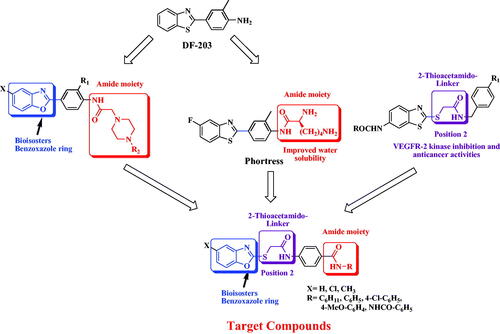
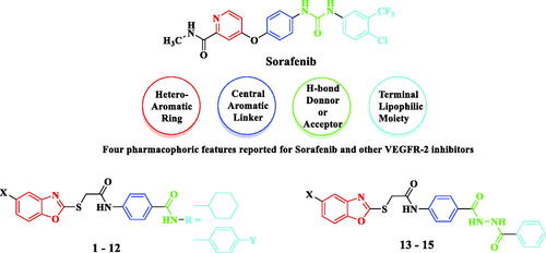
Moreover, the novel benzoxazole series was designed to meet the four main pharmacophoric features reported for sorafenib and other VEGFR-2 inhibitorsCitation46–48. As illustrated in the proposed benzoxazole derivatives (1–15) exhibit pharmacophoric features similar to sorafenib, where the terminal benzoxazole ring could occupy the hinge region of the ATP binding siteCitation47. Also, the central aromatic benzene ring linked via a 2-thioacetamido group could occupy the area between the hinge region and the DFG domain of the activation loopCitation49. In addition, the amide or diamide groups could act as H-bond donors and/or acceptorsCitation50 and finally, the cyclohexyl or phenyl ring represents the terminal hydrophobic moiety that could occupy the allosteric hydrophobic pocket through several hydrophobic interactionsCitation51 ().
Figure 2. Target benzoxazoles fulfilled the pharmacophoric structural features of VEGFR-2 inhibitors.
Considering the aforementioned findings, our group designed and synthesised a new series of benzoxazole-benzamide conjugates linked via a 2-thioacetamido moiety. All targeted compounds were evaluated in-vitro for their anticancer activity against both human breast (MCF-7) and colorectal (HCT-116) cancer cell lines and compared with their cytotoxicity in normal human fibroblasts (WI-38). For further investigation of the potential anticancer mechanism of the synthesised compounds, VEGFR enzymatic inhibition potential was determined, followed by DNA cell cycle analysis for the most active compounds. In addition, the ability of these conjugates to induce cell apoptosis was tested. The level of mitochondrial anti-apoptotic protein Bcl-2 and Bcl-xL in both HCT-116 and MCF-7 cancer cell lines was determined. Finally, molecular docking studies were performed for the synthesised compounds against VEGFR (PDB ID: 4ASD) with sorafenib as a reference ligand.
2. Results and discussion
2.1. Chemistry
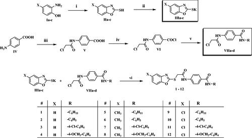
Benzoxazole derivatives 1–15 were synthesised following the general methodologies outlined in Schemes 1 and 2. The key starting materials, 2-mercaptobenzoxazoles IIa-c were synthesised by refluxing the corresponding 2-aminophenol derivatives Ia-c, carbon disulphide, and potassium hydroxide in methanol, according to the reported procedureCitation52. Then, compounds IIa-c were treated with alcoholic KOH to give the corresponding potassium salts, IIIa-c (Scheme 1). On the other hand, 4-aminobenzoic acid IV was reacted with chloroacetyl chloride in DMF to afford the chloroacetamide intermediate V. Then, treatment of compound V by thionyl chloride afforded 4-(2-chloroacetamido)benzoyl chloride VICitation53,Citation54, which was then successively reacted with a set of commercially available amines namely, cyclohexylamine, aniline, 4-chloroaniline, 4-methoxyaniline in acetonitrile and triethylamine (TEA), to get the key intermediates VIIa-d. Finally, compounds VIIa-d were heated with the formerly prepared potassium salts IIIa-c in dry DMF to afford the final target compounds 1–12 (Scheme 1).
Scheme 1 Synthesis of the compounds 1–12; Reagents/conditions: (i) CS2/KOH/CH3OH/reflux 6 h, (ii) KOH/C2H5OH/reflux 4 h, (iii) ClCH2COCl, NaHCO3/DMF/r.t./1h, (iv) SOCl2/1,2-dichloroethane/reflux 4 h, (v) R-NH2/acetonitrile/TEA/r.t. 8 h, (vi) DMF/KI/60 °C/6h.
Scheme 2. Synthesis of the compounds 13–15; Reagents/conditions: (i) CH3OH/conc. H2SO4/reflux 2 h, (ii) NH2-NH2/C2H5OH/reflux 4 h, (iii) acetonitrile/TEA/r.t. 8 h, (vi) DMF/KI/60 °C/6h.
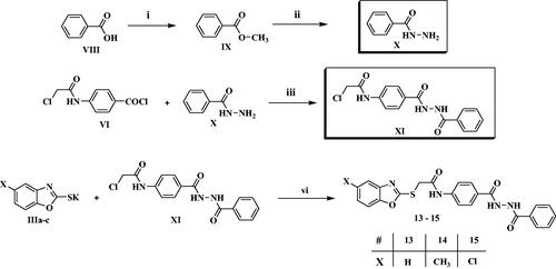
On the other hand, methylbenzoate IX was prepared as reported, by refluxing benzoic acid VIII in methanol in presence of sulphuric acidCitation55,Citation56. Then, refluxing of IX with hydrazine hydrate afforded the corresponding acid hydrazide XCitation57, which was further acylated by VI in acetonitrile and TEA to afford the corresponding derivative XI. As previously, compound IX was finally heated with the formerly prepared potassium salts IIIa-c in dry DMF to afford the final target compounds 13–15 (Scheme 2).
The proposed structures of the final conjugates reported here were in full agreement with their elemental and spectral analysis data. IR spectra of all compounds displayed the absorption bands for the (NH) and (C=O) groups in the 3356–3273 and 1673–1598 cm−1 regions, respectively. Also, compounds 1, 5 and 9 showed additional C-H stretching bands at 2933–2927 cm−1, due to the presence of the aliphatic cyclohexyl group. In addition, 1HNMR spectra for compounds 1, 5 and 9 displayed two signals exchangeable with D2O, referable to the two amidic NH groups at chemical shifts of δ 10.61–10.63 ppm and at δ 8.08 ppm for the acetamido group and benzamido group, respectively. For the remaining compounds, the signals of the two amidic NH groups were in the range of δ 10.68–10.73 ppm and at δ 10.00–10.24 ppm for the acetamido group and benzamido group, respectively. On the other hand, 1HNMR spectra displayed the presence of a singlet peak for the methylene protons of the 2-thioacetamido linker at δ 4.37–4.43 ppm, whereas compounds 5–8 revealed another singlet peak in the aliphatic region referable to the methyl group at δ 2.37–2.28 ppm. Moreover, compounds 4, 8 and 12 displayed an extra singlet signal for the methoxy group at δ 3.72 ppm.
Also, the structures of compounds 13–15 were confirmed by their spectral and elemental analyses. The 1HNMR spectra for compounds 13–15 displayed three singlet signals exchangeable with D2O, one for the acetamido group in the range of δ10.70–10.72 ppm, and two for the acyl hydrazide group at δ10.44 ppm and δ10.39 ppm. Additionally, the spectra showed a singlet signal for the methylene protons of the 2-thioacetamido linker in the range of δ 4.40–4.42 ppm for compounds 13–15 and a singlet signal attributed to the methyl group at δ 2.39 ppm for compound 14.
2.2. Biological evaluation
2.2.1. Anti-proliferative activity against HCT-116 and MCF-7 human cancer cells lines
Recently, benzoxazole derivatives have attracted more attention in drug design, and notably to access compounds with anticancer activity. Several of these derivatives were reported as acting as competitive inhibitors of different tyrosine kinases, with potent cytotoxic activity against various cell linesCitation58,Citation59. Other series of benzoxazole derivatives showed significant potency against colon and breast cancer cell lines and their activity was explained by the potent inhibition of VEGFR enzymesCitation60–62. Thus, in this study a novel series of benzoxazole-benzamide conjugates was initially evaluated for their potential anti-cancer activity against colon cancer cell line (HCT-116), breast cancer cell line (MCF-7) and normal human fibroblasts (WI-38), using the Sulforhodamine B colorimetric (SRB) assayCitation63. Sorafenib as an FDA approved VEGFR-2 inhibitor was utilised as a positive reference compound. The cytotoxic activities were displayed in and and expressed as the median growth inhibitory concentration (IC50).
Figure 3. In vitro anti-proliferative activity of the target compounds 1–15.
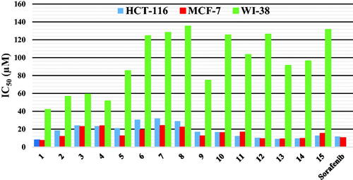
Table 1. In vitro anti-proliferative activity of the compounds 1–15 against HCT-116, MCF-7 human cancer cell lines and W-180 normal cell line, and their corresponding selectivity indices.
Analysing results towards both HCT-116 and MCF-7 cell lines revealed that generally compounds bearing a 5-chlorobenzoxazole moiety (9–12 and 15) showed better cytotoxic activity than their 5-methyl (compounds 5–8 and 14) or their unsubstituted benzoxazole analogs (compounds 2–4 and 13), with the exception of compound 1, bearing an unsubstituted benzoxazole moiety and a cyclohexyl group in its amidic side, and which displayed the best inhibitory activity of these two series, with IC50 values of 7.2 ± 0.01 µM and 7.8 ± 0.015 µM against HCT-116 and MCF7 cell lines, respectively. Concerning the influence of the amide group, a cyclohexyl substituent led globally to more active compounds than a phenyl or a substituted phenyl group (compared compounds 1 to 2–4 or 5 to 6–8 or 9 to 10), except for compound 12 bearing a 4-methoxybenzamide group, which was more active than its cyclohexyl analog 9. Moreover, it is worthy to mention that generally the acyl hydrazide derivatives 13–15 showed higher inhibitory activity than their benzamide analogs towards both cancer cell lines. Thus, as an example, compound 13 showed an IC50 of 9.1 ± 0.005 µM against HCT-116 cells, compared to an IC50 of 18.5 ± 0.005 µM for compound 2.
In addition, results against the MCF-7 cell line showed that compounds 1 and 12–14 exhibited excellent activity with single-digit micromolar IC50 values ranged between 7.2 ± 0.01 and 9.5 ± 0.009 µM, more potent than the reference drug, sorafenib. While compounds 2, 5 and 9 showed good potency with IC50 of 11.7 ± 0.014−12.4 ± 0.007 µM, the remaining compounds had moderate to weak cytotoxic activity with IC50 of 15.3 ± 0.01−24.0 ± 0.011 µM. In a similar way, compounds 1, 12–14 showed a single digit micromolar IC50 values against HCT-116 cells (IC50 range: 7.8 ± 0.015−10.4 ± 0.01 µM), higher than sorafenib that possessed IC50 value of 11.6 ± 1.00 µM. On the other hand, while compounds 11 and 15 showed good potency with IC50 values of 12.2 ± 0.007−12.9 ± 0.014 µM, the remaining compounds had moderate to weak cytotoxic activity with IC50 range of 16.7 ± 0.012−32.0 ± 0.002 µM.
Finally, all tested compounds showed weak cytotoxicity against normal human fibroblasts (WI-38), with IC50 range of 42−135 µM, representing a selectivity index of 2.2 to 13.4, compared to IC50 values against both HCT-116 and MCF-7 cancer cell lines.
These results revealed that some of the novel benzoxazole compounds are very promising candidates as relatively safe cytotoxic agents. Thus, the most active derivatives were submitted for further investigations regarding their potential anti-proliferative mode of action.
2.2.2. VEGFR-2 inhibitory activity
The excellent cytotoxic effects of several benzoxazole derivatives, in particular compound 1, motivated a further exploration of their potential inhibitory activities against VEGFR-2 protein kinase. Representative compounds 1, 9–12 and 15 were selected to determine their potential inhibitory activity. As presented in and , the results revealed that all examined compounds exhibited sub-micromolar IC50 values of VEGFR-2 inhibitory activity. Among all tested compounds, the unsubstituted benzoxazole compound 1, bearing a cyclohexyl group in the amidic side, was the best inhibitor of VEGFR-2 activity with IC50 value of 0.268 µM, more potent than the clinically used kinase inhibitor, sorafenib, which exhibited IC50 value of 0.352 µM, followed by conjugates 11 and 12 with comparable IC50 values of 0.361 µM and 0.385 µM, respectively. On the other hand, compounds 9, 10 and 15 exhibited the least inhibitory activity with IC50 value ranged from 0.597 to 0.704 µM. Finally, the presented results revealed that the VEGFR-2 inhibitory activities were in excellent match with the cytotoxic activities of compounds 1, 11 and 12 suggesting that the anti-proliferative activity might be attributable to VEGFR-2 enzyme inhibition.
Figure 4. Inhibitory activity of 1, 9, 10, 11, 12 and 15 against VEGR-2 Protein Kinase.
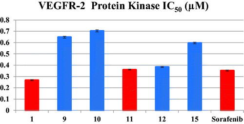
Table 2. Inhibitory activity of 1, 9, 10, 11, 12 and 15 against VEGR-2 Protein Kinase.
2.2.3. Cell cycle analysis
It is clearly known that generally the cytotoxic agents exert their anti-proliferative effect via cell cycle arrest at a specific phase. In the present study, due to the excellent in-vitro anti-proliferative activity of conjugates 1 and 11 against both HCT-116 and MCF-7 cancer cell lines as well as their excellent VEGFR-2 inhibitory activities, cell cycle analysis have been carried out for both compounds. The effect of compounds 1 and 11 on the cell cycle progression in order to determine the phase at which cell cycle arrest takes place in both cancer cell lines was evaluated by a DNA flow cytometry analysis, upon incubation of HCT-116 and MCF-7 cancer cell lines with compounds 1 and 11 at their IC50 concentrations for 24 h ( and and ).
Figure 5. Cell distribution in the subG1, G0/G1, S and G2/M phases for HCT116 cells (B) treated with vehicle control (A), compounds 1 (C) and 11 (D).
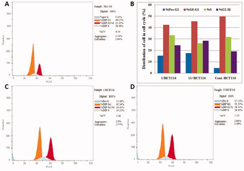
Figure 6. Cell distribution in the subG1, G0/G1, S and G2/M phases for MCF7 cells (B) treated with vehicle control (A), compounds 1 (C) and 11 (D).
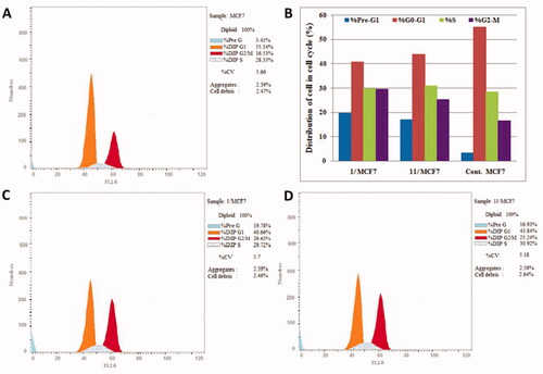
Table 3. Effect of compounds 1, 11 and vehicle control on the cell cycle phases of HCT-116 and MCF-7 cells lines.
The results showed that, for HCT-116 cancer cells lines, the percentage of cells at G2/M phase relatively increased from 18.93% in control to 24.41% and 28.44% after incubation with compounds 1 and 11, respectively. In addition, the percentage of HCT-116 cells in G1 phase was decreased from 49.51% to 42.34% for compound 1 and 45.37% for compound 11 ().
Similarly, the results revealed that, for MCF-7 cancer cell line, the percentage of cells in the G2/M phase was significantly increased from 16.53% to 29.62% for compound 1 and 25.24% for compound 11. In addition, the percentage of MCF-7 cells at G1 phase decreased from 55.14% in control to 40.66% and 43.84% after incubation with compounds 1 and 11, respectively (). These results indicated that compounds 1 and 11 induced cell cycle arrest at G2/M phase. Finally, the upsurge of cell populations in the pre-G1 phase along with the G2-M phase arrest were significant evidence that compounds 1 and 11 induced apoptosis in both HCT-116 and MCF-7 cancer cell lines.
2.2.4. Annexin V-FITC/PI apoptosis test
To determine whether the growth inhibitory action of compounds 1 and 11 is consistent with the induction of apoptosis suggested by the elevated population of pre-G1 in the treated HCT-116 and MCF-7 cells, Annexin V-FITC/PI double staining (AV/PI) apoptosis assay was carried out. The results of this assay revealed that compounds 1 and 11 induced both early and late apoptosis in both HCT-116 and MCF-7 cell lines and the results were outlined in and and .
Figure 7. Effect of compounds 1, 11 and vehicle control on the percentage of annexin V-FITC-positive staining in HCT-116 cell line. The experiments were done in triplicates. The four quadrants identified as: LL, viable; LR, early apoptotic; UR, late apoptotic; UL, necrotic.
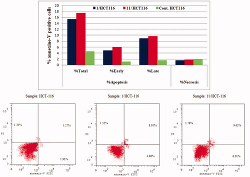
Figure 8. Effect of compounds 1, 11 and vehicle control on the percentage of annexin V-FITC-positive staining in MCF-7 cell lines. The experiments were done in triplicates.
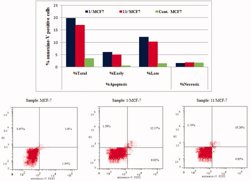
Table 4. Percent of apoptosis and necrosis induced by compounds 1, 11 and vehicle control in HCT-116 and MCF-7 cell lines.
The results revealed that treatment of HCT-116 cells with compound 1 and 11 resulted in an increase in the apoptotic cells percentage for the early apoptosis, from 1.1% for control untreated cells to 4.89% and 6.01%, respectively. In addition, the percentage of apoptotic cells in the late stage was 8.95% to 9.62% compared to control (1.57%). These results revealed that compounds 1 and 11 were able to induce an approximately 3.4-folds and 3.9-folds, respectively, increase in total apoptosis compared to the control for HCT-116 cell line ().
On the other hand, for MCF-7 cancer cell line, the results showed that conjugates 1 and 11 led to an increase in the apoptotic cells percentage for the early apoptosis, from 0.47% for control untreated cells to 6.02% and 4.93%, respectively. In addition, for the late stage, the percentage of apoptotic cells increased from 1.34% for control cells to 12.17% and 10.26%, for compounds 1 and 11, respectively. These results showed that the tested compounds were able to induce an approximately 5.8-fold and 5.0-fold total increase in apoptosis compared to the control, for compounds 1 and 11 respectively (). These results persuaded us to further investigate the effect of compound 1 and 11 on mitochondrial anti-apoptotic biomarkers Bcl-2 and Bcl-xL.
2.2.5. Impact of compounds 1 and 11 on the level of Bcl-2 and Bcl-xL
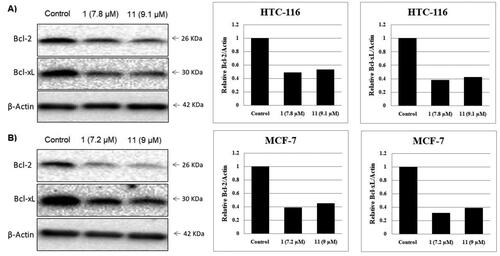
It is well known that the anti-apoptotic proteins Bcl-2 and Bcl-xL are mainly overexpressed in various types of cancer, causing survival of cancer cells and/or drug resistanceCitation7–9. Therefore, inhibition of these proteins expression leads to cancer cell death and has been used as a strategy for anticancer drug developmentCitation23. In this study, the impact of compounds 1 and 11 on Bcl-2 and Bcl-xL expression in HCT-116 and MCF-7 cancer cell lines was examined using Western blot analysis and all the data were normalised to β-actin (). The presented results revealed that benzoxazoles 1 and 11 inhibited Bcl-2 and Bcl-xL expression in both HCT-116 and MCF-7 cancer cell lines in a corresponding manner to their cytotoxic activity and their apoptosis induction ability.
Figure 9. Effect of compounds 1, 11 and vehicle control on anti-apoptotic proteins (Bcl-2 and Bcl-xL) in (A) HCT-116 cancer cells and (B) MCF-7 cancer cells.
2.2.6. Molecular docking
Molecular docking studies are considered as an influential method for interpretation of molecular interactions between the synthesised compounds and the main amino acid residues at the specific binding site of the target receptorCitation64. The activity of the newly synthesised ligands and VEGFR protein interactions at the active binding site was compared according to the docking score values calculated using MOE 2015.10. In the current work, all the synthesised benzoxazole compounds were put through molecular docking studies using MOE software on the VEGFR 3D-structure and using sorafenib as a reference ligand.
The results revealed that, the most biologically active compounds 1 and 11 displayed an excellent docking score (−8.45083 kcal/mol and −8.15044 kcal/mol, respectively) compared to sorafenib docking score (−6.98449 kcal/mol), and both compounds formed direct interactions with many of the amino-acids that sorafenib interacted with (). As shown in , sorafenib had direct interactions with amino-acids Leu840, Glu885, Lys920 and Asp1046 in the active site (). Compound 1 shared sorafenib interactions with amino-acids Leu840 and Asp1046, and additionally exhibited other interactions with amino-acids Lys868, Cys919 and Phe1047 (). On the other hand, compound 11 shared the interaction with only Asp1046 and showed another interaction with amino-acid Lys868 (). Also, compounds 1 and 11 displayed high degrees of superimposition with sorafenib into the VEGFR active site (). Finally, the more interaction formed with amino-acids at the active site by compound 1 than compound 11 strongly support the results of VEGFR enzyme inhibition assay where compound 1 was the most active with IC50 of 0.268 µM compared to that of compound 11 and sorafenib that had IC50s of 0.361 µM and 0.352 µM, respectively ().
Figure 10. Docking of compounds 1, 11 and sorafenib into the VEGFR active site. (A) Interaction of Sorafenib with amino-acids Leu840, Glu885, Lys920 and Asp1046. (B) Interaction of 1 with amino-acids Leu840, Lys868, Cys919, Asp1046 and Phe1047 and superimposition of 1 (shown as cyan sticks) with sorafenib (shown as green sticks). (C) Interaction of 11 with amino-acids Lys868 and Asp1046 and superimposition of 11 (shown as cyan sticks) with sorafenib (shown as green sticks).
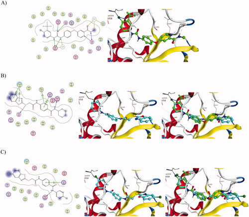
Table 5. Docking energy scores (kcal/mol) obtained from the MOE software for compounds 1–15 and sorafenib.
3. Conclusions
In the current study, a novel series of novel benzoxazole-benzamide conjugates linked via a 2-thioacetamido group (1−15) was designed and synthesised as potential anti-cancer agents with probable inhibitory activity on the VEGFR-2 enzyme and on the expression of anti-apoptotic Bcl-2 and Bcl-xL proteins. The tested compounds were relatively safe against normal human fibroblasts (WI-38) and the cell proliferation of two examined cancer cell lines (HCT-116 and MCF-7) has been notably inhibited by all synthesised compounds with IC50 ranges from 7.8 to 32.0 µM against HCT-116 and from 7.2 to 24.0 µM against MCF-7, as compared to IC50 of 11.6 and 10.5 µM for sorafenib, respectively. In addition, compounds 1, 9, 10, 11, 12 and 15 showed excellent VEGFR-2 inhibitory activity. In particular, benzoxazoles 1 and 11 revealed to be slightly more or equally potent than sorafenib, with IC50 of 0.27, 0.36 µM and 0.35 µM, respectively. Moreover, docking studies showed that the compounds are positioned in a very similar manner to sorafenib into the VEGFR active site. Further mechanistic studies showed that compounds 1 and 11 induced apoptosis and inhibited the expression of anti-apoptotic Bcl-2 and Bcl-xL proteins in both HCT-116 and MCF-7 cancer cell lines. Finally, the high potency of this benzoxazole series suggested that conjugates 1 and 11 could avail as lead compounds for further investigation and optimisation to develop novel anti-proliferative agents, apoptotic inducers and inhibitors of Bcl-2/Bcl-xL expression.
4. Experimental
4.1. Chemistry
4.1.1. General
Melting points (°C) of the synthesised compounds were uncorrected and were measured using Electrothermal Stuart 5MP3. Follow-up of reactions was performed using TLC plates of silica gel 60 F254 (Merck). The NMR spectrometric analyses have been recorded using Bruker-Avance 400 NMR spectrometer (400 MHz for 1HNMR and100 MHz for 13CNMR) in deuterated dimethylsulphoxide (DMSO-d6). Chemical shifts (δH) were reported relative to the solvent (DMSO-d6). Mass spectra were recorded on Finnigan Mat SSQ 7000 mode EI 70 eV at the micro analytical unit, Cairo University, Cairo, Egypt. Schimadzu FT-IR 8400S spectrophotometer has been used for functional group analysis at the micro analytical unit, Cairo University, Cairo, Egypt. Elemental analyses were performed at the Regional Centre for Microbiology and Biotechnology, Al-Azhar University, Cairo, Egypt.
4.1.2. General methodology for preparation of the target compounds 1-12
In DMF (10 ml), a mixture of potassium salts IIIa-c (0.001 mol) and the convenient 4-(2-chloroacetamido)-N-(substituted) phenyl benzamide VIIa-d (0.001 mol), and KI (0.001 mol) was heated at 60 °C for 6 h. After completion of the reaction, the mixture was poured on crushed ice. The formed precipitates were filtered, dried, and recrystallized from methanol to afford the corresponding final target compounds 1–12.
4.1.3. General methodology for preparation of the target compounds 13-15
In DMF (10 ml), a mixture of potassium salts IIIa-c (0.001 mol) and N-(4-(2-benzoyl-hydrazine-1-carbonyl)phenyl)-2-chloroacetamide XI (0.001 mol), and KI (0.001 mol) was heated at 60 °C for 6 h. After completion of the reaction, the mixture was poured on crushed ice. The formed precipitates were filtered, dried, and recrystallized from methanol to afford the corresponding final target compounds 13–15.
Full characterisation (1HNMR, 13CNMR, IR, Mass spectrum and elemental analysis) data for novel compounds 1–15 have been presented in the Supplementary Materials.
4.2. Biological evaluation
All the procedures of the experiment utilised for biological evaluation in this article were performed as previously described; cytotoxicityCitation63,Citation65, VEGFR-2 inhibitory activityCitation14, cell cycle analysisCitation66, Annexin V-FITC/PI apoptosis assayCitation67, and anti-apoptotic markers (Bcl-2, and Bcl-xL)Citation68,Citation69. All procedures were mentioned in detail in the Supplementary Materials.
4.3. Molecular docking
Virtual Molecular Docking studies were carried out using Molecular Operating Environment (MOE®) version 2015.10. The RCSB: Protein Data Bank was utilised to retrieve the crystal structure of Vascular Endothelial Growth Factor Receptor (VEGFR) co-crystallized with sorafenib (PDB ID: 4ASD) Citation70. The downloaded protein was used for the docking study as a receptor and sorafenib was used as a reference drug.
Supplemental Material
Download PDF (1.9 MB)Acknowledgements
The researcher Radwan El-Haggar is funded by a postdoctoral mission from the Ministry of Higher Education of the Arab Republic of Egypt.
Disclosure statement
No potential conflict of interest was reported by the author(s).
References
- Siegel RL, Miller KD, Jemal A. Cancer statistics, 2020. CA Cancer J. Clin 2020;70:1587–30.
- Fabbro D, Parkinson D, Matter A. Protein tyrosine kinase inhibitors: new treatment modalities? Curr Opin Pharmacol 2002;2:374–81.
- Wu C, Wang M, Tang Q, et al. Design, synthesis, activity and docking study of sorafenib analogs bearing sulfonylurea unit. Molecules 2015;20:19361–71.
- Baudino TA. Targeted cancer therapy: the next generation of cancer treatment. Curr Drug Discov Technol 2015;12:3–20.
- Topcul M, Cetin I. Endpoint of cancer treatment: targeted therapies, Asian Pac. Asian Pac J Cancer Prev 2014;15:4395–403.
- Qin S, Li A, Yi M, et al. Recent advances on anti-angiogenesis receptor tyrosine kinase inhibitors in cancer therapy. J Hematol Oncol 2019;12:27–37.
- Edlich F. BCL-2 proteins and apoptosis: recent insights and unknowns. Biochem Biophys Res Commun 2018;500:26–34.
- Hata AN, Engelman JA, Faber AC. The BCL2 family: key mediators of the apoptotic response to targeted anticancer therapeutics. Cancer Discov 2015;5:475–87.
- Marone M, Ferrandina G, Macchia G, et al. Bcl-2, Bax, Bcl-x(L) and Bcl-x(S) expression in neoplastic and normal endometrium. Oncology 2000;58:161–8.
- Lugano R, Ramachandran M, Dimberg A. Tumor angiogenesis: causes, consequences, challenges and opportunities. Cell Mol Life Sci 2020;77:1745–70.
- Folkman J. Angiogenesis in cancer, vascular, rheumatoid and other disease. Nat Med 1995;1:27–31.
- Karamysheva AF. Mechanisms of angiogenesis. Biochemistry 2008;73:751–62.
- Kerbel RS. Tumor angiogenesis: past, present and the near future. Carcinogenesis 2000;21:505–15.
- Ahmed MF, Santali EY, El-Haggar R. Novel piperazine-chalcone hybrids and related pyrazoline analogs targeting VEGFR-2 kinase; design, synthesis, molecular docking studies, and anticancer evaluation. J. Enzy. Inhib. Med. Chem 2021;36:307–18.
- Modi SJ, Kulkarni VM. Vascular Endothelial Growth Factor Receptor (VEGFR-2)/KDR inhibitors: medicinal chemistry perspective. Med Drug Discov 2019;2:100009.
- Potashman MH, Bready J, Coxon A, et al. Design, synthesis, and evaluation of orally active benzimidazoles and benzoxazoles as vascular endothelial growth factor-2 receptor tyrosine kinase inhibitors. J Med Chem 2007;50:4351–73.
- Yuan X, Yang Q, Liu T, et al. Design, synthesis and in vitro evaluation of 6-amide-2-aryl benzoxazole/benzimidazole derivatives against tumor cells by inhibiting VEGFR-2 kinase. Eur J Med Chem 2019;179:147–65.
- DR, Green Means to an end: apoptosis and other cell death mechanisms. New York: Cold Spring Harbor, Cold Spring Harbor Laboratory Press; 2011.
- Spierings D, McStay G, Saleh M, et al. Connected to death: the (unexpurgated) mitochondrial pathway of apoptosis. Science 2005;310:66–7.
- Wang B, Xu A. Aryl hydrocarbon receptor pathway participates in myocardial ischemia reperfusion injury by regulating mitochondrial apoptosis. Med Hypotheses 2019;123:2–5.
- Modugno M, Banfi P, Gasparri F, et al. Mcl-1 antagonism is a potential therapeutic strategy in a subset of solid cancers. Exp Cell Res 2015;332:267–77.
- Placzek WJ, Wei J, Kitada S, et al. A survey of the anti-apoptotic Bcl-2 subfamily expression in cancer types provides a platform to predict the efficacy of Bcl-2 antagonists in cancer therapy. Cell Death Dis 2010;1:e40.
- Eldehna WM, Abo-Ashour MF, Al-Warhi T, et al. Development of 2-oxindolin-3-ylidene-indole-3-carbohydrazide derivatives as novel apoptotic and anti-proliferative agents towards colorectal cancer cells. J Enzy Inhib Med Chem 2021;36:319–28.
- Sabt A, Abdelhafez OM, El-Haggar RS, et al. Novel coumarin-6-sulfonamides as apoptotic anti-proliferative agents: synthesis, in vitro biological evaluation, and QSAR studies. J Enzyme Inhib Med Chem 2018;33:1095–107.
- Chen G, Liu Z, Zhang Y, et al. Synthesis and anti-inflammatory evaluation of novel benzimidazole and imidazopyridine derivatives. ACS Med Chem Lett 2013;4:69–74.
- Kakkar S, Narasimhan B. A comprehensive review on biological activities of oxazole derivatives. BMC Chem 2019;13:1–24.
- Mohamed MS, Rashad AE, Adbel-Monem M, Fatahalla SS. New anti-inflammatory agents. Z Naturforsch C J Biosci 2007;62:27–31.
- Veerasamy R, Roy A, Karunakaran R, Rajak H. Structure-activity relationship analysis of benzimidazoles as emerging anti-inflammatory agents: an overview. Pharmaceuticals 2021;14:663–93.
- Seth K, Garg SK, Kumar R, et al. Chakraborti, 2-(2-Arylphenyl)benzoxazole As a Novel Anti-Inflammatory Scaffold: Synthesis and Biological Evaluation. ACS Med Chem Lett 2014;5:512–6.
- Balaswamy G, Pradeep P, Srinivas K, Rajakomuraiah T. Synthesis, characterization and anti-microbial activity of new series of benzoxazole derivatives. Int J Chem Sci 2012;10:1830–6.
- Gamba E, Mori M, Kovalenko L, et al. Identification of novel 2-benzoxazolinone derivatives with specific inhibitory activity against the HIV-1 nucleocapsid protein. Eur J Med Chem 2018;145:154–64.
- Rida SM, Ashour FA, El-Hawash SA, et al. Synthesis of some novel benzoxazole derivatives as anticancer, anti-HIV-1 and antimicrobial agents. Eur J Med Chem 2005;40:949–59.
- Zeyrek CT, Arpacı ÖT, Arısoy M, Onurdağ FK. Synthesis, antimicrobial activity, density functional modelling and molecular docking with COVID-19 main protease studies of benzoxazole derivative: 2-(p-chloro-benzyl)-5-[3-(4-ethly-1-piperazynl) propionamido]-benzoxazole. J Mol Struct 2021;1237:130413.
- Bhole RP, Chikhale RV, Wavhale RD, et al. Design, synthesis and evaluation of novel enzalutamide analogues as potential anticancer agents. Heliyon 2021;7:e06227.
- Bradshaw TD, Westwell AD. The development of the antitumour benzothiazole prodrug, Phortress, as a clinical candidate. Curr Med Chem 2004;11:1009–21.
- Dadashpour S, Kucukkilinc TT, Ercan A, et al. Synthesis and anticancer activity of benzimidazole/benzoxazole substituted triazolotriazines in hepatocellular carcinoma. Anticancer Agents Med Chem 2019;19:2120–9.
- Desai S, Desai V, Shingade S. In-vitro Anti-cancer assay and apoptotic cell pathway of newly synthesized benzoxazole-N-heterocyclic hybrids as potent tyrosine kinase inhibitors. Bioorg Chem 2020;94:103382.
- Dhadda S, Kumar A, Kamlesh R, et al. Benzothiazoles: From recent advances in green synthesis to anti-cancer potential. Sustain Chem Phar 2021;24:100521.
- Eldehna WM, El Hassab MA, Abo-Ashour MF, et al. Development of isatin-thiazolo[3,2-a]benzimidazole hybrids as novel CDK2 inhibitors with potent in vitro apoptotic anti-proliferative activity: Synthesis, biological and molecular dynamics investigations. Bioorg Chem 2021;110:104748.
- El-Hady HA, Abubshait SA. Synthesis and anticancer evaluation of imidazolinone and benzoxazole derivatives. Arab J Chem 2017;10:S3725–S3731.
- El-Hameed RHA, Fatahala SS, Sayed AI. Synthesis of some novel benzimidazole derivatives as anticancer agent, and evaluation for CDK2 inhibition activity. Med Chem 2022;18:1–11.
- Mantzourani C, Gkikas D, Kokotos A, et al. Synthesis of benzoxazole-based vorinostat analogs and their antiproliferative activity. Bioorg Chem 2021;114:105132.
- Osmaniye D, Korkut Çelikateş B, Sağlık BN, et al. Synthesis of some new benzoxazole derivatives and investigation of their anticancer activities. Eur J Med Chem 2021;210:112979.
- Xiang P, Zhou T, Wang L, et al. Novel benzothiazole, benzimidazole and benzoxazole derivatives as potential antitumor agents: synthesis and preliminary in vitro biological evaluation. Molecules 2012;17:873–83.
- Khajondetchairit P, Phuangsawai O, Suphakun P, et al. Design, synthesis, and evaluation of the anticancer activity of 2-amino-aryl-7-aryl-benzoxazole compounds. Chem Biol Drug Des 2017;90:987–94.
- Eskander RN, Tewari KS. Incorporation of anti-angiogenesis therapy in the management of advanced ovarian carcinoma-mechanistics, review of phase III randomized clinical trials, and regulatory implications. Gynecol Oncol 2014;132:496–505.
- Lee K, Jeong KW, Lee Y, et al. Pharmacophore modeling and virtual screening studies for new VEGFR-2 kinase inhibitors. Eur J Med Chem 2010;45:5420–7.
- Xie QQ, Xie HZ, Ren JX, et al. Pharmacophore modeling studies of type I and type II kinase inhibitors of Tie2. J Mol Graph Model 2009;27:751–8.
- Machado VA, Peixoto D, Costa R, et al. Synthesis, antiangiogenesis evaluation and molecular docking studies of 1-aryl-3-[(thieno[3,2-b]pyridin-7-ylthio)phenyl]ureas: Discovery of a new substitution pattern for type II VEGFR-2 Tyr kinase inhibitors. Bioorg Med Chem 2015;23:6497–509.
- Wang Z, Wang N, Han S, et al. Dietary compound isoliquiritigenin inhibits breast cancer neoangiogenesis via VEGF/VEGFR-2 signaling pathway. PLoS One 2013;8:e68566.
- Dietrich J, Hulme C, Hurley LH. The design, synthesis, and evaluation of 8 hybrid DFG-out allosteric kinase inhibitors: a structural analysis of the binding interactions of Gleevec, Nexavar, and BIRB-796. Bioorg Med Chem 2010;18:5738–48.
- Kaul S, Kumar A, Sain B, Bhatnagar AK. Simple and Convenient One‐Pot Synthesis of Benzimidazoles and Benzoxazoles using N,N‐Dimethylchlorosulfitemethaniminium Chloride as Condensing Agent. Synthetic Commun 2007;37:2457–60.
- Alsaif NA, Dahab MA, Alanazi MM, et al. New quinoxaline derivatives as VEGFR-2 inhibitors with anticancer and apoptotic activity: design, molecular modeling, and synthesis. Bioorg Chem 2021;110:104807.
- Alanazi MM, Elkady H, Alsaif NA, et al. New quinoxaline-based VEGFR-2 inhibitors: design, synthesis, and antiproliferative evaluation with in silico docking, ADMET, toxicity, and DFT studies. RSC Adv 2021;11:30315–28.
- Alanazi MM, Eissa IH, Alsaif NA, et al. Design, synthesis, docking, ADMET studies, and anticancer evaluation of new 3-methylquinoxaline derivatives as VEGFR-2 inhibitors and apoptosis inducers. J Enzyme Inhib Med Chem 2021;36:1760–82.
- Alsaif NA, Taghour MS, Alanazi MM, et al. Discovery of new VEGFR-2 inhibitors based on bis([1, 2, 4]triazolo)[4,3-a:3',4'-c]quinoxaline derivatives as anticancer agents and apoptosis inducers. J Enzyme Inhib Med Chem 2021;36:1093–114.
- El-Zahabi MA, Sakr H, El-Adl K, et al. Design, synthesis, and biological evaluation of new challenging thalidomide analogs as potential anticancer immunomodulatory agents. Bioorg Chem 2020;104:104218.
- Chikhale R, Thorat S, Choudhary RK, et al. Design, synthesis and anticancer studies of novel aminobenzazolyl pyrimidines as tyrosine kinase inhibitors. Bioorg Chem 2018;77:84–100.
- Stevens MF, McCall CJ, Lelieveld P, et al. Structural studies on bioactive compounds. 23. Synthesis of polyhydroxylated 2-phenylbenzothiazoles and a comparison of their cytotoxicities and pharmacological properties with genistein and quercetin. J Med Chem 1994;37:1689–95.
- Ghoshal T, Patel TM. Anticancer activity of benzoxazole derivative (2015 onwards): a review. Fut J Pharm Sci 2020;6:94–113.
- Omar AME, AboulWafa OM, El-Shoukrofy MS, Amr ME. Benzoxazole derivatives as new generation of anti-breast cancer agents. Bioorg Chem 2020;96:103593.
- Peng F, Liu D, Zhang Q, et al. VEGFR-2 inhibitors and the therapeutic applications thereof: a patent review (2012-2016). Expert Opin Ther Pat 2017;27:987–1004.
- Skehan P, Storeng R, Scudiero D, et al. New colorimetric cytotoxicity assay for anticancer-drug screening. J Natl Cancer Inst 1990;82:1107–12.
- Meng XY, Zhang HX, Mezei M, Cui M. Molecular docking: a powerful approach for structure-based drug discovery. Curr Comput Aided Drug Des 2011;7:146–57.
- Omar AM, El-Araby ME, Abdelghany TM, et al. Introducing of potent cytotoxic novel 2-(aroylamino)cinnamamide derivatives against colon cancer mediated by dual apoptotic signal activation and oxidative stress. Bioorg Chem 2020;101:103953.
- Wang J, Lenardo MJ. Roles of caspases in apoptosis, development, and cytokine maturation revealed by homozygous gene deficiencies. J Cell Sci 2000;113:753–7.
- Lo KK, Lee TK, Lau JS, et al. Luminescent biological probes derived from ruthenium(II) estradiol polypyridine complexes. Inorg Chem 2008;47:200–8.
- Aborehab NM, Elnagar MR, Waly NE. Gallic acid potentiates the apoptotic effect of paclitaxel and carboplatin via overexpression of Bax and P53 on the MCF-7 human breast cancer cell line. J Biochem Mol Toxicol 2021;35:e22638.
- Burnette WN. "Western blotting": electrophoretic transfer of proteins from sodium dodecyl sulfate-polyacrylamide gels to unmodified nitrocellulose and radiographic detection with antibody and radioiodinated protein A. Anal Biochem 1981;112:195–203.
- Available from: http://www.rcsb.org/