
Abstract
The global outbreak of the COVID-19 pandemic provokes scientists to make a prompt development of new effective therapeutic interventions for the battle against SARS-CoV-2. A new series of N-(5-nitrothiazol-2-yl)-carboxamido derivatives were designed and synthesised based on the structural optimisation principle of the SARS-CoV Mpro co-crystallized WR1 inhibitor. Notably, compound 3b achieved the most promising anti-SARS-CoV-2 activity with an IC50 value of 174.7 µg/mL. On the other hand, compounds 3a, 3b, and 3c showed very promising SARS-CoV-2 Mpro inhibitory effects with IC50 values of 4.67, 5.12, and 11.90 µg/mL, respectively. Compound 3b docking score was very promising (−6.94 kcal/mol) and its binding mode was nearly similar to that of WR1. Besides, the molecular dynamics (MD) simulations of compound 3b showed its great stability inside the binding pocket until around 40 ns. Finally, a very promising SAR was concluded to help to design more powerful SARS-CoV-2 Mpro inhibitors shortly.
Graphical Abstract
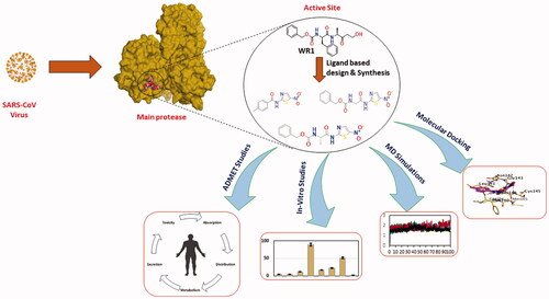
A new series of N-(5-nitrothiazol-2-yl)-carboxamido derivatives were designed and synthesised based on the structural optimisation principle.
In vitro antiviral activities against SARS-CoV-2 using SARS-CoV-2 cell-based inhibitory assay.
The anticipated inhibitory effects of the synthesised compounds (3a–g) towards the SARS-CoV-2 Mpro enzyme were emphasised by using the SARS-CoV-2 Mpro assay.
Molecular docking studies, molecular dynamics simulations for 100 ns, and MM-GBSA calculations were carried out for the newly synthesised compounds (3a–g) compared to the co-crystallized inhibitor (WR1).
ADMET and toxicity in silico studies were applied for the designed derivatives.
Finally, our interesting work rationale helped to conclude a very promising structure-activity relationship (SAR) finding.
HIGHLIGHTS
1. Introduction
The COVID-19 global outbreak is attributed to SARS-CoV-2Citation1. Owing to its overwhelming expansion and spreading, the virus caused an unprecedented global health crisis. Subsequently, the World Health Organisation (WHO) officially claimed that COVID-19 is pandemic in March 2020. SARS-CoV-2 has reached over 170 countries and has adversely impacted over 235 million individuals with a death toll nearing 5.2 million as of 22 November 2021Citation1,Citation2. Besides, the incubation period of SARS-CoV-2 is nearly 2–14 days and can be extended up to 24 days. The virus's long incubation period, and its possible asymptomatic nature, could be in charge of infections spreading. The fast rise in COVID-19 cases increases the need for effective interventionsCitation3–6.
Furthermore, the virus belongs to the Coronaviridae family and generally coronaviruses can be classified to four genera, gamma-coronavirus (γ-CoV), delta-coronavirus (δ-CoV), alpha-coronavirus (α-CoV), and beta-coronavirus (β-CoV). Both α- and β-species mainly hit mammals, while γ- and δ- species hit birdsCitation7. Notably, it was confirmed that SARS‐CoV‐2 shares almost 80% of the genome with SARS‐CoVCitation8. Infection by SARS-CoV-2 is transmitted mainly through human-to-human contact from respiratory droplets. The viral infection varies in severity from asymptomatic to threatening fatal disease. Consequently, the most common symptoms include headache, fever, non-productive cough, fatigue, and dyspnoea. Patients with severe disease may develop viral pneumonia, hypoxia, and acute respiratory distress. So, intubation and mechanical ventilation are requiredCitation1. Additionally, neurological symptoms including skeletal muscle injury, acute cerebrovascular diseases, consciousness impairment, and loss of smell and/or taste could be manifested by SARS-CoV-2 infectionCitation9,Citation10.
Additionally, coronaviruses belong to RNA viruses [single-stranded positive-sense (+)] that are distinctly prevalent in wildlife and humans. Notably, coronaviruses have the most enormous known RNA genomes. Hence, the virus’s two encoded overlapping open-reading frames are translated into the two polyproteins named; pp1a and pp1ab. So, these polyproteins are processed further to give rise to four structural proteins and sixteen non-structural proteins (nsps)Citation11. Subsequently, the virus replicase polyprotein is processed by two distinct cysteine proteases; the papain-like protease (PLpro) and the main protease (Mpro)Citation12,Citation13. The proteolytic refining of the sixteen nsps by PLpro and 3CLpro is crucial for virus maturation and replication, and therefore PLpro and 3CLpro emerged as key druggable targetsCitation14–18.
For the sake of achieving rapid therapeutic interventions, a handful set of repurposed drugsCitation19,Citation20 like chloroquine, hydroxychloroquine, and remdesivir, has been used frequently for COVID-19 treatmentCitation3,Citation4. Although remdesivir, which gained urgent approval, hydroxychloroquine and nafamostat are viewed as outstanding therapeutic candidates, their low clinical effects and adverse side effects warrant the search for more effective and safer treatmentsCitation21. Several SARS-CoV-2 druggable targets were elucidated such as Mpro, spike (S) protein, papain-like protease (PLpro), and RNA-dependent RNA polymerase. The viral Mpro is regarded as an outstanding target for druggabilityCitation3,Citation22.
The Mpro enzyme is one of the best coronavirus drug targets due to the resemblance in their active site and mechanisms with β-Coronaviruses from previous epidemics; SARS-CoV and MERS-CoV (Middle East respiratory syndrome coronavirus)Citation23. Mpro is a preserved drug target without a human homolog, hence lowering the possibility of accidentally targeting host proteins. Therefore, Mpro is perceived as a potential target for broad-spectrum drug developmentCitation23,Citation24. It is worth mentioning that findings propose that SARS-CoV-2 possesses the power to utilise human angiotensin-converting enzyme 2 (ACE2) receptors in the seek of cell entrance as displayed in Citation7,Citation16.
Figure 1. Schematic diagram showing SARS-CoV-2 host, its transmission, and the virus Mpro as a promising druggable target of interest in an infected cell.

Despite all efforts and attempts to find a treatment for SARS-CoV-2 infection, rising issues of COVID-19 mortality and morbidity are still encountered globally. Although vaccines have been developed, efficient and safe drugs are urgently neededCitation5,Citation10,Citation25.
Lately, a new synthetic nucleoside derivative prodrug, named molnupiravir, was approved in the U.K for COVID-19 treatment. Molnupiravir acts by copying errors during RNA virus replicationCitation26. It is an active orally RdRp inhibitor with reasonable pharmacokinetic features. It has gained significant attention for its capability to inhibit the spreading of SARS-CoV-2, with a remarkable reduction in the viral load and quick recovery timeCitation27. A single-dose administration of molnupiravir produces a mean Cmax of 13.2 ng/mL and tmax between 0.25 and 0.75 h with a biological t1/2 of 7 h. It was suggested that molnupiravir has no accumulative toxicity and that was assured by its area under the plasma concentration versus time following multiple doses, increases with no accumulation in a dose-proportional mannerCitation27. Moreover, molnupiravir could exhibit rapid onset, a wide therapeutic window, and fewer side effects with good tolerability and safety profile. Hence, it can be considered a very promising therapeutic intervention against SARS-CoV-2Citation27. Additionally, the oral antiviral drug, named PF-07321332, was developed by Pfizer For COVID-19 treatment as well. PF-07321332 acts as an active Mpro inhibitor of the virusCitation26. Protease inhibitors act by interrupting the protease enzyme cutting, thus, the polypeptide processing to smaller protein is blocked. PF-07321332 is co-administered with ritonavir in low doses as a booster to enhance the PF-07321332 bloodstream levelsCitation28. The combination of ritonavir/PF-07321332 was marketed as paxlovidCitation26.
In recent months, many researchers disclosed the discovery of potent inhibitors for SARS-CoV-2 using molecular docking and dynamics in silico approachesCitation19,Citation29–36. Moreover, the literature revealed that some novel chemically synthesised compounds were designed and evaluated biologically as outstanding inhibitors of SARS-CoV-2 MproCitation37–40. Obviously, the N-heterocyclic scaffolds commonly exhibit a pivotal function and exert an advanced biological activity against SARS-CoV-2. Hence, a promising therapeutic intervention for COVID-19 treatment can be acquiredCitation25. Therefore, in the current work, we aimed to synthesise a series of N-heterocyclic scaffold derivatives that have the same pharmacophoric features of SARS-CoV Mpro native inhibitor (N3) as depicted in Scheme 1. Thus, the virus Mpro was targeted revealing the potential of the synthesised compounds as promising candidates for COVID-19 treatment using both in vitro and in-silico approaches for their assessment.
Scheme 1. Chemical synthesis of the designed target compounds (3a–g) attempted to combat COVID-19.

1.1. The rationale for work design
WR1 is the three-letter code of the native inhibitor of SARS-CoV Mpro downloaded from PDB with ID 2OP9Citation41. Observing the native inhibitor (WR1) binding mode at SARS-CoV Mpro, we can conclude that it could be stabilised within its binding pocket via the following essential pharmacophoric features ();
Figure 2. The rationale work design shows the identification of the essential pharmacophores acquired by the SARS-CoV Mpro co-crystallized inhibitor.

H-bond donor (NH) to compose an H-bond with Cys145 amino acid.
H-bond acceptor (CO) to compose an H-bond with Glu166 amino acid.
Aromatic moiety to occupy the hydrophobic groove composed of the amino acids; Met49 and Met165.
Additional moiety (3-hydroxy propanone) to interact with His163, Cys145, or Phe140 amino acids.
On the other hand, it is worth mentioning that the inhibitor-binding site is located at Cys–His dyad which composes the catalytic cleft located between the SARS-CoV-2 Mpro domains I and II. Herein, the authors analysed the pharmacophoric features of SARS-CoV Mpro co-crystallized inhibitor (WR1) to synthesise a new series of compounds (3a–g) using the ligand-based design approachCitation42 and based on the structural optimisation principle. In addition, taking into account the close structural similarity within the two strains of SARS-CoV (1 and 2)Citation4,Citation10, we dedicated our efforts to synthesising a novel series of N-(5-nitrothiazol-2-yl)-carboxamido derivatives as significant inhibitors of SARS-CoV-2 Mpro (), where;
Figure 3. Schematic representation describing the achievement of the previously identified pharmacophoric features of SARS-CoV Mpro inhibitor in the newly designed drug candidates (3a–g).

We kept the H-bond donor moiety (amidic group) such as the co-crystallized inhibitor (WR1).
We kept the H-bond acceptor moiety (benzyl carbamate) such as the co-crystallized inhibitor (WR1) except for compound 3a.
We modified the aromatic ring that fits within the hydrophobic groove composed of the amino acids; Met49 and Met165 to other different moieties with different sizes (methyl, isopropyl, benzyl, pyrrole, or indole substituents) in compounds 3c, 3d, 3e, 3f, and 3g, respectively. Also, we removed this moiety in both compounds 3a and 3b.
We replaced the previously mentioned additional moiety (3-hydroxy propanone) that interacts with His163, Cys145, or Phe140 with 5-nitrothiazole moiety which was extracted from nitazoxanide () which was later approved to possess potent antiviral activities against hepatitis B and C, influenza A, and coronaviruses. Recently, nitazoxanide was evaluated against SARS-CoV-2 through in vitro assessment which confirmed its promising activity (EC50 of 2.12 μM)Citation43.
Figure 4. The replacement of the 3-hydroxy propanone moiety of the co-crystallized SARS-CoV Mpro inhibitor that interacts with His163, Cys145, or Phe140 with 5-nitrothiazole moiety which was extracted from the potent anti-SARS-CoV-2, nitazoxanide.

Based on the aforementioned rationale, we were able to assess the impact of the discussed modifications on the potential of the synthesised candidates to get a lead compound and obtain a reasonable structure-activity relationship (SAR) which could aid medicinal chemists to design more promising anti-SARS-CoV-2 drug candidates soon as well.
2. Results and discussion
2.1. Chemistry
Compounds 3a–g were synthesised by treating N-acyl benzotriazoles (1a–g) with 5-nitrothiazol-2-amine (2) at room temperature in the presence of triethylamine in acetonitrile for 1 h following the reported methodologiesCitation44,Citation45.
2.2. In vitro studies
2.2.1. SARS-CoV-2 inhibitory assay
To investigate the anti-SARS-CoV-2 activity in Vero E6 cells, the cytotoxicity of the tested compounds was assessed in Vero E6 cells via MTT test and the results unravelled that the cytotoxic concentration 50 (CC50) values were 1466 µg/mL (3a), 1853 µg/mL (3b), 2118 µg/mL (3c), 1204 µg/mL (3d), 2040 µg/mL (3e), 2802 µg/mL (3f), and 1626 µg/mL (3g) (). Furthermore, the antiviral activities were estimated using the dose-response curves. The result showed that the concentrations that induce inhibition to 50% of the investigated cells (IC50) by the tested compounds were 1377 µg/mL (3a), 174.7 µg/mL (3b), 698 µg/mL (3c), 1285 µg/mL (3e), and 1252 µg/mL (3f) (). For all tested compounds, the IC50 values were estimated by plotting log inhibitory concentrations (X-axis) against normalised response (Y-axis), (variable slope) utilising GraphPad Prism software (version 5.01) nonlinear regression analysis. However, compounds (3d) and (3g) displayed IC50 values higher than their corresponding CC50 values neglecting their applicability as anti-SARS-CoV-2. Consequently, compound 3b showed the best selectivity index (SI = CC50/IC50) with a SI = 10, followed by 3c with a SI value of 3.
Figure 5. Cytotoxicity concentration 50 (CC50) of the newly designed and synthesised SARS-CoV Mpro analogs (3a–g) on Vero E6 cells. Besides, inhibitory concentration (IC50) to estimate the antiviral activity against SARS-CoV-2 [hCoV-19/Egypt/NRC-03/2020 (Accession Number on GSAID: EPI_ISL_430820)] using Vero E6 cells.
![Figure 5. Cytotoxicity concentration 50 (CC50) of the newly designed and synthesised SARS-CoV Mpro analogs (3a–g) on Vero E6 cells. Besides, inhibitory concentration (IC50) to estimate the antiviral activity against SARS-CoV-2 [hCoV-19/Egypt/NRC-03/2020 (Accession Number on GSAID: EPI_ISL_430820)] using Vero E6 cells.](/cms/asset/b28cc87b-e97a-43d6-b434-d7db206fec07/ienz_a_2105322_f0005_c.jpg)
2.2.2. SARS-CoV-2 Mpro inhibitory assay (cell-based)
The anticipated inhibitory effects of the synthesised derivatives (3a-g) towards the SARS-CoV-2 Mpro enzyme were emphasised by using the SARS-CoV-2 Mpro assay. Out of the synthesised compounds, compounds 3a, 3b, and 3c unravelled so outstanding SARS-CoV-2 Mpro inhibitory effects with IC50 values of 4.67, 5.12, and 11.90 µg/mL, respectively, as displayed in as well as the Supplementary Data (Supplementary Table 1). It is worth mentioning that among these promising compounds, compounds 3a and 3b fulfilled the best inhibitory activity against SARS-CoV-2 Mpro with very promising IC50 values.
Figure 6. Mpro inhibitory concentration 50 (IC50) against SARS-CoV-2 for the synthesised compounds (3a–g), where compounds 3a, 3b, and 3c unravelled so outstanding SARS-CoV-2 Mpro inhibitory effects with IC50 4.67, 5.12, and 11.90 µM, respectively. *p ˂ 0.05 compared to 3a. #p ˂ 0.05 compared to 3b. &p ˂ 0.05 compared to 3c. $p ˂ 0.05 compared to 3d. @P ˂ 0.05 compared to 3e. !p ˂ 0.05 compared to 3g.

2.3. In silico studies
2.3.1. Molecular docking studies
At the beginning of the docking process, to assure the accuracy of the docking protocol, the MOE program was validated. So, the program validation was initiated by the native ligand (WR1) re-docking against the SARS-CoV Mpro target receptorCitation46–48. A valid docking protocol was ensured by getting a low RMSD value (1.33 Å) between the re-docked conformer and the co-crystallized conformer of WR1 as shown in Citation49,Citation50.
Figure 7. 3D diagram unveiling the native ligand WR1 (Green), and redocked WR1 (Violet) superimposition at SARS-CoV Mpro with PDB: 2OP9 for MOE program validation.

WR1, as a native co-crystallized ligand, formed hydrogen bonds with Phe140 and His163 through the hydroxyl group of the oxopentan-2-yl moiety of WR1 at distances 2.79 and 2.91 Å, respectively. However, the docked WR1 formed hydrogen bonds with Ser144, Cys145, and Gly143 through the carbonyl group of the oxopentan-2-yl moiety of WR1 at distances 2.91, 2.94, and 2.87 Å, respectively. Moreover, the carbamate moiety of WR1 interacted with Glu166 via a hydrogen bond at a distance of 2.92 Å, but the amide moiety of WR1 formed a hydrogen bond with Asn142 and Cys145 at distances 2.95 and 3.49 Å, respectively ().
Figure 8. Native co-crystallized WR1 inside SARS-CoV Mpro active site with PDB: 2OP9. The red dashed lines stand for hydrogen bonds.

So, by analysing the docking depicted in and of our synthesised compounds (3a–g) against Mpro pockets of SARS-CoV, taking into consideration the pharmacophoric features discussed before, we can conclude the following:
Figure 9. 3D pictures of the synthesised compounds representing the binding interactions and positioning at the SARS-CoV Mpro pocket, with the co-crystallized redocked ligand (WR1). H-bonds were described by red dashed lines while H-pi bonds by black ones.

Table 1. Binding interaction scores, RMSD, amino acids, and bond types of the synthesised compounds (3a–g) inside the SARS-CoV-2 Mpro pocket of the co-crystallized WR1 inhibitor.
The redocked co-crystallized ligand, WR1, unveiled binding energy of −6.52 kcal/mol. It forms only one hydrogen bond with Glu166 through its carbamate moiety at a distance of 2.98 Å. However, taking them as representative examples with high anticipated intrinsic activity, compound 3a has a binding interaction score of −5.43 kcal/mol towards Mpro pockets of SARS-CoV. The amide nitrogen of compound 3a forms a hydrogen bond with Phe140 at a distance of 3.18 Å, whereas the nitro group forms a hydrogen bond with Cys145 at a distance of 3.05 Å. Moreover, the oxygen of the amide group of compound 3a interacts with Glu166 through a hydrogen bond at a distance of 3.12 Å. Moreover, compound 3b has a binding interaction score of −6.94 kcal/mol towards Mpro pockets of SARS-CoV. The phenyl ring of compound 3 b forms a pi-H bond with Glu 166 at a distance of 3.63 Å. Whereas, the oxygen of the amide group of compound 3b binds with Cys145 via hydrogen bond at a distance of 3.03 Å. Furthermore, compound 3c has a binding interaction score of −6.07 kcal/mol towards Mpro pockets of SARS-CoV-2. The nitro group and the oxygen of amide moiety at the thiazole ring of compound 3c were capable of composing H-bond with the amino acids; Cys145 and His163 at 3.39 and 3.14 Å, respectively, the two main amino acids composing the SARS-CoV-2 Mpro catalytic dyadCitation51 indicating anticipated significant intrinsic activity against SARS-CoV-2. Besides, the carbamate nitrogen of compound 3c forms H-bond with Phe144 with a distance of 3 Å, whereas the phenyl ring of Compound 3c forms a pi-H bond with Glu166 with a distance of 3.98 Å. Moreover, the 2 D interactions of the newly designed hits (3a–g) were described in the Supplementary Data (Supplementary Table 2).
2.3.2. Molecular dynamics (MD) simulations
To record the behaviour of the examined candidates inside the binding pocket of SARS-CoV during a time of 100 ns and using the same criteria for the physiological environment, MD simulations were performed accordingly. All of the seven docked complexes along with the co-crystallized WR1 inhibitor—as a standard—were subjected to MD simulations for 100 ns.
2.3.2.1. RMSD and RMSF analysis
To compare the degree of deviation for the complexed protein structure related to its initial native form quantitatively, the RMSD was studied. This helps to investigate the system’s overall stability through the simulation time.
The RMSD of the eight complexes showed good stability behaviours all over the simulation time with RMSD values in the range of (0.7–1.3) Å ().
Figure 10. The RMSD of the Cα atoms of the complexes (3a–g and WR1) for the SARS-CoV protein against the time of simulation (100 ns).

The Root Mean Square Fluctuation (RMSF) is useful to show the local changes that occur in the protein structure. In addition, it clarifies the degree of the protein residues’ flexibility through the simulation. The RMSF of the eight complexes was reported in the Supplementary Data (Supplementary Figure 1). The residue from 0 to 301 represents chain A, and residues from 302 to 602 represent chain B of the dimer. The most fluctuation was within the 0–3 Å range, the only exception is for terminal Ala0, Ser1, and Ser301 from both chains were found to fluctuate at around 3.10–3.30 Å.
Additionally, snapshots at 0, 50, and 100 ns for 3a-2OP9, 3b-2OP9, 3c-2OP9, and WR1-2OP9 complexes were represented in the Supplementary Data (Supplementary Figure 2).
The RMSD of ligands within the protein's active site was described against the time of simulation (). Most compounds showed stability inside the protein's active site during the simulation except for compounds 3b and 3g.
Figure 11. The RMSD of ligands (3a–g and WR1) for the SARS-CoV protein, respectively, against the time of simulation (100 ns).

Compound 3a moved around 3–4 Å from its original site and moved deeper inside the active site; the fluctuation at ∼55–57 and 92–95 ns is due to losing interaction with residue Glu166. Moreover, compound 3b was still stable inside the active pocket till around 40 ns before it lost its interactions and entirely moved out of the active site. This may recommend a great conformational change within the examined protein due to the interaction with compound 3b which may explain its superior antiviral activityCitation52. Compounds 3c, 3d, 3e, 3f, and WR1 behave nearly in the same way as compound 3a, and the compounds moved deeper inside the active site than their initial position by around 8, 3, 4, 6, and 4 Å, respectively. Compound 3g was not stable; it started to fluctuate from the beginning of the simulation and moved by 4 Å from its original site, and at around 28 ns and moved further by 4 Å from its new position up to about 75 ns, where it lost its interaction and pushed out the active site.
2.3.2.2. Histogram and heat map analyses
Histograms for the SARS-CoV protein-ligand contacts of the selected four complexes during the simulation time (100 ns) are described in .
Figure 12. Histogram for the interactions between the tested ligand towards the SARS-CoV protein during the 100 ns of the simulation for (A) 3a, (B) 3b, (C) 3c, and (D) WR1.

Regarding 3a-complex, Glu166 contributed ∼90%, besides Gly143, Ser144, and Cys145 contributed (∼10–45%) of the interactions as H-bonding; however, Leu167 and Pro168 formed the hydrophobic interactions mainly. Also, Asn142 and Gln189 were the main members contributing to the H2O-bridges H-bonds, and also no ionic bonds were recorded. Obviously, Glu166 was the most participating amino acid in the interactions through hydrogen bonds ().
Moreover, Thr26, Asn142, Ser144, Gly143, Gln189, Cys145, Gln192, His163, Thr190, and Glu166 formed the main H-bonding for 3b-complex; besides, His41 (∼35%), and Met165 amino acids formed the hydrophobic interactions. Ionic interactions were formed mainly through His41 (∼40%); and Glu166 and Thr26 amino acids formed mainly the water bridges hydrogen bonds. Notably, His41 amino acid was the most contributing one in the interactions through hydrophobic-, ionic-, and H2O bridges H- bonds ().
Furthermore, the histogram of 3c-complex showed that Asn142, Gly143, and Gln189 amino acids formed >35% of the hydrogen bonds; whereas His41 (>90%), Met49 (⁓40%), and Leu27 contributed to the hydrophobic interactions mainly. Ionic interactions were only observed through a small contribution of Asp187 amino acid. Moreover, the H2O bridges H-bonds were formed through Thr26, His164, and Gln189 mainly. His41 amino acid was the principal amino acid that contributed to the binding fraction as well ().
Finally, the WR1-complex histogram -as a reference standard- represented that the principal amino acids for H-bonds were Gln189 (⁓55%), Gly143, and Glu166 (>35%); and the main members for hydrophobic interactions were His41 (∼30%), Met49, and Met165 amino acids. Also, the ionic interactions were only observed through small contributions of Phe140 and Glu166 amino acids; and Glu166 (>40%) and Gln189 amino acids formed mainly the water bridges hydrogen bonds. It was clear that both Glu166 and Gln189 were the most types that contributed to the binding fraction through H- and H2O bridges H- bonds ().
The heat maps refer to the total number of contacts of 3a, 3b, 3c, and WR1 within the SARS-CoV active pocket concerning the simulation time are depicted in .
Figure 13. Heat map for SARS-CoV protein-ligand contacts all over the 100 ns of simulation for (A) 3a, (B) 3b, (C) 3c, and (D) WR1.

It was obvious that the principal interactions for 3a within the SARS-CoV active site were through Glu166 (>90%), Gly143 (>50%), and Ser144 (>50%) amino acids all over the simulation time (). However, the binding residues for 3b within the SARS-CoV active site were His41 (>95%) and Glu166 (>50%) amino acids throughout the 100 ns of simulation (). At the same time, His41 (>95%) and Gln189 (>70%) were the main amino acid residues for the interactions with 3c within SARS-CoV binding pocket throughout the simulation time (). Furthermore, the main binding residues to WR1 were observed to be Glu166 (>90%) and Gln189 (>80%) at the time of simulation (). This concludes the great importance of Glu166, Gln189, and His41 amino acids for the interactions with the expected inhibitors within the binding pocket of SARS-CoV.
Moreover, the previously reported Glu166 residue to be critical in the ligand-binding inside the active pocket of SARS-CoV MproCitation17 was used for distance measurements (Supplementary Data, Supplementary Figure 3). Besides, the histograms and heat maps for compounds 3d, 3e, and 3f were provided in the Supplementary Data (Supplementary Figure 4).
2.3.2.3. Analysis of ligand properties
Ligand properties include the RMSD, Intramolecular H-bonds (intraHB), Radius of Gyration (rGyr), Molecular Surface Area (MolSA), Polar Surface Area (PSA), and Solvent Accessible Surface Area (SASA), as depicted in .
Figure 14. Ligand properties during the 100 ns of simulation for (A) 3a, (B) 3b, (C) 3c, and (D) WR1.

The RMSD and rGyr for 3a-complex were observed to be within the range of (0.6–1.2) and (3.8–4) Å with equilibrium values around 0.9 and 3.92 Å, respectively. Also, no intraHB was observed during the 100 ns of simulation and the MolSA range was within (236–243.5 Å2) and showed small fluctuations during the 100 ns of simulation reaching an equilibrium at about 240 Å2. Moreover, the SASA was within the (50–200 Å2) range and showed fluctuations after 90 ns with an equilibrium around 140 Å2. Moreover, its PSA was between 156 and168 Å2 with the equilibrium at 161 Å2 ().
Furthermore, for the 3b-complex, the RMSD was (0.8–2.4 Å) and achieved an equilibrium of around 1.6 Å. The rGyr was in between (4–5.5 Å) with an equilibrium around 4.8 Å. The intraHB appeared as a small band at about 65 ns only. Both MolSA and SASA were within the (288–312) and (100–300) Å2 range and showed equilibrium around 304 and 180 Å2, respectively. The SASA showed fluctuations at 45 ns and persisted up to the end and the PSA was within the (195–225 Å2) range with a small fluctuation at 50 ns ().
Furthermore, for 3c-complex, the RMSD and rGyr were within the range of (0.6–1.6) and (3.4–4.4) Å with observed equilibrium values around 0.6 and 3.8 Å, respectively. Notably, the intraHB was observed through the 100 ns of simulation and increased in the second half as well. The MolSA fluctuated between (280 and 315 Å2) with an equilibrium at 305 Å2, while the SASA was within (60–240 Å2) where its fluctuations decreased after the beginning of the simulation with an equilibrium around 110 Å2. On the other hand, the PSA fluctuations were within the (190–212 Å2) range with the equilibrium at 204 Å2 ().
Finally, the co-crystallized inhibitor (WR1-complex) showed an RMSD within the (0.5–3.5 Å) range with large fluctuations all over the 100 ns of simulation and the equilibrium was at 2 Å. Also, the rGyr was within the range of (4–4.8 Å) with more fluctuations from 40 ns to the end of the 100 ns of simulation and the equilibrium was observed around 4.5 Å. Moreover, the intraHB appeared from the beginning until the end of the 100 ns. The MolSA fluctuations were within the (360–405 Å2) range and got their equilibrium at 395 Å2. At the same time, the SASA appeared as large fluctuations (80–320 Å2) along the simulation time with an equilibrium at 160 Å2. Furthermore, its PSA fluctuations were within the (100–180 Å2) range and achieved equilibrium at 150 Å2 ().
Based on the above, we can conclude that both compounds 3b and 3c were greatly similar to the WR1 inhibitor in the intraHB presence indicating corresponding similar binding behaviours. Also, the properties of their ligand were superior to those of WR1 which recommend a preferable binding affinity and consequently a promising intrinsic activity as expected.
2.3.3. MM-GBSA calculations
The Coulomb, Hydrogen-bonding, Covalent-binding, Generalised Born electrostatic solvation, Lipophilic, and Van der Waals energies were calculated using the mean MM-GBSA binding energy supported by SchrodingerCitation30,Citation53. All the got results are depicted in .
Table 2. MM-GBSA energies (kcal/mol) for complexes (3a–g and WR1) of SARS-CoV.
As it can be seen from , the WR1 has the highest MM-GBSA binding energy of −60.82 kcal/mol. Compounds 3d and 3e showed similar binding energy of −51.13 and −51.86 Kcal/mol, respectively. 3e also showed a similar H-bond energy and lipophilic energy to WR1. Other compounds have binding energies from −42 to −48 kcal/mol which is outstanding for these compounds' mechanism of action to be presented as potent SARS-CoV Mpro inhibitors. Notably, compound 3b showed significant binding energy (−44.50 kcal/mol) relative to the co-crystallized WR1 inhibitor (−60.82 kcal/mol). On the other hand, it showed superior covalent binding energy (2.72) compared to the reference docked inhibitor with (1.95).
2.3.4. Prediction of pharmacokinetic and physicochemical properties
The pharmacokinetic and physicochemical properties of the synthesised derivatives 3a–g were described using SwissADME (the online web tool) as depicted in . Concerning their physicochemical properties, all of the synthesised compounds are from moderately soluble to soluble in water and thus much fewer concerns may be encountered in drug formulations. It was suggested that for any drug to be absorbed, it should be available at the absorption site in solution formCitation54.
Table 3. Physicochemical and ADMET studies of the novel candidates 3a–g and WR1.
Besides, concerning the ADME results, except for compounds 3a and WR1, the other synthesised compounds attain unfortunately low GIT absorption due to their poor lipophilicity. So oral route may not be suitable for these compounds if administered in their current form. All of the synthesised compounds do not cross the blood-brain barrier (BBB), hence these compounds may not encounter CNS side effectsCitation55. Fortunately, all of the synthesised compounds are not substrates for P-glycoprotein (Pgp-), so they may be not susceptible to this efflux mechanism. Besides, compounds 3a–c exhibit less inhibiting power towards the most common hepatic metabolising enzymes (CYP 1A2, CYP3A4, CYP2C9, CYP2C19, and CYP2D6) among other synthesised compounds. Moreover, Lipinski’s ruleCitation56 is not violated by all synthesised compounds, so assuring their advantage as drug members. Notably, compound 3a may be utilised as a lead compound for future optimizations.
Moreover, the toxicity of the synthesised candidates could be predicted using the pkCSM descriptors algorithm protocol. Except for compounds 3a,f, all other candidates do not experience Ames toxicity, and so they could not be considered mutagenic agentsCitation57. Besides, all the synthesised candidates do not exhibit a cardiotoxic effect since they are non-inhibitors of hERG ICitation58. Additionally, except for compound 3f, all of the synthesised derivatives could be regarded as non-inhibitors of hERG II, hence the cardiac arrhythmia threat may be avoidedCitation59. Also, compound 3a is non-hepatotoxic. Finally, compounds 3b,c,f show feasible tolerability due to their oral rat chronic toxicity (in silico) relative to lower values.
2.4. Structure–activity relationship (SAR) study
According to in vitro results, acylation of amino thiazole with amino acid enhanced antiviral activity. Notably, the activity was inversely proportional to the size of the substituent at the α-position ()Citation60. So, a bulky substituent at α-position diminished the activity of synthesised compounds against SARS-CoV. However, direct acylation of the aminothiazole with an aromatic ring didn’t improve the antiviral activity.
Figure 15. SAR studies of the newly designed targets (3a–g) as SARS-CoV-2 Mpro inhibitors. The red rectangle refers to the H-bond acceptor moiety and the green circle refers to the moiety that fits into the hydrophobic groove.

Therefore, based on both the in vitro ( and ) and the in silico ( and , and ) results, we can conclude the following interesting points describing the recommended structure-activity relationship (SAR) of the examined candidates (3a–g) as depicted in :
Compound 3b with no hydrophobic side chain (either aliphatic or aromatic) showed the best anti-SARS-CoV-2 (174.7 µg/mL) and almost the SARS-CoV-2 Mpro inhibition (5.12 µg/mL) activities as well. Its docking score (−6.94 kcal/mol) was very promising compared to other candidates and its binding mode was nearly similar to that of the native co-crystallized WR1. This may be attributed to its good penetration throughout the cells of SARS-CoV-2.
Compound 3d designed with the isopropyl hydrophobic side chain showed a superior docking score (−7.01 kcal/mol) compared to that of WR1 (−6.52 kcal/mol). Also, its ΔG binding energy calculated from the MM-GBSA (−51.13 kcal/mol) was promising compared to that of WR1 (−60.82 kcal/mol). The MD simulations of compound 3d showed that it moved deeper inside the active site of SARS-CoV than its initial position by around 3 Å indicating a stable behaviour as well. However, its SARS-CoV-2 inhibitory activity was higher than its corresponding CC50 value with a weak SARS-CoV-2 Mpro inhibition (88.84 µg/mL) performed through a cell-based induced assay. This may be explained by expecting the very poor penetration of compound 3d throughout the viral cells which inversely affected its antiviral activity. Therefore, a suitable formulation for compound 3d is required soon to confirm the recommended hypothesis.
Compound 3a with the smallest size showed a weak activity against SARS-CoV-2 (1377 µg/mL) but a highly promising SARS-CoV-2 Mpro inhibition (4.67 µg/mL). This indicates that the Mpro inhibitory activity is inversely proportional to the size of the substituent at the α-position.
Compound 3c having a simple methyl hydrophobic side chain showed superior SARS-CoV-2 inhibitory activity (698 µg/mL) with a promising Mpro inhibition (11.90 µg/mL). It showed very significant values of the binding score and ΔG binding energy (−6.07 and −45.65 kcal/mol, respectively) with almost the same binding mode as the native co-crystallized WR1 inhibitor.
Compound 3g with the largest hydrophobic indole side chain was not stable during the MD simulations. It started to fluctuate from the beginning of the simulation and moved away from its original position till it lost its interaction and was pushed out of the active site. At the same time, its SARS-CoV-2 inhibitory activity was higher than its corresponding CC50 value and showed a very weak inhibition towards the SARS-CoV-2 Mpro (51.37 µg/mL). This confirms again that the Mpro inhibition is inversely related to the size of the substituent at the α-position.
Both compounds 3e and 3f with benzyl and pyrrolidine side chains were observed to be weak members against SARS-CoV-2 with IC50 values of 1285 and 1252 µg/mL, respectively. However, their SARS-CoV-2 Mpro inhibitory activities were moderate with IC50 values of 16.57 and 22.37 µg/mL, respectively. This may explain their good docking scores, binding modes, ΔG binding energies, and MD results towards SARS-CoV Mpro as a target receptor.
All the designed derivatives bound both Glu166 and Cys145 amino acids which are crucial for the inhibition of the SARS-CoV Mpro active site.
Furthermore, a multiple linear regression model was established to assess the correlation between the two independent variables (anticipated Log P and docking score) and the dependent variable (IC50 values) as shown in . It was revealed that R2 was 0.49. Thus, in other words, we can conclude that nearly 49% of the IC50 values’ variability could be elucidated by the independent Log P and docking scores entire set.
Table 4. The IC50 values (µg/mL), logP, and docking scores of the examined candidates for the construction of the multiple linear regression model.
3. Conclusion
Owing to COVID-19 global expansion and overwhelming spread with the rising death toll, scientists and researchers are committed to developing new effective drugs as fast as possible. So, in this presented work, a novel wave of N-(5-nitrothiazol-2-yl)-carboxamido derivatives (3a–g) was designed and chemically synthesised based on the fundamental pharmacophoric features of the co-crystallized inhibitor WR1 of SARS-CoV. Compound 3b was the superior anti-SARS-CoV-2 candidate with an IC50 of 174.7 µg/mL. Moreover, the drug candidates 3a, 3b, and 3c experienced potential SARS-CoV-2 Mpro inhibition with IC50 of 4.67, 5.12, and 11.90 µg/mL, respectively. Hence, the attained results extremely assured our designed rationale and comply with the attained computational insights using molecular docking and dynamics simulations which declared the strong anticipated activities for these drug candidates. The promising compounds 3a, 3b, and 3c displayed binding interactions of −5.43, −6.94, and −6.07 kcal/mol, respectively. Furthermore, the presented work shed light on the SAR of the synthesised derivatives 3a–g pointing out a structural modification that could enhance activity against COVID-19 for future design. Obviously, the activity was inversely proportional to the size of the substituent at the α-position. So, a bulky substituent at α-position diminished the activity of synthesised compounds against SARS-CoV. Therefore, based on the above, compound 3b with no hydrophobic side chain (either aliphatic or aromatic) showed the best anti-SARS-CoV-2 (174.7 µg/mL) and almost the SARS-CoV-2 Mpro inhibition (5.12 µg/mL) activities as well. Its docking score (−6.94 kcal/mol) was very promising compared to other candidates and its binding mode was nearly similar to that of the native co-crystallized WR1. This may be attributed to its good penetration throughout the cells of SARS-CoV-2. Finally, most investigated compounds, particularly compound 3b, showed feasible tolerability in ADMET studies.
4. Materials and methods
4.1. Chemistry
4.1.1. General
All materials were purchased from commercial suppliers and used with no extra purification. The final compounds' purities were elucidated by tandem mass spectrometry (LC/MS) using a gradient elution system (acetonitrile/water 5/95/95/5, 5 min, 0.05% formic acid) on Ascentis Express Peptide C18 column, and UV detection (254 nm). The final compounds' purities were 95% or greater. A Bruker NMR 400 MHz Avance III spectrometer operating at 100 MHz for 13C NMR and 400 MHz for 1H NMR was utilised for NMR spectra recording. Chemical shifts are given relative to tetramethylsilane (TMS) in part per million (ppm), and coupling constants J are given in Hertz. HPLC-HRMS analyses were carried out using Agilent (Santa Clara, CA) 1200 series binary pump (G1312B), and columns waters XTerra MS C18 (3.5 um; 2.1 × 150 mm) + Phenomenex C18 security guard column (2 × 4 mm) on gradient elution mobile phase using 0.2% acetic acid in H2O/methanol; wavelength = 254 nm. Elemental analyses were established at Microanalytical Centre, Faculty of Science, Cairo University, Egypt using Manual Elemental Analyser Heraeus (Germany) and Automatic Elemental Analyser CHN Model 2400 Perkin Elmer (USA).
4.1.2. General procedure for the synthesis of compounds 3a–3g
N-Acyl benzotriazoles (1 equiv, 0.2 mmol) were added to a stirred solution of 5-nitrothiazol-2-amine (1.1 equiv, 0.22 mmol) and triethylamine (1.1 equiv, 0.22 mmol) in acetonitrile (4 ml). The reaction mixture was stirred for 1 h. The solvent was evaporated, and the residue was acidified with HCl (2 N). The precipitated solid was filtered, washed with HCl (2 N), water, and dried to obtain the desired products. All the NMR analysis data of the target compounds (3a–g) was added to the Supplementary Data (SI1).
4.1.2.1. 4-Methyl-N-(5-nitrothiazol-2-yl)benzamide (3a)
 Yellow microcrystals (96%). 1H NMR (500 MHz, DMSO-d6) δ 3.47 (s, 1H), 8.69 (s, 1H), 8.05–8.03 (m, 2H), 7.38 (d, J= 8.0 Hz, 2H), 2.40 (s, 3H); 13C NMR (125 MHz, DMSO-d6) δ 166.7, 163.2, 144.5, 143.1, 142.5, 129.8, 129.1, 128.4, 21.6. LC/MS m/z: 264 [M + H+]. Anal. Calcd. for C11H9N3O3S: C, 50.18; H, 3.45; N, 15.96. Found: C, 50.22; H, 3.37; N, 15.93.
4.1.2.2. Benzyl (2-((5-nitrothiazol-2-yl)amino)-2-oxoethyl)carbamate (3b)
 Yellow microcrystals (92%). 1H NMR (500 MHz, DMSO-d6) δ 13.19 (s, 1H), 8.61 (s, 1H), 7.76 (t, J= 5.1 Hz, 1H), 7.35–7.29 (m, 5H), 5.04 (s, 3H), 4.01 (d, J= 4.4 Hz, 2H); 13C NMR (125 MHz, DMSO-d6) δ 170.7, 162.0, 157.0, 143.2, 137.4, 137.3, 128.8, 128.3, 128.3, 66.2, 44.0. LC/MS m/z: 337 [M + H+]. Anal. Calcd. for C13H12N4O5S: C, 46.43; H, 3.60; N, 16.66. Found: C, 46.48; H, 3.55; N, 16.71.
4.1.2.3. Benzyl (S)-(1-((5-nitrothiazol-2-yl)amino)-1-oxopropan-2-yl)carbamate (3c)
 Yellow microcrystals (94%). 1H NMR (500 MHz, DMSO-d6) δ 13.25 (s, 1H), 8.63 (s, 1H), 7.90 (d, J= 6.2 Hz, 1H), 7.41–7.11 (m, 5H), 5.01 (s, 2H), 4.33 (s, 1H), 1.30 (d, J= 6.6 Hz, 3H); 13C NMR (125 MHz, DMSO-d6) δ 174.1, 162.1, 156.3, 143.1, 142.5, 137.2, 128.8, 128.3, 128.3, 66.1, 50.6, 17.6. LC/MS m/z: 351 [M + H+]. Anal. Calcd. for C14H14N4O5S: C, 48.00; H, 4.03; N, 15.99. Found: C, 48.11; H, 4.09; N, 15.88.
4.1.2.4. Benzyl (S)-(3-methyl-1-((5-nitrothiazol-2-yl)amino)-1-oxobutan-2-yl)carbamate (3d)
 Yellow microcrystals (91%). 1H NMR (500 MHz, DMSO-d6) δ 13.27 (s, 1H), 8.60 (s, 1H), 7.81 (d, J= 7.6 Hz, 1H), 7.42–7.13 (m, 5H), 5.00 (s, 2H), 4.15 (t, J= 7.3 Hz, 1H), 2.05–1.99 (m, J= 13.5, 6.5 Hz, 1H), 0.86 (t, J= 6.4 Hz, 6H); 13C NMR (125 MHz, DMSO-d6) δ 173.2, 161.6, 156.8, 143.1, 142.5, 137.2, 128.8, 128.3, 128.3, 66.2, 60.7, 30.3, 19.4, 18.7. LC/MS m/z: 379 [M + H+]. Anal. Calcd. for C16H18N4O5S: C, 50.79; H, 4.79; N, 14.81. Found: C, 50.87; H, 4.73; N, 14.89.
4.1.2.5. Benzyl (S)-(1-((5-nitrothiazol-2-yl)amino)-1-oxo-3-phenylpropan-2-yl)carbamate (3e)
 Brownish yellow microcrystals (95%). 1H NMR (500 MHz, DMSO-d6) δ 13.27 13.43 (s, 1H), 8.62 (s, 1H), 7.97 (d, J= 7.8 Hz, 1H), 7.39–7.12 (m, 10H), 4.93 (s, 2H), 4.57–4.50 (m, 1H), 3.05 (dd, J= 13.6, 3.9 Hz, 1H), 2.87–2.76 (m, 1H); 13C NMR (125 MHz, DMSO-d6) δ 173.1, 161.9, 156.5, 143.1, 142.5, 137.5, 137.1, 129.7, 128.8, 128.6, 128.3, 128.1, 127.1, 66.0, 56.8, 37.0. LC/MS m/z: 427 [M + H+]. Anal. Calcd. for C20H18N4O5S: C, 56.33; H, 4.25; N, 13.14. Found: C, 56.41; H, 4.29; N, 13.21.
4.1.2.6. Benzyl (S)-2-((5-nitrothiazol-2-yl)carbamoyl)pyrrolidine-1-carboxylate (3f)
 Yellow microcrystals (93%). 1H NMR (500 MHz, DMSO-d6) δ 13.29 (s, 1H), 8.59 (s, 1H), 7.57–6.91 (m, 5H), 5.13–4.81 (m, 2H), 4.56–4.41 (m, 1H), 3.57–3.35 (m, 2H), 2.31–2.14 (m, 1H), 1.99–1.70 (m, 3H); 13C NMR (125 MHz, DMSO-d6) δ 173.4, 173.1, 161.9, 154.5, 153.7, 143.1, 143.0, 142.5, 137.2, 136.8, 128.8, 128.5, 128.3, 128.1, 128.0, 127.7, 66.7, 60.1, 59.6, 47.5, 47.0, 31.3, 30.3, 24.5, 23.7. LC/MS m/z: 377 [M + H+]. Anal. Calcd. for C16H16N4O5S: C, 51.06; H, 4.28; N, 14.89. Found: C, 51.13; H, 4.22; N, 14.75.
4.1.2.7. Benzyl (S)-(3-(1H-indol-3-yl)-1-((5-nitrothiazol-2-yl)amino)-1-oxopropan-2-yl)carbamate (3g)
 Brown microcrystals (90%). 1H NMR (500 MHz, DMSO-d6) δ 13.46 (s, 1H), 10.84 (s, 1H), 8.60 (s, 1H), 7.87 (d, J= 7.2 Hz, 1H), 7.68 (d, J= 7.8 Hz, 1H), 7.44–7.07 (m, 7H), 7.02 (t, J= 7.3 Hz, 1H), 6.93 (t, J= 7.3 Hz, 1H), 4.93 (s, 2H), 4.51–4.56 (m, 1H), 3.17 (dd, J= 14.4, 5.0 Hz, 1H), 3.00 (dd, J= 13.7, 9.8 Hz, 1H); 13C NMR (125 MHz, DMSO-d6) δ 173.6, 162.0, 156.4, 143.1, 142.4, 137.1, 136.5, 128.8, 128.3, 128.2, 127.4, 124.8, 121.4, 119.0, 118.7, 111.8, 109.3, 66.1, 56.0, 27.7. LC/MS m/z: 466 [M + H+]. Anal. Calcd. for C22H16N5O5S: C, 56.77; H, 4.11; N, 15.05. Found: C, 56.84; H, 4.16; N, 15.02.
4.2. In vitro studies
4.2.1. MTT assay
It was performed to calculate the newly synthesised candidates’ minimum concentrations that cause 50% toxicity to the cells (CC50). First, the newly synthesised derivatives were dissolved in ddH2O with 10% DMSO and then diluted with Dulbecco's Modified Eagle's Medium (DMEM) to the desired concentrations. The MTT assay method was performed with minor changes using VERO-E6 cells (ready for the virus propagation) to be applied in other experiments. The complete methodology was elucidated in the Supplementary Data (SI2).
4.2.2. Inhibitory concentration 50 (IC50)
The IC50 for each examined compound (3a–g) which is equivalent to the minimum concentration to inhibit the virus infectivity by 50% compared to the virus control was calculatedCitation61. The full methodology was depicted in the Supplementary Data (SI3).
4.2.3. SARS-CoV-2 Mpro assay (cell-based)
The Mpro activity was investigated using the 3CL Protease Assay Kit. The applied protocol and methodology were depicted in the Supplementary Data (SI4). Herein, the present assay was established to assess the newly synthesised candidates (3a–g) inhibitory effects on the SARS-CoV-2 Mpro enzyme as a recommended mechanism of action.
4.3. In silico studies
4.3.1. Docking studies
The activity of synthesised derivatives (3a–g) against SARS-CoV Mpro, was investigated via molecular docking employing the MOE 2019 suiteCitation62–65. It was utilised to reveal the interactions of the aforementioned synthesised candidates towards SARS-CoV Mpro. Thereby, molecular docking was carried out to rationalise the mechanism of action for the synthesised derivatives as SARS-CoV Mpro inhibitorsCitation66.
4.3.1.1. Preparation of the synthesized candidates 3a–g
The synthesised candidates were chemically drawn by PerkinElmer ChemOffice Suite 2019 version 19.0.0.22 and then prepared for docking as described in the default procedureCitation67–72. The synthesised derivatives (3a–g) and the co-crystallized WR1 inhibitor were inserted into the same database (MDB file) and saved to be ready for SARS-CoV Mpro docking.
4.3.1.2. Preparation of SARS-CoV Mpro receptor
The X-ray structure of SARS-CoV Mpro was obtained from the protein data bank online web (PDB entry: 2OP9)Citation41. The target receptor was protonated, corrected for errors, and minimised energetically to be prepared for docking as discussed in detailCitation73–78.
4.3.1.3. Docking of the synthesized candidates to SARS-CoV Mpro target
The docking step was carried out and the docking protocol (general) was utilised to comply with the previously described methodologiesCitation79–84 to investigate poses with the most acceptable RMSD, scores, and interactionsCitation85–88.
4.3.2. Molecular dynamics (MD) simulations
The desmond package of Schrödinger LLCCitation89 was used to apply the MD simulationsCitation90,Citation91. Moreover, the Schrodinger thermal_mmgbsa.py python script was used to measure the MM-GBSA energies for all examined complexesCitation29,Citation92,Citation93. The full MD methodology was described in the Supplementary Data (SI5).
4.3.3. MM-GBSA calculations
The Schrodinger thermal_mmgbsa.py python script was used to perform the average MM-GBSA binding energiesCitation30,Citation53. Also, the Coulomb, Covalent-binding, Hydrogen-bonding, Generalised Born electrostatic solvation, Lipophilic, and Van der Waals energies were calculated. The methodology was depicted in the Supplementary Data (SI6).
4.3.4. Prediction of pharmacokinetic and physicochemical properties
The pharmacokinetic and physicochemical investigation is an outstanding step in identifying novel candidates from a hit to a drugCitation94–96. So, the Swiss Institute of Bioinformatics (SIB) supplies the free Swiss ADME evaluating the physicochemical, pharmacokinetic, and ADME parameters of the synthesised candidates could be predicted as well. Chemical structures of the synthesised derivatives (3a–g) and the co-crystallized ligand WR1 were transformed to SMILES, then submitted for further calculationsCitation97,Citation98. Moreover, the toxicity features of the synthesised candidates were evaluated employing the pkCSM protocolCitation99,Citation100.
5. Statistical analysis
The results were represented as mean ± SD. One-way analysis of variance (ANOVA) followed by a Tukey–Kramer multiple comparison test. Then, the Kruskal-Wallis test followed by a Dunn's multiple comparison test was used for statistical comparison of parametric and nonparametric data, respectively.
Author contributions
Mohamed Elagawany and Ahmed A. Al-Karmalawy: conceptualization. Mohamed Elagawany, Ayman Abo Elmaaty, Bahaa Elgendy, and Ahmed A. Al-Karmalawy: formal Analysis. Mohamed Elagawany, Eman Y. Santali, and Ahmed A. Al-Karmalawy: funding acquisition. Ayman Abo Elmaaty, Ahmed Mostafa, Noura M. Abo Shama, Eman Y. Santali, and Ahmed A. Al-Karmalawy: methodology. Mohamed Elagawany and Ahmed A. Al-Karmalawy: project administration and resources. Ayman Abo Elmaaty and Ahmed A. Al-Karmalawy: software. Ahmed A. Al-Karmalawy: supervision. Ayman Abo Elmaaty and Ahmed A. Al-Karmalawy: validation. Mohamed Elagawany, Ayman Abo Elmaaty, Ahmed Mostafa, Eman Y. Santali, Bahaa Elgendy, and Ahmed A. Al-Karmalawy: writing—original draft. Mohamed Elagawany, Ayman Abo Elmaaty, Ahmed Mostafa, and Ahmed A. Al-Karmalawy: writing—review and editing. All authors approved the final version of the manuscript.
Supplemental Material
Download PDF (2.3 MB)Acknowledgements
The authors would like to extend their sincere appreciation to Taif University ResearchSupporting project number (TURSP-2020/330). Eman Y. Santali would like to thank TaifUniversity, Taif, Saudi Arabia, for providing computer facilities for running MD simulations.Mohamed Elagawany would like to thank the Science and Technology Development Fund(Egypt) for financial support (STDF-TDG Grant 43324).
Disclosure statement
No potential conflict of interest was reported by the author(s).
Additional information
Funding
References
- Ghosh AK, Brindisi M, Shahabi D, et al. Drug development and medicinal chemistry efforts toward SARS‐coronavirus and Covid‐19 therapeutics. Chem Med Chem 2020;15:907–32.
- Worldmeter COVID-19 live update. https://www.worldometers.info/coronavirus/.
- Amin SA, Banerjee S, Gayen S, Jha T. Protease targeted COVID-19 drug discovery: what we have learned from the past SARS-CoV inhibitors? Eur J Med Chem 2021;215:113294.
- Sarhan AA, Ashour NA, Al‐Karmalawy AA. The journey of antimalarial drugs against SARS-CoV-2: review article. Inf Med Unlocked 2021;24:100604.
- Shehata MM, Mahmoud SH, Tarek M, et al. In silico and in vivo evaluation of SARS-CoV-2 predicted epitopes-based candidate vaccine. Molecules 2021;26:6182.
- Roshdy WH, khalifa MK, San JE, et al. SARS-CoV-2 Genetic diversity and lineage dynamics of in Egypt. medRxiv 2022;2022.01.05.22268646.
- Chellasamy G, Arumugasamy SK, Govindaraju S, Yun K. Analytical insights of COVID-19 pandemic. Trends Anal Chem 2020;133:116072.
- Caldaria A, Conforti C, Di Meo N, et al. COVID-19 and SARS: differences and similarities. Dermatol Ther 2020;33:e13395.
- Baig AM. Updates on what ACS reported: emerging evidences of COVID-19 with nervous system involvement. ACS Chem Neurosci 2020;11:1204–5.
- Al-Karmalawy AA, Soltane R, Abo Elmaaty A, et al. Coronavirus disease (COVID-19) control between drug repurposing and vaccination: a comprehensive overview. Vaccines 2021;9:1317.
- Forni D, Cagliani R, Clerici M, Sironi M. Molecular evolution of human coronavirus genomes. Trends Microbiol 2017; 25: 35–48.
- Fehr AR, Perlman S. Coronaviruses: an overview of their replication and pathogenesis. Coronaviruses 2015;1282:1–23.
- Ratia K, Saikatendu KS, Santarsiero BD, et al. Severe acute respiratory syndrome coronavirus papain-like protease: structure of a viral deubiquitinating enzyme. Proc Nat Acad Sci USA 2006;103:5717–22.
- Graham RL, Sparks JS, Eckerle LD, et al. SARS coronavirus replicase proteins in pathogenesis. Virus Res 2008; 133: 88–100.
- Kanjanahaluethai A, Chen Z, Jukneliene D, Baker SC. Membrane topology of murine coronavirus replicase nonstructural protein 3. Virology 2007;361:391–401.
- Alnajjar R, Mostafa A, Kandeil A, Al-Karmalawy AAJH. Molecular docking, molecular dynamics, and in vitro studies reveal the potential of angiotensin II receptor blockers to inhibit the COVID-19 main protease. Heliyon 2020;6:e05641.
- El-Masry R, Al-Karmalawy AA, Alnajjar RA, et al. Newly synthesized series of oxoindole-oxadiazole conjugates as potential anti-SARS-CoV-2 agents: in silico and in vitro studies. New J Chem 2022;46:5078–90.
- Ashour NA,AEA, Sarhan AA, Elkaeed EB, et al. Systematic review of the global intervention for SARS-CoV-2 combating: from drugs repurposing to molnupiravir approval. Drug Des Dev Ther 2022;16:685–715.
- Elmaaty AA, Alnajjar R, Hamed MI, et al. Revisiting activity of some glucocorticoids as a potential inhibitor of SARS-CoV-2 main protease: theoretical study. RSC Adv 2021;11:10027–42.
- Al-Karmalawy AA, Khattab M. J N J o C. Molecular modelling of mebendazole polymorphs as a potential colchicine binding site inhibitor. New J Chem 2020;44:13990–6.
- Lee JY, Shin YS, Jeon S, et al. Design, synthesis and biological evaluation of 2-aminoquinazolin-4 (3H)-one derivatives as potential SARS-CoV-2 and MERS-CoV treatments. Bioorg Med Chem Lett 2021; 39:127885.
- Zaki AA, Al-Karmalawy AA, El-Amier YA, Ashour A. J N J o C. Molecular docking reveals the potential of Cleome amblyocarpa isolated compounds to inhibit COVID-19 virus main protease. New J Chem 2020;44:16752–8.
- Sheik Amamuddy O, Verkhivker GM, Tastan Bishop OZ. Impact of early pandemic stage mutations on molecular dynamics of SARS-CoV-2 Mpro. J Chem Inf Model 2020;60:5080–102.
- Joshi RS, Jagdale SS, Bansode SB, et al. Discovery of potential multi-target-directed ligands by targeting host-specific SARS-CoV-2 structurally conserved main protease. J Biomol Struct Dyn 2021;39:3099–114.
- Singh VK, Chaurasia H, Kumari P, et al. Design, synthesis, and molecular dynamics simulation studies of quinoline derivatives as protease inhibitors against SARS-CoV-2. J Biomol Struct Dyn 2021;12:1–24.
- Ashour NA, Elmaaty AA, Sarhan AA, et al. A systematic review of the global intervention for SARS-CoV-2 combating: from drugs repurposing to molnupiravir approval. Drug Des Dev Ther 2022;16:685–715.
- Imran M, Kumar Arora M, Asdaq SMB, et al. Discovery, development, and patent trends on molnupiravir: a prospective oral treatment for COVID-19. Molecules 2021;26:5795.
- Ahmad B, Batool M, Ain QU, et al. Exploring the binding mechanism of PF-07321332 SARS-CoV-2 protease inhibitor through molecular dynamics and binding free energy simulations. Int J Mol Sci 2021;22:9124.
- Elmaaty AA, Darwish KM, Khattab M, et al. In a search for potential drug candidates for combating COVID-19: computational study revealed salvianolic acid B as a potential therapeutic targeting 3CLpro and spike proteins. J Biomol Struct Dyn 2021;30:1–28.
- Elmaaty A, Hamed M, Ismail M, et al. Computational insights on the potential of some NSAIDs for Treating COVID-19: priority set and lead optimization. Molecules 2021;26:3772.
- Mahmoud DB, Ismail WM, Moatasim Y, et al. Delineating a potent antiviral activity of Cuphea ignea extract loaded nano-formulation against SARS-CoV-2: in silico and in vitro studies. J Drug Deliv Sci Technol 2021;66:102845.
- Mahmoud A, Mostafa A, Al-Karmalawy AA, et al. Telaprevir is a potential drug for repurposing against SARS-CoV-2: computational and in vitro studies. Heliyon 2021; 7:e07962.
- Kandeil A, Mostafa A, Kutkat O, et al. Bioactive polyphenolic compounds showing strong antiviral activities against severe acute respiratory syndrome coronavirus 2. Pathogens 2021;10:758.
- Zaki AA, Ashour A, Elhady SS, et al. Calendulaglycoside A showing potential activity against SARS-CoV-2 main protease: molecular docking, molecular dynamics, and SAR studies. J Tradit Complement Med 2022;12:16–34.
- Soltane R, Chrouda A, Mostafa A, et al. Strong inhibitory activity and action modes of synthetic maslinic acid derivative on highly pathogenic coronaviruses: COVID-19 drug candidate. Pathogens 2021;10:623.
- Al-Karmalawy AA, Alnajjar R, Dahab M, et al. Molecular docking and dynamics simulations reveal the potential of anti-HCV drugs to inhibit COVID-19 main protease. Pharm Sci 2021;9:10.
- Dai W, Zhang B, Jiang X-M, et al. Structure-based design, synthesis and biological evaluation of peptidomimetic aldehydes as a novel series of antiviral drug candidates targeting the SARS-CoV-2 main protease. BioRxiv 2020;368(6497):1331–5.
- Stille JK, Tjutrins J, Wang G, et al. Design, synthesis and in vitro evaluation of novel SARS-CoV-2 3CLpro covalent inhibitors. Eur J Med Chem 2021;229:114046.
- Mohamed NM, Eltelbany RF. Synthetic coumarin derivatives as SARS‐CoV‐2 major protease inhibitors: design, synthesis, bioevaluation and molecular docking. Chem Sel 2021;6:13616–26.
- Qiao J, Li Y-S, Zeng R, et al. SARS-CoV-2 Mpro inhibitors with antiviral activity in a transgenic mouse model. Science 2021;371:1374–8.
- Goetz D, Choe Y, Hansell E, et al. Substrate specificity profiling and identification of a new class of inhibitor for the major protease of the SARS coronavirus. Biochemistry 2007;46:8744–52.
- Ezz Eldin RR, Saleh MA, Alotaibi MH, et al. Ligand-based design and synthesis of N'-Benzylidene-3,4-dimethoxybenzohydrazide derivatives as potential antimicrobial agents; evaluation by in vitro, in vivo, and in silico approaches with SAR studies. J Enzyme Inhibit Med Chem 2022;37:1098–119.
- Stachulski AV, Taujanskas J, Pate SL, et al. Therapeutic potential of nitazoxanide: an appropriate choice for repurposing versus SARS-CoV-2? ACS Infect Dis 2021;7:1317–31.
- El Khatib M, Elagawany M, Todadze E, et al. Microwave-assisted regiospecific synthesis of pseudohalohydrin esters. Synlett 2012;23:1384–8.
- Ibrahim MA, Elagawany M, Ibrahim TS. Green and catalyst-free synthesis of olsalazine analogs. Green Chem Lett Rev 2016;9:91–5.
- El Gizawy HA, Boshra SA, Mostafa A, et al. Pimenta dioica (L.) Merr. bioactive constituents exert anti-SARS-CoV-2 and anti-inflammatory activities: molecular docking and dynamics, in vitro, and in vivo studies. Molecules 2021;26:5844.
- Ghanem A, Emara HA, Muawia S, et al. Tanshinone IIA synergistically enhances the antitumor activity of doxorubicin by interfering with the PI3K/AKT/mTOR pathway and inhibition of topoisomerase II: in vitro and molecular docking studies. New J Chem 2020;44:17374–81.
- Alesawy MS, Al‐Karmalawy AA, Elkaeed EB, et al. Design and discovery of new 1,2,4‐triazolo[4,3‐c]quinazolines as potential DNA intercalators and topoisomerase II inhibitors. Archiv Der Pharmazie 2021;354:e2000237.
- Eliaa SG, Al-Karmalawy AA, Saleh RM, Elshal MF. Empagliflozin and doxorubicin synergistically inhibit the survival of triple-negative breast cancer cells via interfering with the mTOR pathway and inhibition of calmodulin: in vitro and molecular docking studies. ACS Pharmacol Transl Sci 2020;3:1330–8.
- Khattab M, Al‐Karmalawy AA. Revisiting activity of some nocodazole analogues as a potential anticancer drugs using molecular docking and DFT calculations. Front Chem 2021;9:92.
- Jin Z, Du X, Xu Y, et al. Structure of M pro from SARS-CoV-2 and discovery of its inhibitors. Nature 2020;582:289–93.
- Hammoud MM, Khattab M, Abdel-Motaal M, et al. Synthesis, structural characterization, DFT calculations, molecular docking, and molecular dynamics simulations of a novel ferrocene derivative to unravel its potential antitumor activity. J Biomol Struct Dyn 2022;8:1–18.
- Al-Karmalawy AA, Dahab MA, Metwaly AM, et al. Molecular docking and dynamics simulation revealed the potential inhibitory activity of ACEIs against SARS-CoV-2 targeting the hACE2 receptor. Front Chem 2021;9:661230.
- Savjani KT, Gajjar AK, Savjani JK. Drug solubility: importance and enhancement techniques. Int Sch Res Not 2012;2012:1–10.
- Alavijeh MS, Chishty M, Qaiser MZ, Palmer AM. Drug metabolism and pharmacokinetics, the blood-brain barrier, and central nervous system drug discovery. NeuroRx 2005;2:554–71.
- Lipinski CA, Lombardo F, Dominy BW, Feeney PJ. Experimental and computational approaches to estimate solubility and permeability in drug discovery and development settings. Adv Drug Deliv Rev 1997;23:3–25.
- Levy DD, Zeiger E, Escobar PA, et al. Recommended criteria for the evaluation of bacterial mutagenicity data (Ames test). Mutat Res/Genet Toxicol Environ Mutagen 2019;848:403074.
- Roy S, Mathew M. Fluid flow modulates electrical activity in cardiac hERG potassium channels. J Biol Chem 2018;293:4289–303.
- Sanguinetti MC. HERG1 channel agonists and cardiac arrhythmia. Curr Opin Pharmacol 2014;15:22–7.
- Möhring PC, Coville NJ. The influence of cyclopentadienyl ring substituent steric and electronic effects on the ethylene-α-olefin copolymerisation behaviour of (CpR) 2ZrCl2ethylalumoxane catalysts. J Mol Catal A Chem 1995;96:181–95.
- Marques NP, Lopes CS, Marques NCT, et al. A preliminary comparison between the effects of red and infrared laser irradiation on viability and proliferation of SHED. Lasers in Med Sci 2019;34:465–71.
- Chemical Computing Group Inc. Molecular operating environment (MOE). Montreal: Chemical Computing Group Inc.; 2016.
- Mahmoud A, Kotb E, Alqosaibi AI, et al. In vitro and in silico characterization of alkaline serine protease from Bacillus subtilis D9 recovered from Saudi Arabia. Heliyon 2021;7:e08148.
- Al-Karmalawy A, Ma C, Taghour MS, et al. Design and synthesis of new quinoxaline derivatives as potential histone deacetylase inhibitors targeting hepatocellular carcinoma: in silico, in vitro and SAR studies. Front Chem 2021;9:725135.
- Khalifa MM, Al-Karmalawy AA, Elkaeed EB, et al. Topo II inhibition and DNA intercalation by new phthalazine-based derivatives as potent anticancer agents: design, synthesis, anti-proliferative, docking, and in vivo studies. J Enzyme Inhibit Med Chem 2022;37:299–314.
- Elshal M, Eid N, El-Sayed I, et al. Concanavalin-A shows synergistic cytotoxicity with tamoxifen via inducing apoptosis in estrogen receptor-positive breast cancer. Pharm Sci 2021;28(1):76–85.
- Zaki AA, Al‐Karmalawy AA, Khodir AE, et al. Isolation of cytotoxic active compounds from Reichardia tingitana with investigation of apoptosis mechanistic induction: in silico, in vitro, and SAR studies. S Afr J Bot 2022; 144:115–23.
- Taher RF, Al ‐Karmalawy AA, Abd El Maksoud AI, et al. Two new flavonoids and anticancer activity of Hymenosporum flavum: in vitro and molecular docking studies. J Herbmed Pharmacol 2021;10:443–58.
- Diab RT, Abdel-Sami ZK, Abdel-Aal EH, et al. Design and synthesis of a new series of 3,5-disubstituted-1,2,4-oxadiazoles as potential colchicine binding site inhibitors: antiproliferative activity, molecular docking, and SAR studies. New J Chem 2021;45:21657–69.
- Aziz MA, Shehab WS, Al-Karmalawy AA, et al. Design, synthesis, biological evaluation, 2d-QSAR modeling, and molecular docking studies of novel 1H-3-indolyl derivatives as significant antioxidants. Int J Mol Sci 2021;22:10396.
- Elebeedy D, Badawy I, Elmaaty AA, et al. In vitro and computational insights revealing the potential inhibitory effect of Tanshinone IIA against influenza A virus. Computers Biol Med 2022;141:105149.
- El-Azab MF, Al-Karmalawy AA, Antar SA, et al. A novel role of nano selenium and sildenafil on streptozotocin-induced diabetic nephropathy in rats by modulation of inflammatory, oxidative, and apoptotic pathways. Life Sci 2022;303:120691.
- Shoala T, Al-Karmalawy AA, Germoush MO, et al. Nanobiotechnological approaches to enhance potato resistance against potato leafroll virus (PLRV) using glycyrrhizic acid ammonium salt and salicylic acid nanoparticles. Horticulturae 2021;7:402.
- Samra RM, Soliman AF, Zaki AA, et al. Bioassay-guided isolation of a new cytotoxic ceramide from Cyperus rotundus L. S Afr J Bot 2021;139:210–6.
- El-Shershaby MH, El-Gamal KM, Bayoumi AH, et al. The antimicrobial potential and pharmacokinetic profiles of novel quinoline-based scaffolds: synthesis and in silico mechanistic studies as dual DNA gyrase and DHFR inhibitors. New J Chem 2021;45:13986–4004.
- El‐Helby AGA, Sakr H, Eissa IH, et al. Benzoxazole/benzothiazole‐derived VEGFR‐2 inhibitors: design, synthesis, molecular docking, and anticancer evaluations. Archiv Pharma 2019;352:1900178.
- Mahmoud DB, Bakr MM, Al-karmalawy AA, et al. Scrutinizing the feasibility of nonionic surfactants to form isotropic bicelles of curcumin: a potential antiviral candidate against COVID-19. AAPS Pharm Sci Tech 2021;23:44.
- Hazem RM, Antar SA, Nafea YK, et al. Pirfenidone and vitamin D mitigate renal fibrosis induced by doxorubicin in mice with Ehrlich solid tumor. Life Sci 2022; 288:120185.
- Raslan MA, F. Taher R, Al-Karmalawy AA, et al. Cordyline fruticosa (L.) A. Chev. leaves: isolation, HPLC/MS profiling and evaluation of nephroprotective and hepatoprotective activities supported by molecular docking. New J Chem 2021;45:22216–33.
- El-Shershaby MH, Ghiaty A, Bayoumi AH, et al. From triazolophthalazines to triazoloquinazolines: a bioisosterism-guided approach toward the identification of novel PCAF inhibitors with potential anticancer activity. Bioorg Med Chem 2021;42:116266.
- Elia SG, Al-Karmalawy AA, Nasr MY, Elshal MF. Loperamide potentiates doxorubicin sensitivity in triple-negative breast cancer cells by targeting MDR1 and JNK and suppressing mTOR and Bcl-2: in vitro and molecular docking study. J Biochem Mol Toxicol 2022;36:e22938.
- El‐Helby AGA, Sakr H, Eissa IH, et al. Design, synthesis, molecular docking, and anticancer activity of benzoxazole derivatives as VEGFR‐2 inhibitors. Archiv Pharma 2019;352:1900113.
- Salem MA, Aborehab NM, Al-Karmalawy AA, et al. Potential valorization of edible nuts by-products: exploring the immune-modulatory and antioxidants effects of selected nut shells extracts in relation to their metabolic profiles. Antioxidants 2022;11:462.
- Belal A, Elanany MA, Santali EY, et al. Screening a panel of topical ophthalmic medications against MMP-2 and MMP-9 to investigate their potential in keratoconus management. Molecules 2022;27:3584.
- Munikrishnappa CS, Suresh Kumar GV, Bhandare RR, et al. Multistep synthesis and screening of heterocyclic tetrads containing furan, pyrazoline, thiazole and triazole (or oxadiazole) as antimicrobial and anticancer agents. J Saudi Chem Soc 2022;26:101447.
- Ghanem A, Al-Karmalawy AA, Abd El Maksoud AI, et al. Rumex vesicarius L. extract improves the efficacy of doxorubicin in triple-negative breast cancer through inhibiting Bcl2, mTOR, JNK1 and augmenting p21 expression. Inform Med Unlocked 2022;29:100869.
- El-Naggar AM, Hassan AMA, Elkaeed EB, et al. Design, synthesis, and SAR studies of novel 4-methoxyphenyl pyrazole and pyrimidine derivatives as potential dual tyrosine kinase inhibitors targeting both EGFR and VEGFR-2. Bioorg Chem 2022;123:105770.
- Hammoud MM, Nageeb AS, Morsi MA, et al. Design, synthesis, biological evaluation, and SAR studies of novel cyclopentaquinoline derivatives as DNA intercalators, topoisomerase II inhibitors, and apoptotic inducers. New J Chem 2022;46:11422–36.
- Release S. 3: Desmond molecular dynamics system, DE Shaw Research, New York, NY, 2017. In: Maestro-Desmond interoperability tools. New York, NY: Schrödinger; 2017.
- Elebeedy D, Elkhatib WF, Kandeil A, et al. Anti-SARS-CoV-2 activities of tanshinone IIA, carnosic acid, rosmarinic acid, salvianolic acid, baicalein, and glycyrrhetinic acid between computational and in vitro insights. RSC Adv 2021;11:29267–86.
- El-Demerdash A, Al-Karmalawy AA, Abdel-Aziz TM, et al. Investigating the structure–activity relationship of marine natural polyketides as promising SARS-CoV-2 main protease inhibitors. RSC Adv 2021;11:31339–63.
- Hamed MIA, Darwish KM, Soltane R, et al. β-Blockers bearing hydroxyethylamine and hydroxyethylene as potential SARS-CoV-2 Mpro inhibitors: rational based design, in silico, in vitro, and SAR studies for lead optimization. RSC Adv 2021;11:35536–58.
- Elmaaty AA, Darwish KM, Chrouda A, et al. In silico and in vitro studies for benzimidazole anthelmintics repurposing as VEGFR-2 antagonists: novel mebendazole-loaded mixed micelles with enhanced dissolution and anticancer activity. ACS Omega 2022;7:875–99.
- Gaber AA, El‐Morsy AM, Sherbiny FF, et al. Pharmacophore‐linked pyrazolo [3, 4‐d] pyrimidines as EGFR‐TK inhibitors: synthesis, anticancer evaluation, pharmacokinetics, and in silico mechanistic studies. Archiv Pharma 2021;31:e2100258.
- Nafie MS, Arafa K, Sedky NK, et al. Triaryl dicationic DNA minor-groove binders with antioxidant activity display cytotoxicity and induce apoptosis in breast cancer. Chemico-Biol Interact 2020;324:109087.
- Gad EM, Nafie MS, Eltamany EH, et al. Discovery of new apoptosis-inducing agents for breast cancer based on ethyl 2-amino-4,5,6,7-tetra hydrobenzo[b]thiophene-3-carboxylate: synthesis, in vitro, and in vivo activity evaluation. Molecules 2020;25:2523.
- Daina A, Michielin O, Zoete V. SwissADME: a free web tool to evaluate pharmacokinetics, drug-likeness and medicinal chemistry friendliness of small molecules. Sci Rep 2017;7:42717–3.
- Soltan MA, Elbassiouny N, Gamal H, et al. In silico prediction of a multitope vaccine against Moraxella catarrhalis: reverse vaccinology and immunoinformatics. Vaccines 2021;9:669.
- Pires DE, Blundell TL, Ascher DB. pkCSM: predicting small-molecule pharmacokinetic and toxicity properties using graph-based signatures. J Med Chem 2015;58:4066–72.
- Soltan MA, Eldeen MA, Elbassiouny N, et al. Proteome based approach defines candidates for designing a multitope vaccine against the Nipah virus. Int J Mol Sci 2021;22:9330.