
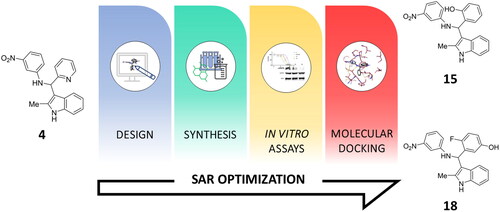
Abstract
EGFR is a protein kinase whose aberrant activity is frequently involved in the development of non-small lung cancer (NSCLC) drug resistant forms. The allosteric inhibition of this enzyme is currently one among the most attractive approaches to design and develop anticancer drugs. In a previous study, we reported the identification of a hit compound acting as type III allosteric inhibitor of the L858R/T790M double mutant EGFR. Herein, we report the design, synthesis and in vitro testing of a series of analogues of the previously identified hit with the aim of exploring the structure-activity relationships (SAR) around this scaffold. The performed analyses allowed us to identify two compounds 15 and 18 showing improved inhibition of double mutant EGFR with respect to the original hit, as well as interesting antiproliferative activity against H1975 NSCLC cancer cells expressing double mutant EGFR. The newly discovered compounds represent promising starting points for further hit-to-lead optimisation.
GRAPHICAL ABSTRACT
Introduction
The Epidermal Growth Factor Receptor (EGFR) is a protein tyrosine kinase of pivotal importance for the development of anticancer drugsCitation1. Its function depends on the strictly regulated shift between active and inactive conformations, which can be distinguished by the different positions of key structural elements such as the DFG-motif and the αC helixCitation2–5. Primary mutations in the EGFR sequence such as the L858R are known to be involved in tumour development and drug resistance, especially in non-small-cell lung cancer (NSCLC)Citation6,Citation7, accounting for more than 80% of highly fatal lung cancersCitation8,Citation9. Indeed, L858R promotes the active conformation of the αC helix, destabilising the inactive stateCitation2,Citation3. Although a number of ATP-competitive inhibitors of EGFR has been approved by the Food and Drug Administration (FDA) so far, the onset of secondary mutations, such as the T790M at the gatekeeper makes most of these drugs ineffective as it alters enzymatic activity and drug binding, by increasing affinity of the protein for ATPCitation10. Affinity for ATP, however, reverts to levels comparable to wild-type EFGR in the concurrent presence of activating mutations such as L858R, which reduces the effectiveness of first- and second-generation ATP-competitive EGFR inhibitors. Likewise, the C797S mutation abrogates the activity of several irreversible inhibitors, as it removes the covalent reactive cysteineCitation11. For these reasons, the discovery of allosteric inhibitors of EGFR, and, in particular, of the more clinically relevant dru g resistant forms, has become of great interest as a possible way for circumventing drug resistanceCitation12,Citation13.
In a recent study, a high throughput docking screening performed on a set of commercially available molecules led to the identification of a hit candidate (compound 4 in ) acting as type III allosteric inhibitor of T790M/L858R double mutant EGFR, with an EC50 of 6.2 ± 0.5 μMCitation14. Of note, the compound showed also anti-proliferative activity against three NSCLC cell lines exhibiting different EGFR status, the observed IC50 values being 14.5 ± 0.3 μM (wt EGFR H1299 cell line), 19 ± 2 μM (mutant EGFR H1650 cell line), and 24 ± 2 μM (double mutant H1975 cell line)Citation14.
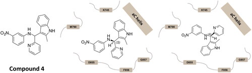
Figure 1. Chemical structure of the hit compound 4, and the binding modes predicted on EGFR by dockingCitation14.
Following our interest in developing allosteric inhibitors of protein kinasesCitation14–17, in this work, we sought to explore the structure-activity relationships (SAR) around this hit compound by designing, synthesising and in vitro testing a library of seventeen analogues bearing structural modifications at both the phenyl, pyridine and indole rings. Two of the designed compounds were found to be significantly more active than the parent hit against double mutant EGFR. The two compounds were further characterised for their anti-proliferative activity against the H1975 NSCLC cell line expressing the double mutant.
Results
Molecular design
Starting from the structure of the hit compound 4, we explored chemical modifications in all three aromatic rings (). These modifications were inspired by the docking complexes of compound 4 in complex with mutant EGFRCitation14.
Table 1. Chemical structures of the synthesised compounds.
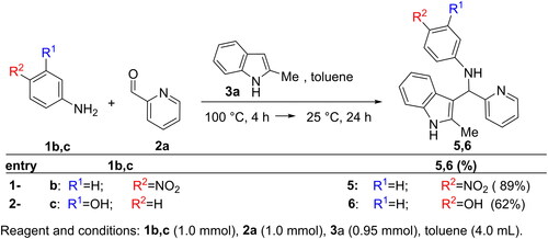
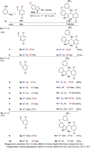
Firstly, the nitro group in the meta position was moved to the para position (5) or substituted with a hydroxyl group (6). The indole moiety in 4 was then substituted with a 5-fluorine atom, providing compound 7. Removal of the 2-methyl group of the indole ring provided 8. The latter compound was further substituted with a methyl ester residue at the positions 6 (9) or 5 (10). As for modifications of the pyridine ring, an m-fluorine atom or a o-hydroxyl group were added, resulting in compound 11 or 12, respectively. Attempts to introduce an additional fluorine atom on compound 12 failed. Then, the nitrogen atom of the pyridine residue was removed (13) and the resulting phenyl ring was substituted with an o-hydroxyl (15) or a m-hydroxyl group (16). Synthesis of the o-hydroxyl-m-fluoro derivative afforded compound 17. Replacement of the hydroxyl group of compound 17 with a methoxy group led to compound 14. Likewise, synthesis of the o-fluoro-m-hydroxyl derivative afforded compounds 18 and 20.
Chemical synthesis
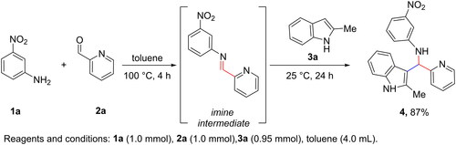
The model compound of our studies (4, Scheme 1) was prepared via an aza Friedel-Craft (AFC) one pot process. As shown in Scheme 1, the synthesis of 4 involves an aniline derivative (1a, meta nitro aniline), an aromatic aldehyde (2a, picolinaldehyde) and an indole ring (3a, 2-methylindole): these are the three structural units object of our investigation. Furthermore, the reaction proceeds through condensation of 1a and 2a followed by the ACF reaction of indole 3a and the imine intermediate avoiding the use of any catalystCitation18,Citation19.
Scheme 1. Synthesis of hit compound 4.
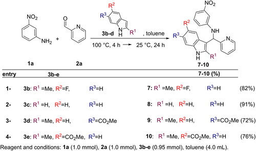
The generality of the multicomponent ACF process was tested on a range of substituted anilines 1, indoles 3 and aldehydes 2 (Schemes 2–4). Furthermore, good yields of inhibitors 5,6 were reached when aldehyde 2a and indole 3a were reacted with anilines 1b,c bearing functional groups whit different electronic properties (Scheme 2). A similar behaviour was observed when indoles 3b-e were employed in the presence of aniline 1a and aldehyde 2a (synthesis of 7–10, Scheme 3). Also the use of picolinaldehydes 2b,c -bearing respectively a fluorine and a hydroxy residue- or benzaldeyde 2d or 2-methoxy, 6-Fluoro benzaldehyde 2e did not affect the reaction outcome (synthesis of inhibitors 11–14, Scheme 3). On the other hand, the modulation of the electronic properties of hydroxy aldehyde 2f,i was crucial to promote the reaction pathway (synthesis of 15–20). Thus, the OH residue in 2f,i was O-acetylated prior to the ACF reactionCitation20. In turn, the final inhibitors 15–20 were isolated after cleavage of the acetyl moiety under basic conditionsCitation21.
Scheme 2. Modification at the aromatic aniline-ring: synthesis of 5 and 6.
Scheme 3. Modification at the indole residue: synthesis of 7–10.
Scheme 4. Synthesis of inhibitors 11–20.
Biological evaluation
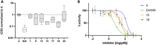
The synthesised compounds were tested in vitro by non-radioactive kinase assay, using compounds 4 and EAI045 as references (Table S1). As shown in , a number of compounds showed comparable or better activity than 4. In particular, compounds 15 and 18 showed significantly improved activity with respect to 4 (p < 0.0001). All other tested compounds were inactive on mutant EGFR (IC50 > 200 µM).
Figure 2. Activity of compounds in kinase assays. (A) Box and whiskers plot (min to max) of normalised IC50 values, relative to compound 4, from five independent experiments. (B) Dose-response curves of the indicated compounds from one representative experiment.
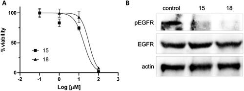
Compounds 15 and 18 were then tested in cell proliferation assays on the H1975 lung cancer cell line expressing the double mutant L858R/T790M EGFR. Both compounds inhibited cell proliferation and EGFR phosphorylation at micromolar concentrations ().
Figure 3. Activity of compounds 15 and 18 in H1975 cells. (A) Cell viability was measured by MTS assay. (B) EGFR phosphorylation was assessed by Western blot after 8-h incubation with the compounds (10 µM). Total EGFR and actin are shown for loading control.
Molecular docking
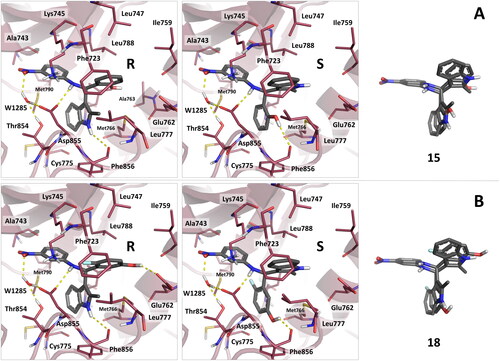
Docking calculations of both the stereoisomers of the best compounds 15 and 18 were firstly performed with Glide Citation22, starting from the crystal structure of T790M EGFR in complex with EAI001 (PDB code: 5D41)Citation12. In the predicted complexes, the nitrophenylamine moiety of both the compound stereoisomers is involved in a well-defined network of interactions, while the indole and hydroxyphenyl groups explored different orientations within the type III allosteric pocket. In particular, the nitro group of 15 and 18 hydrogen bonds with a water, the importance of this molecule for the binding of type III allosteric ligands to mutant T790M EGFR being previously assessedCitation23. In turn, the water molecule hydrogen bonds with the side chain of Thr854 and Asp855 (), the latter residue interacting with the amine group of the investigated compounds. The phenyl ring was predicted to establish hydrophobic interactions with the side chain of Ala743, Leu788 and Met790. The hydroxyphenyl and indole moieties of the R and S stereoisomers of compounds 15 and 18 were predicted to accommodate differently within the allosteric pocket of EGFR, albeit their superimposition revealed overlapping structural features and similar pattern of interactions with protein residues (). In particular, the hydroxyphenyl moiety of 15S and 18S was predicted to accommodate near the side chain of Phe856 and Met766, the phenyl ring establishing favourable hydrophobic interactions, and the hydroxyl group hydrogen bonding to the backbone carbonyl of Phe856, similarly to what previously observed ()Citation14. In the R stereochemistry, the latter interaction is performed by the amino group of the indole instead of the hydroxyphenyl. In the 15S and 18S complexes, the indole portion of the compounds binds in proximity to Leu747 and Ile759, the amino group hydrogen bonds with the side chain of Glu766 (). Besides, this interaction is mapped by the hydroxyl groups at the ortho and meta positions in the 15R and 18R complexes (), respectively. The introduction at these positions of functional groups unable to establish h-bond interactions resulted in lack of activity (e.g. compounds 13 and 14), suggesting that these hydrogen bonds are required for activity. Substitutions with a fluorine in the phenyl moiety are tolerated, when a hydroxyl group is present at the ortho and meta positions (e.g. compounds 17, 18 and 20) ().
Figure 4. Binding mode predicted for compounds 15 and 18 by docking. (A) reports the binding mode predicted for the R and S stereoisomers of compound 15. (B) reports the binding mode predicted for the R and S stereoisomers of compound 18. The superimposition of the R and S stereoisomers of 15 and 18 in their predicted binding mode is also reported. As can be observed, the stereoisomers of the compounds show similar pattern of interactions, with good superimposition of their aromatic and h-bond donating groups.
To investigate the effect of protein flexibility on the predicted docking poses Citation24, we also performed Induced Fit Docking (IFD) Citation25,Citation26 calculations. Remarkably, IFD provided results similar to those obtained by docking with Glide. Altogether, the results of these analyses confirmed that the two stereoisomers of these compounds present good complementarity with the allosteric site of mutant EGFR. The additional hydrogen bonds formed by 15 and 18 may explain their higher activity. The 3D coordinates of the complexes of compounds 15 and 18 with double mutant EGFR are available upon request to the authors.
Conclusions
In this study, a series of compounds were designed, synthesised, and in vitro tested to explore the SAR of a class of previously discovered type III allosteric inhibitors of double mutant EGFRCitation14. The performed studies allowed to identify two derivatives with significantly higher inhibitory activity with respect to the starting hit compound 4. The best compounds identified from the in vitro assays on EGFR (i.e. 15 and 18) were also tested for their antiproliferative activity towards the H1975 NSCLC cancer cell lines expressing the double mutant, obtaining satisfactory results. Docking analyses were finally performed by means of Glide and the Induced Fit Protocol to help explaining their higher activity and rationalise the SAR of the investigated compounds. The results of this study allowed to highlight the importance of hydrogen-bonding groups at key positions of the investigated scaffold, providing interesting structural clues for further hit to lead optimisation in the allosteric pocket. Despite remarkable on-target biological activity, these hits still display off-target toxicity in EGFR-non-dependent cells (Figure S1), indicating the need for further refinement. However, the compounds are small-in-size and present a rather low decorated scaffold, which represent key assets for further medicinal chemistry optimisation, especially in the relatively uncharted context of type III allosteric inhibitors.
Supplemental Material
Download PDF (398 KB)Disclosure statement
No potential conflict of interest was reported by the author(s).
Additional information
Funding
References
- Salomon DS, Brandt R, Ciardiello F, Normanno N. Epidermal growth factor-related peptides and their receptors in human malignancies. Crit Rev Oncol/Hematol. 1995;19(3):183–232.
- Eck MJ, Yun CH. Structural and mechanistic underpinnings of the differential drug sensitivity of EGFR mutations in non-small cell lung cancer. Biochim Biophys Acta. 2010;1804(3):559–566.
- Martin-Fernandez ML, Clarke DT, Roberts SK, Zanetti-Domingues LC, Gervasio FL. Structure and dynamics of the EGF receptor as revealed by experiments and simulations and its relevance to non-small cell lung cancer. Cells. 2019;8(4):316.
- Sutto L, Gervasio FL. Effects of oncogenic mutations on the conformational free-energy landscape of EGFR kinase. Proc Natl Acad Sci U S A. 2013;110(26):10616–10621.
- Liu B, Bernard B, Wu JH. Impact of EGFR point mutations on the sensitivity to gefitinib: insights from comparative structural analyses and molecular dynamics simulations. Proteins. 2006;65(2):331–346.
- Normanno N, De Luca A, Bianco C, Strizzi L, Mancino M, Maiello MR, Carotenuto A, De Feo G, Caponigro F, Salomon DS. Epidermal growth factor receptor (EGFR) signaling in cancer. Gene. 2006;366(1):2–16.
- Paez JG, Jänne PA, Lee JC, Tracy S, Greulich H, Gabriel S, Herman P, Kaye FJ, Lindeman N, Boggon TJ, et al. EGFR mutations in lung cancer: correlation with clinical response to gefitinib therapy. Science. 2004;304(5676):1497–1500.
- Zarogoulidis K, Zarogoulidis P, Darwiche K, Boutsikou E, Machairiotis N, Tsakiridis K, Katsikogiannis N, Kougioumtzi I, Karapantzos I, Huang H, et al. Treatment of non-small cell lung cancer (NSCLC). J Thorac Dis. 2013;5:S389–S396.
- Alexander M, Kim SY, Cheng H. Update 2020: management of non-small cell lung cancer. Lung. 2020;198(6):897–907.
- Westover D, Zugazagoitia J, Cho BC, Lovly CM, Paz-Ares L. Mechanisms of acquired resistance to first- and second-generation EGFR tyrosine kinase inhibitors. Ann Oncol. 2018;29(suppl_1):i10–i19.
- Wheeler DL, Dunn EF, Harari PM. Understanding resistance to EGFR inhibitors—impact on future treatment strategies. Nat Rev Clin Oncol. 2010;7(9):493–507.
- Jia Y, Yun CH, Park E, Ercan D, Manuia M, Juarez J, Xu C, Rhee K, Chen T, Zhang H, Palakurthi S, et al. Overcoming EGFR T790M and C797S resistance with mutant-selective allosteric inhibitors. Nature. 2016;534(7605):129–132.
- Tripathi SK, Biswal BK. Allosteric mutant-selective fourth-generation EGFR inhibitors as an efficient combination therapeutic in the treatment of non-small cell lung carcinoma. Drug Discov Today. 2021;26(6):1466–1472.
- Caporuscio F, Tinivella A, Restelli V, Semrau MS, Pinzi L, Storici P, Broggini M, Rastelli G. Identification of small-molecule EGFR allosteric inhibitors by high-throughput docking. Future Med Chem. 2018;10(13):1545–1553.
- Rastelli G, Anighoro A, Chripkova M, Carrassa L, Broggini M. Structure-based discovery of the first allosteric inhibitors of cyclin-dependent kinase 2. Cell Cycle. 2014;13(14):2296–2305.
- Christodoulou MS, Caporuscio F, Restelli V, Carlino L, Cannazza G, Costanzi E, Citti C, Lo Presti L, Pisani P, Battistutta R, et al. Probing an allosteric pocket of CDK2 with small molecules. ChemMedChem. 2017;12(1):33–41.
- Carlino L, Christodoulou MS, Restelli V, Caporuscio F, Foschi F, Semrau MS, Costanzi E, Tinivella A, Pinzi L, Lo Presti L, et al. Structure-activity relationships of hexahydrocyclopenta[c]quinoline derivatives as allosteric inhibitors of CDK2 and EGFR. ChemMedChem. 2018;13(24):2627–2634.
- Bonandi E, Perdicchia D, Colombo E, Foschi F, Marzullo P, Passarella D. C3 Aza-alkylation of indoles. Org Biomol Chem. 2020;18(32):6211–6235.
- Christodoulou MS, Beccalli EM, Foschi F, Giofrè S. Pd-catalyzed domino reactions involving alkenes to access substituted indole derivatives. Synthesis. 2020;52(19):2731–2760.
- Papis M, Loro C, Penso M, Broggini G, Foschi F. Synthesis of morpholino nucleosides starting from enantiopure glycidol. Org Chem Front. 2022;9(11):2949–2954.
- Quici S, Casoni A, Foschi F, Armelao L, Bottaro G, Seraglia R, Bolzati C, Salvarese N, Carpanese D, Rosato A. Folic acid-conjugated europium complexes as luminescent probes for selective targeting of cancer cells. J Med Chem. 2015;58(4):2003–2014.
- Friesner RA, Banks JL, Murphy RB, Halgren TA, Klicic JJ, Mainz DT, Repasky MP, Knoll EH, Shaw DE, Shelley M, Perry JK, et al. Glide: a new approach for rapid, accurate docking and scoring. 1. Method and assessment of docking accuracy. J Med Chem. 2004;47(7):1739–1749.
- Tinivella A, Rastelli G. Investigating the selectivity of allosteric inhibitors for mutant T790M EGFR over wild type using molecular dynamics and binding free energy calculations. ACS Omega. 2018; 3(12):16556–16562.
- Zhao Z, Xie L, Bourne PE. Structural insights into characterizing binding sites in epidermal growth factor receptor kinase mutants. J Chem Inf Model. 2019;59(1):453–462.
- Sherman W, Day T, Jacobson MP, Friesner RA, Farid R. Novel procedure for modeling ligand/receptor induced fit effects. J Med Chem. 2006;49(2):534–553.
- Sherman W, Beard HS, Farid R. Use of an induced fit receptor structure in virtual screening. Chem Biol Drug Des. 2006;67(1):83–84.