
Abstract
A series of novel quinoline-O-carbamate derivatives was rationally designed for treating Alzheimer’s disease (AD) by multi-target-directed ligands (MTDLs) strategy. The target compounds were synthesised and evaluated by AChE/BuChE inhibition and anti-inflammatory property. The in vitro activities showed that compound 3f was a reversible dual eeAChE/eqBuChE inhibitor with IC50 values of 1.3 µM and 0.81 µM, respectively. Moreover, compound 3f displayed good anti-inflammatory property by decreasing the production of IL-6, IL-1β and NO. In addition, compound 3f presented significant neuroprotective effect on Aβ25-35-induced PC12 cell injury. Furthermore, compound 3f presented good stabilities in artificial gastrointestinal fluids, liver microsomes in vitro and plasma. Furthermore, compound 3f could improve AlCl3-induced zebrafish AD model by increasing the level of ACh. Therefore, compound 3f was a promising multifunctional agent for the treatment of AD.
Introduction
Alzheimer’s disease (AD) is the main cause of dementia and is quickly becoming one of the most expensive, lethal, and burdening disease of this centuryCitation1. At present, more than 50 million people worldwide are affected and the figure of the AD patients will triple by 2050Citation2.
The pathologic changes of AD are convoluted and multilayered, these abnormal changes include the low levels of acetylcholine (ACh), loss of synapses, neuroinflammation, aberrant accumulation of amyloid-β (Aβ) oligomer, neurofibrillary tangles within neurons and elevated oxidative stressCitation3. Presently, the primary therapeutic options for AD treatment are acetylcholinesterase inhibitors (AChEIs), such as donepezil, rivastigmine, galantamine, as well as the N-methyl-D-aspartate (NMDA) receptor antagonist, memantine, which can improve memory and cognitive function to a certain extent but cannot halt or reverse the progression of this diseaseCitation4,Citation5. Due to the complexity of AD and the involvement of multi-systems in the process of AD, the multi-target-directed ligands (MTDLs) approach, that is one molecule hits two or more AD-relevant complementary targets and produces synergistic effect on the disease network, has been developed in both symptomatic and disease-modifying efficienciesCitation5,Citation6.
The “cholinergic hypothesis” states that loss of cholinergic function is the important biomarkers of AD. It has been verified that the high levels of AChE facilitate the hydrolysation of ACh lead to memory deficit in the early stages of ADCitation7,Citation8. Moreover, AChE produces promotion in Aβ deposition in the form of senile plaques in the brain of afflicted individualsCitation9. BuChE is an enzyme closely related to AChE and serves as a co-regulator of cholinergic neurotransmission by hydrolysing ACh. It is fact that the AChE level in the brain declines progressively as the progress of AD goes, while BChE level soars to 165% of the normal level in the late stages of AD, this implies BChE takes over the hydrolysis of ACh in late ADCitation10. An increased BuChE activity also plays an important role in Aβ-aggregation during the early stages of senile plaque formationCitation11. Thus, dual AChE/BuChE inhibition have been documented as effective management for the treatment of AD by an increase in the availability of ACh in the brain regions and decrease in the Aβ deposition, such as rivastigmine.
Investigations suggest that inflammation clearly occurs in pathologically vulnerable regions of the AD brain, which related to microglia, astrocytes and inflammatory factors. In the AD brain, damaged neurons and neurites and highly insoluble Aβ oligomer and neurofibrillary tangles provide obvious stimuli for inflammation, such as tumour necrosis factor (TNF-α), IL-6, IL-1β and NO. AD inflammation significantly contributed to AD pathogenesisCitation12–14. Therefore, to develop anti-inflammatory agent could provide an effective approach to slow the progression or delay the onset of AD.
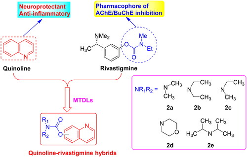
Quinoline core (), a bicyclic structure in which a pyridinic ring is fused to a benzene moiety, occurs in several natural compounds (Cinchona alkaloids) and in drugs, with a broad range of biological activities including antimicrobial, antifungal, antitumor, antiviral, anticholesterol and anti-Alzheimer’s diseaseCitation15,Citation16. The 8-hydroxy quinoline derivatives clioquinol and PBT2 (), promising multifunctional agents, have been investigated for their neuroprotective effect in AD and have entered clinical trials for ADCitation17. Other quinoline derivatives also show AChE/BuChE inhibitory potency, antioxidant activity, metal chelators, anti-inflammatory potency and inhibition effects on Aβ aggregationCitation18–20. Moreover, rivastigmine is a dual AChE/BuChE inhibitor approved by FDA for the treatment of mild to moderate ADCitation21. The carbamate moiety is the pharmacophore, and many carbamate derivatives have been developed as multifunctional agents anti-AD by our group and other groupsCitation22–25. Herein, we plan to introduce carbamate moiety into the promising quinoline skeleton to obtain novel quinoline-rivastigmine hybrids by MTDLs (). The synthesised target compounds are evaluated through in vitro and in vivo and hope to get a promising multifunctional agent to treat AD.
Figure 1. The structure of quinoline, clioquinol and PBT2.
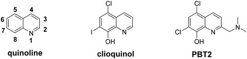
Figure 2. The design strategy of quinoline-rivastigmine hybrids.
Results and discussion
Chemistry
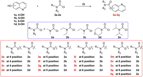
The synthetic route of target derivatives 3a-3q were summarised in Scheme 1. Briefly, the starting material 1a–1d (4-hydroxyquinoline 1a, 5-hydroxyquinoline 1b, 6-hydroxyquinoline 1c, and 8-hydroxyquinoline 1d) were treated with excessive amounts of dimethylcarbamoyl chloride (2a), diethylcarbamyl chloride (2b), N-ethyl-N-methylcarbamoyl chloride (2c), 4-morpholinecarbonyl chloride (2d), and diisopropylcarbamoyl chloride (2e), respectively, in the presence of K2CO3 in CH3CN at 60–65 °C to obtain the target compounds 3a–3q. All the target compounds were purified by chromatography, and the analytical and spectroscopic data confirmed their structures, as detailed in the Supporting Information.
Scheme 1. Synthesis of target compounds 3a-3q. Reagents and conditions: (i) 1.3 equivalent N,N-disubstituted carbamoyl chlorides (2a-2e), 1.5 equivalent K2CO3, CH3CN, 65 °C, for 6–10 h.
Biological activity
Inhibition effects on AChE and BuChE
The inhibitory activities of the synthesised compounds against eeAChE (from electric eel) and eqBuChE (from equine serum) were tested by the modified Ellman’s methodCitation26,Citation27. Rivastigmine and donepezil were used as positive compounds. As displayed in , both the carbamate fragments and the position of carbamate fragment significantly influenced eeAChE/eqBuChE inhibitory potency. Overall speaking, compounds 3a–3d with carbamate fragments at 4-position of quinoline showed significantly selective eqBuChE inhibitory potency, especially, compound 3d showed the best eqBuChE inhibitory potency with IC50 value of 0.87 µM; When the carbamate fragments at 5-position of quinoline, the compounds 3e–3h were potent dual AChE/BuChE inhibitors, particularly, compound 3f showed excellent dual AChE/BuChE inhibitory activity with IC50 values of 1.3 µM and 0.81 µM, respectively; When the carbamate fragments at 6-position of quinoline, compounds 3i–3l showed moderate selective eeAChE inhibitory potency, particularly, compound 3k showed moderate eeAChE inhibitory activity with IC50 value of 10.3 µM; When the carbamate fragments at 8-position of quinoline, compounds 3m–3q were potent selective eeAChE inhibitors, particularly, compound 3m showed good eeAChE inhibitory activity with IC50 value of 6.5 µM.
Table 1. AChE/BuChE inhibitory activities by target compounds 3a–3q, rivastigmine, and donepezil.
As for the inhibitory effect of carbamate fragment, when the carbamate fragment at the 4-position of quinoline, compound 3a-3d were selective eqBuChE inhibitors, compound 3a with dimethylcarbamoyl fragment showed good eqBuChE inhibitory potency with IC50 value of 2.83 µM. When the dimethylcarbamoyl of 3a was replaced with diethylcarbamoyl to get compound 3b, the eqBuChE inhibitory potency increased to 1.06 µM. When the dimethylcarbamoyl of 3a was replaced with N-ethyl-N-methylcarbamoyl to get compound 3c, the eqBuChE inhibitory potency increased to 1.92 µM. When the dimethylcarbamoyl of 3a was replaced with 4-morpholinecarbonyl to obtain compound 3d, the eqBuChE inhibitory potency increased to 0.87 µM. When the carbamate fragment at the 5-position of quinoline, compound 3e with dimethylcarbamoyl fragment, compound 3f with diethylcarbamoyl fragment, and compound 3g with N-ethyl-N-methylcarbamoyl fragment were good dual eeAChE/eqBuChE inhibitors with IC50 values of 4.5 µM/0.94 µM, 1.3 µM/0.81 µM and 3.2 µM/2.23 µM, respectively, while compound 3h with 4-morpholinecarbonyl showed moderate eeAChE inhibitory activity (IC50 = 12.9 µM) and good eqBuChE inhibitory potency (IC50 = 1.76 µM). When the carbamate fragment at the 6-position of quinoline, compounds 3i-3l showed moderate eeAChE inhibitory activities (ranging from 10.3 µM to 17.6 µM) and weak eqBuChE inhibitory activities. When the carbamate fragment at the 8-position of quinoline, compounds 3m-3q showed moderate to good eeAChE inhibitory activities (ranging from 6.5 µM to 14.8 µM) and weak eqBuChE inhibitory activities. Compound 3m with dimethylcarbamoyl fragment showed good eeAChE inhibitory potency (IC50 = 6.5 µM). When the dimethylcarbamoyl of 3m was replaced with diethylcarbamoyl and N-ethyl-N-methylcarbamoyl, respectively, to get compounds 3n and 3o, respectively, the eeAChE presented slightly decrease with IC50 values of 9.7 µM and 8.2 µM, respectively. While, when the dimethylcarbamoyl of 3m was replaced with 4-morpholinecarbonyl and diisopropylcarbamoyl, respectively, to get compounds 3p and 3q, respectively, the eeAChE sharply decreased to 12.6 µM and 14.8 µM, respectively.
Furthermore, the representative compounds (3b, 3d, 3e and 3f) with good eeAChE or eqBuChE inhibitory activity were re-evaluated using hAChE and hBChE. As shown in , overall speaking, the selective eqBuChE inhibitors 3b and 3d showed good huBuChE inhibitory potency with IC50 values of 5.39 µM and 9.46 µM, respectively and weak eeAChE inhibitory potency. The dual eeAChE/eqBuChE inhibitors 3e and 3f showed good huBuChE inhibitory potency and hAChE inhibitory potency, particularly, compound 3f presented good dual hAChE/hBuChE inhibitory potency with IC50 values of 5.6 µM and 2.34 µM, respectively, deserving for further investigation.
Anti-inflammatory property
According to the ChEs inhibitory activity, compounds 3a, 3c, 3e, 3f, 3k, 3m and 3n were selected to tested the anti-inflammatory potency by measuring the production of inflammatory mediators IL-6, IL-1β and NO in LPS-induced BV-2 cellsCitation28,Citation29.
Cytotoxicity of compounds 3a, 3c, 3e, 3f, 3k, 3m and 3n on BV-2 cells. Firstly, the cytotoxicity of compounds 3a, 3c, 3e, 3f, 3k, 3m and 3n were tested using CCK8 assay. As displayed in , the cell viability did not show obvious change after adding compounds 3a, 3c, 3e, 3f, 3k, 3m, and 3n at 2 µM.
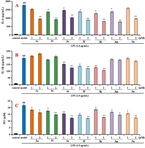
Evaluation the production of IL-6, IL-1β and NO in LPS-stimulated BV-2 cells. As shown in , when BV-2 cells were exposed to 1 µg/mL LPS for 24h, the levels of IL-6, IL-1β and NO significantly increased (p < 0.01), suggesting that the inflammatory model of BV-2 cells was successfully created and worked well. As displayed in , compounds 3a, 3c, 3e, 3f, 3k, 3m and 3n (at 1 µM and 2µM, respectively) could significantly decreased the levels of IL-6 compared with model group. As displayed in , compounds 3e, 3f and 3k could significantly decreased the levels of IL-1β compared with model group, while compounds 3a, 3c, 3m and 3n did not obviously decrease the level of IL-1β compared with model group. As displayed in , compounds 3a, 3c, 3e, 3f, 3k, 3m and 3n could significantly decreased the release volume of NO. The results exhibited that compounds 3e, 3f, 3k showed significant anti-inflammatory activity by decreasing the production of inflammatory mediators IL-6, IL-1β and NO.
Figure 3. The cell viability of compounds 3a, 3c, 3e, 3f, 3k, 3m, and 3n on the BV-2 cells, which was determined using MTT assay. The data are expressed as the mean ± SD by three independent experiments.
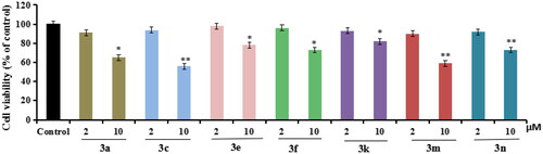
Figure 4. Effects of compounds 3a, 3c, 3e, 3f, 3k, 3m, and 3n on the production of IL-6, IL-1β and NO in LPS-stimulated BV-2 cells. (A) Effects of compounds 3a, 3c, 3e, 3f, 3k, 3m, and 3n on the production of IL-6; (B) Effects of compounds 3a, 3c, 3e, 3f, 3k, 3m, and 3n on the production of IL-1β; (C) Effects of compounds 3a, 3c, 3e, 3f, 3k, 3m, and 3n on NO release. Data were expressed as mean ± SD through three independent experiments. ##p < 0.01 vs control; **p < 0.01, *p < 0.05 vs LPS-induced model group.
hAChE/hBuChE reversibility of inhibition by 3f
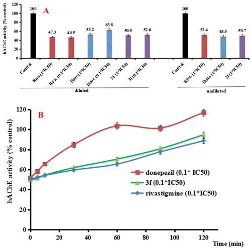
According to results from the AChE/BuChE inhibition and anti-inflammatory property, compound 3f presented good dual AChE/BuChE inhibitory activity and significant anti-inflammatory potency. Thus, compound 3f was selected to perform further investigation. In order to determine whether compound 3f was a reversible hAChE and hBuChE inhibitor, the recovery of hAChE and hBuChE inhibitor inhibition after dilution were performed (Citation26. For the hAChE reversibility of inhibition (), when donepezil, rivastigmine and compound 3f diluted to 0.1 × IC50, respectively, the hAChE activity increased to 10.6%, −0.8% and 1.7%, respectively. Moreover, the recovery of hAChE inhibitors inhibition after dilution was carried out with time monitoring. As shown in , the hAChE activity restored to 103.6% by treating with 0.1× IC50 donepezil at 60 min, and treatment with 0.1 × IC50 rivastigmine and compound 3f, hAChE activity restored to 88.7% and 94.7% at 120 min, respectively. The results showed that compound 3f was a reversible hAChE inhibitor.
Figure 5. (A) hAChE recovery after preincubation of compound 3f diluted to 1× or 0.1 × IC50, compared to donepezil (done.) diluted, rivastigmine (riva.) diluted and undiluted inhibition. (B) hAChE recovery of donepezil, 3f diluted to 0.1 × IC50, were monitored with time at room temperature for 120 min. Data were expressed as the mean ± SD by three independent experiments.
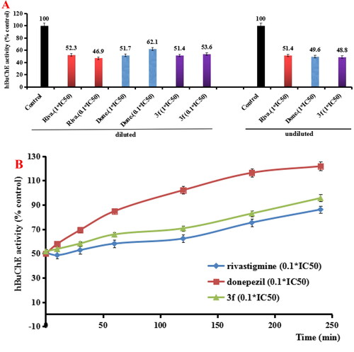
For the hBuChE reversibility of inhibition, as shown in , compared to controls, the 0.1 × IC50 of donepezil, rivastigmine and compound 3f increased the hBuChE activity to 8.3%, 1.2% and 3.1%, respectively. Furthermore, the recovery of hBuChE inhibitor inhibition after dilution with time monitoring was shown in . When treating with 0.1× IC50 donepezil, rivastigmine and compound 3f for 120 min, the hBuChE activity restored to 102.1%, 62.3% and 70.7%, respectively. As time went, the hBuChE activity restored to 86.2%, 121.8% and 95.6%, respectively at 240 min, suggesting that compound 3f was a reversible hBuChE inhibitor. Therefore, the above results showed that compound 3f was a reversible AChE and BChE inhibitor.
Figure 6. (A) hBuChE recovery after preincubation of compound 3f diluted to 1× or 0.1 × IC50, compared to donepezil (done.) and rivastigmine (riva.) diluted, and undiluted inhibition. (B) hBuChE recovery of donepezil, rivastigmine and 3f diluted to 0.1 × IC50, were monitored with time at room temperature for 240 min. Data were expressed as the mean ± SD by three independent experiments.
Enzyme kinetic study
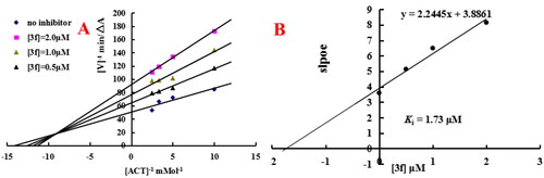
In order to determine the mechanism of quinoline-O-carbamate derivatives, the representative compound 3f was chose to perform Lineweaver-Burk double reciprocal plot by plotting the initial velocities of the substrate at y-axis and increasing concentrations substrate at x-axisCitation27. As displayed in , graphical analysis displayed both increasing slopes (decreased Vmax) and intercepts (higher Km) at increasing concentration of compound 3f. As displayed in , replots of the slope versus concentration of compound 3f obtained an estimate of the inhibition constant and the Ki value was 1.73 µM. The results suggested that compound 3f was a mixed-type of AChE inhibitor and was able to simultaneously bind both catalytic active site (CAS) and peripheral anionic site (PAS) of AChE.
Figure 7. Enzyme kinetic study on the mechanism of eeAChE inhibition by compound 3f. (A) Overlaid Lineweaver-Burk reciprocal plots of AChE initial velocity at increasing acetylthiocholine concentration in the absence and in the presence of 3f were determined. (B) The plots of slope versus the concentration of 3f for determining the inhibition constants Ki.
Molecular modelling studies of 3f
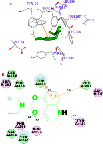
The possible interacting mechanism of compound 3f with hAChE (PDB code: 4ey4) and hBuChE (PDB code: 4tpk) was carried out using AUTODOCK 4.2 package, respectivelyCitation22,Citation23. It had been reported that amino acid residues Tyr72, Asp74, Trp86, Tyr124, Trp286, Tyr337, Phe295 and Phe297 were the key active site residues of hAChECitation30. As displayed in 3f-hAChE complex (), the carbonyl group interacted with Phe295 (2.5 Å) and Arg296 (1.6 Å) via two intramolecular hydrogen bonding, respectively. The polar H of quinoline interacted with Tyr124 (1.9 Å) via one intermolecular hydrogen bonding. The benzene ring of quinoline interacted with Trp286 (5.6 Å) and Phe297 (3.9 Å) via one π-π interaction and one σ-π interaction, respectively. Besides, some hydrophobic interactions were presented between compound 3f and residues (such as Trp286, Tyr341, Phe338, Leu289, and Asp74). The above interactions offered a possible mechanism for its high AChE inhibitory activity.
Figure 8. (A) Compound 3f (green stick) acted on residues in the binding site of hAChE (PDB code: 4ey4). (B) D docking model of 3f with hAChE.
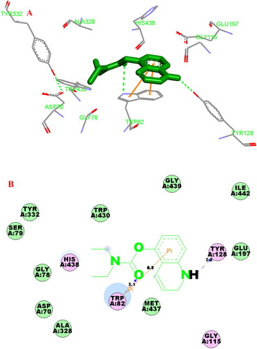
It had been verified that residues Trp82, His438, Val328, Phe329, Trp231, Val288, Gln119, Thr120 and Gly121 were considered to be an PDB entry 4tpk active site residues of hBuChECitation31. In the hBuChE −3f (), the carbonyl group interacted with Trp82 via one intramolecular hydrogen bonding (3.1 Å). The polar H of quinoline interacted with Tyr128 via one intermolecular hydrogen bonding (2.2 Å). The benzene ring of quinoline interacted with Trp82 via two π-π interactions (3.3 Å and 4.5 Å). Moreover, some hydrophobic interactions were presented between compound 3f and residues (such as Trp82, Tyr332, Glu197, Gly115, Tyr128, Trp430, His438 and Ala328). Thus, the above observed phenomenon could provide a reasonable binding mechanism for potent BChE inhibitory activity of 3f.
Figure 9. (A) Compound 3f (green stick) acted on residues in the binding site of hBuChE (PDB code: 4tpk). (B) 2 D docking model of 3f with hBuChE.
Molecular dynamics (MD) simulations
The stability of docked binding pose of the compound 3f-AChE complex and 3f-BuChE were analysed by molecular dynamics simulation analysis using Amber 16Citation24. shows that the root means square deviations (RMSD) analysis of compound 3f with the amino acid residues of AChE and BuChE, respectively. The results demonstrated that RMSDs of all the replicas for the six simulated systems presented relatively stable fluctuations after 50 ns of the MSMD simulations, suggesting that the six simulated systems basically reach equilibrium. The binding free energies of compound 3f towards hAChE and hBuChE calculated using MM-PBSA were displayed in , and have values of −30.59 and −37.65 kcal/mol, respectively, which were mainly contributed by Van der Waals forces, electrostatic interactions and non-polar solvation energiesCitation24. Furthermore, shows the key residues and interaction modes of compound 3f with hAChE and hBuChE, respectively. In , the O atom O-carbamate fragment formed one intermolecular hydrogen bonding with the key amino acid Tyr124 (2.6 Å). displays the interactions modes of 3f with hBuChE, the carbonyl group of carbamate fragment interacted with key amino acid residue Gly116 (2.9 Å), Gly117 (2.9 Å) and Ser198 (2.9 Å) via one intermolecular hydrogen bonding interactions, respectively.
Figure 10. (A) RMSD analysis of compound 3f with hAChE (PDB code: 4ey4) (black stick); (B) RMSD analysis of compound 3f with hBuChE (PDB code: 4tpk) (black stick).
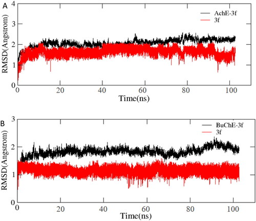
Figure 11. (A) The docking model for 3f into protein crystal structure of hAChE (PDB code: 4ey4). (B) The docking model for 3f into protein crystal structure of hBuChE (PDB code: 4tpk).
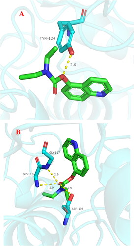
Table 2. The binding free energy and components of 3f-AChE and 3f-BuChE (kcal/mol).
Neuroprotective effect
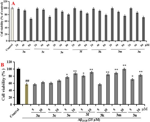
The neuroprotective effect of compounds 3a, 3c, 3e, 3f, 3k, 3m, and 3n on Aβ25–35-induced PC12 cells injury were evaluated using CCK-8 assayCitation32. As presented in , compounds 3a, 3c, 3e, 3f, 3k, 3m, and 3n did not show any cytotoxicity at concentration below 40 µM, indicating a widely safe range. Furthermore, as shown in , when PC12 cells were exposed to 25 µM Aβ25-35, the cell viability sharply declined to 57.3% (p < 0.01) compared with untreated group. When the PC12 cells was treated with compounds 3a, 3c, 3e, 3f, 3k, 3m, and 3n, respectively, the results showed that compounds 3a and 3c with carbamate fragment at 4 positin of quinoline did not show significant neuroprotective effects on Aβ25-35-induced PC12 cells injury, compound 3k with carbamate fragment at 6 positin of quinoline showed significant neuroprotective effects at 10 µM, compounds 3e and 3f with carbamate fragment at 5 positin of quinoline displayed remarkable neuroprotective effects at 5 and 10 µM, respectively, compounds 3m and 3n with carbamate fragment at 8 positin of quinoline showed significant neuroprotective effects at 5 and 10 µM, respectively.
Figure 12. Cell viability was tested by MTT assay. (A) Cytotoxicity of compounds 3a, 3c, 3e, 3f, 3k, 3m, and 3n on PC12 cells. (B) Attenuation of Aβ25-35-induced PC12 cell injury by compounds 3a, 3c, 3e, 3f, 3k, 3m, and 3n. values were expressed as mean ± SD by three independent experiments. ##p < 0.01 vs control; **p < 0.01, *p < 0.05 vs Aβ25-35 group.
Stability studies of 3f
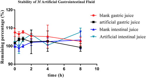
Compound 3f was selected to investigate the stabilities by artificial gastrointestinal conditions and (human and rat) liver microsomesCitation33. As displayed in , the remaining amount of compound 3f in blank gastric fluid, blank intestinal fluid, artificial gastric fluid and artificial intestinal fluid was 102.6%, 103.4%, 99.2% and 107.7%, respectively, after 8 h incubation, suggesting that compound 3f was stable in both artificial intestinal and gastric fluids.
Figure 13. Stability of compound 3f in artificial gastrointestinal fluids (n = 3).
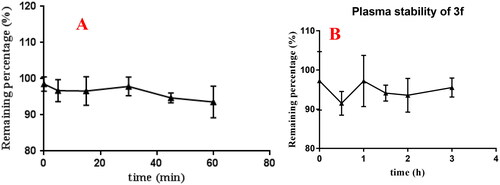
The stability of compound 3f was also performed using rat liver microsomes assay. According to the results from , after 60 min incubation, the remaining amount of 3f in rat liver microsomes was 104.4%. Futher, the plasma stability of compound 3f was also performed using rat plasma. As displayed in , the remaining amount of 3f was 95.5% after 3 h incubation in rat plasma.
Figure 14. (A) rat liver microsomes stabilities of compound 3f (n = 3); (B) the plasma stability of compound 3f (n = 3).
The above results showed that compound 3f presented good stabilities in artificial gastrointestinal fluids and moderate stabilities in liver microsomes.
Effect of 3f on AlCl3-induced zebrafish AD
AlCl3-induced zebrafish AD model has been widely used for high throughput screening of potential novel anti-AD leading compoundsCitation34,Citation35.
Acute toxicity. Firstly, the safety profile of compound 3f were investigated with seven concentrations (0.65, 1.3, 2.6, 5.2, 10.3, 20.5 and 40.9 µM). As displayed in , when the concentration increased to 40.9 µM, the percentage survival was 100% after 4h of post fertilisation (hpf), while all the zebrafish were dead for 24 hpf. When the concentration under 20.5 µM, the survival rate of other concentrations were 100% during 120 hpf. The results showed that the maximum non-lethal concentration (MNLC) of 3f was 20.5 µM. And based on the MNLC, six concentrations of 3f (0.65, 1.3, 2.6, 5.2, 10.3 and 20.5 µM) were selected to further investigate the acute toxicity. As shown in , the pericardial oedema/body length did not produce significant change at five concentrations of 3f, while the swim bladder area significantly decreased after treating with 5.2 µM 3f. So, three concentrations of 3f (0.65, 1.3 and 2.6 µM) were selected to further investigate the liver toxicity. As listed in , both the liver florescence area and median fluorescence intensity did not produce obvious change at three concentrations, suggesting that compound 3f was safe at 2.6 µM.
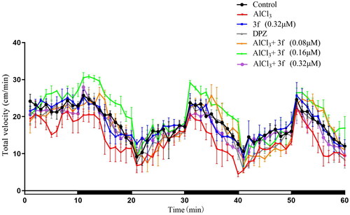
Effects of 3f on AlCl3-Induced Zebrafish AD Model. Based on the above results, compound 3f with three concentrations (0.08, 0.16 and 0.32 µM) were performed to evaluate the dyskinesia and reaction ././Lenovo/AppData/Local/youdao/dict/Application/7.5.2.0/resultui/dict/?keyword = capacity of AlCl3-induced zebrafish AD. The total velocity were shown in , compound 3f at dose of 0.16 µM could improve AlCl3-induced zebrafish AD model compared with donepezil.
Figure 15. The acute toxicity of compound 10c. (A) The percentage survival (%); (B) morphologic change; (C) changes of pericardial edema/body length; (D) changes of swim bladder area; (E) changes in the liver; (F) changes of liver fluorescence area; (G) changes of median fluorescence intensity.
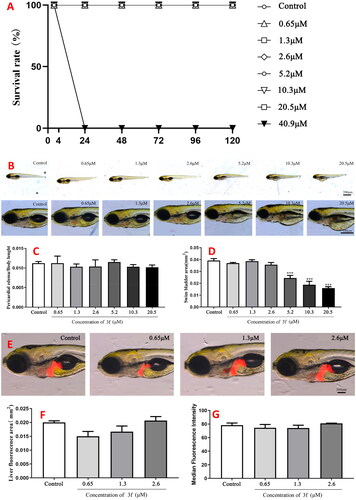
Figure 16. The total velocity of compound 3f on AlCl3-induced zebrafish AD model (n = 60). And the white and dark stripes on x-axis represent periods of light and dark, respectively.
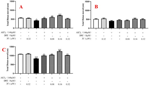
More specifically, for the distance moved, in the dark environment (), when zebrafish were treated with 140 µM AlCl3, the distance moved significantly declined to 447.4 cm (p < 0.05) compared with untreated group (582.9 cm). When treatment with 8 µM donepezil, the distance moved increased to 543.8 cm (p < 0.05). When treatment with compound 3f (0.08, 0.16 and 0.32 µM), the distance moved was 607.3 cm (p < 0.01), 709.4 cm (p < 0.001) and 521.7 cm, respectively. While, in the light environment (), when zebrafish were exposed to 140 µM AlCl3, the distance did not show statistical significance between the untreated group, model group, and tested compound group. Furthermore, under alternating dark light stimuli condition (), when treatment with compound 3f (0.08, 0.16 and 0.32 µM), the distance moved was 1029.2 cm (p < 0.01), 1241.5 cm (p < 0.001) and 1022.8 cm (p < 0.01), respectively, which was better than 8 µM donepezil (999.4 cm). The results showed that in the dark or alternating dark light stimuli condition, 8 µM donepezil could improve distance moved in the AlCl3-induced zebrafish AD model. Moreover, under the dark condition, light condition, and alternating dark light condition, respectively, 0.16 µM compound 3f presented the best distance moved.
Figure 17. The total distance moved of compound 3f on AlCl3-induced zebrafish AD model. (A) Under the dark environment; (B) Under the light environment; (C) Under alternating dark light stimuli condition. #p < 0.05 and ###p < 0.001 vs untreated group. *p < 0.05, **p < 0.01 and **p < 0.001 vs model group.
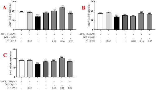
For the velocity, in the dark environment (), when zebrafish were treated with 140 µM AlCl3, the velocity significantly declined to 13.8 cm/min (p < 0.001) compared with untreated group (19.1 cm/min). When treatment with 8 µM donepezil, the velocity increased to 18.5 cm/min (p < 0.001). When treatment with compound 3f (0.08, 0.16 and 0.32 µM), the velocity was 21.3 cm/min (p < 0.001), 24.7 cm/min (p < 0.001), and 17.3 cm/min (p < 0.001), respectively. In the light condition (), when treatment with compound 3f (0.08, 0.16 and 0.32 µM), the velocity were 14.7 cm/min, 19.3 cm/min (p < 0.001) and 17.6 cm/min (p < 0.01), respectively. In alternating dark light stimuli condition (), when treatment with compound 3f (0.08, 0.16 and 0.32 µM), the velocity were 17.5 cm/min (p < 0.001), 22.3 cm/min (p < 0.001) and 17.0 cm/min (p < 0.001), respectively, which was better than donepezil (16.7 cm/min). The results showed that 8 µM donepezil significantly improved velocity in the AlCl3-induced zebrafish AD model under dark or alternating dark light stimuli condition. Moreover, under the dark, light and alternating dark light stimuli condition, respectively, 0.16 µM compound 3f presented the best velocity.
Figure 18. The total velocity of compound 3f on AlCl3-induced zebrafish AD model. (A) Under the dark environment; (B) under the light environment; (C) under alternating dark light stimuli condition. #p < 0.05 and ###p < 0.001 vs untreated group. *p < 0.05, **p < 0.01 and **p < 0.001 vs model group.
To sum up, compound 3f could significantly improve dyskinesia and reaction ././Lenovo/AppData/Local/youdao/dict/Application/7.5.2.0/resultui/dict/?keyword = capacity of AlCl3-induced zebrafish AD model, which was better than donepezil, and the optimal dose was 0.16 µM.
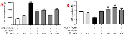
3.Mechanisms of compound 3f on AlCl3-induced AD in zebrafish model. After the behavioural experiments, the 168hpf zebrafish were killed and ground, the tissue were used to test the AChE activity and ACh levels. As shown in , when zebrafish were exposed to 140 µM AlCl3, the AChE activity sharply increased to 147908.7 nmol/min/g (p < 0.001) compared with untreated zebrafish (40018.7 nmol/min/g). When AD zebrafish model were treated with 8 µM donepezil, the AChE activity significantly decreased to 93540.7 nmol/min/g (p < 0.001). When treatment with compound 3f (0.08, 0.16 and 0.32 µM), the AChE activity were 98095.7 nmol/min/g (p < 0.001), 64663.2 nmol/min/g (p < 0.001) and 102433.6 nmol/min/g (p < 0.001), respectively. Furthermore, shows that when zebrafish were treated with 140 µM AlCl3, the ACh activity sharply decreased to 7.3 (p < 0.01) compared with untreated group (13.9 pmol/L). When treatment with 8 µM donepezil, the ACh activity significantly increased to 14.0 pmol/L (p < 0.01). When treatment with compound 3f (0.08, 0.16 and 0.32 µM), the ACh activity were 16.4 pmol/L (p < 0.001), 18.4 pmol/L (p < 0.001) and 15.4 pmol/L (p < 0.01), respectively. The results suggested that compound 3f could improve the levels of ACh in AD zebrafish model by inhibiting AChE activity, and the optimal dose of compound 3f was 0.16 µM.
Figure 19. (A) The AChE activity of 3f on AlCl3-induced zebrafish AD model. (B) The ACh level of 3f on AlCl3-induced zebrafish AD model. ##p < 0.01 and ###p < 0.001 vs untreated group. **p < 0.01 and **p < 0.001 vs model group.
Conclusion
Alzheimer’s disease (AD) was a chronic, progressive brain neurodegenerative disorder. Up to now, there was no effective drug to halt or reverse the progress of AD. Given the complex pathogenesis of AD, the MTDLs strategy was considered as the promising approach. Herein, a series of novel quinoline-O-carbamate derivatives was rationally designed for treating AD by fusing quinoline and rivastigmine. The synthesised target compounds were synthesised and evaluated by AChE/BuChE inhibition and anti-inflammatory property. The in vitro biological activities revealed that compound 3f was a reversible dual eeAChE/eqBuChE inhibitor with IC50 values of 1.3 µM and 0.81 µM, respectively. Compound 3f also showed good anti-inflammatory property by declining the production of inflammatory mediators IL-6, IL-1β and NO. In addition, compound 3f presented significant neuroprotective effect on Aβ1-42-induced PC12 cell injury. Moreover, compound 3f possessed favourable stabilities in artificial gastrointestinal fluids, liver microsomes in vitro and plasma. Furthermore, compound 3f significantly improved dyskinesia and reaction ././Lenovo/AppData/Local/youdao/dict/Application/7.5.2.0/resultui/dict/?keyword = capacity of AlCl3-induced zebrafish AD model by increasing the level of ACh. Therefore, compound 3f was a promising multifunctional agent for the treatment of AD, deserving for further investigation.
Experimental section
Chemistry
Unless otherwise noted, all the chemicals and solvents were obtained from Shanghai Titan Scientific Co.,Ltd. and Sigma-Aldrich, and were used without further purification. All the reactions were monitored by thin-layer chromatography (TLC) using silica gel GF254 plates, and crude products were purified by column chromatography using silica gel (230–400 mesh) purchased from Qingdao Haiyang Chemical Co. Ltd. (China). The purity of the target compounds was determined by high-performance liquid chromatography (HPLC) analysis to be over 96%. HPLC analysis was carried out on a Waters X-Bridge C18 column (4.6 mm × 150 mm, 5 µm) at a flow ratio of 0.8 ml/min. The 1H NMR and 13C NMR spectra of the synthetic compounds were recorded using TMS as the internal standard on a Varian INOVA at 400 and 100 MHz, respectively. The high-resolution mass spectra was obtained using a Waters Xevo G2-XS-Qtof mass spectrometer.
General procedures for the synthesis of quinoline-O-carbamate derivatives
To a solution of the starting material 1 (4-hydroxyquinoline 1a, 5-hydroxyquinoline 1b, 6-hydroxyquinoline 1c, and 8-hydroxyquinoline 1d, respectively) (1.0 mmol), and anhydrous K2CO3 (1.7 mmol) in anhydrous CH3CN (6 ml), N,N-disubstituted carbamoyl chlorides 2a–2e (1.5 mmol) was added dropwise in ice-water bath. The mixture was naturally heated to room temperature and then heated to 65 °C for 6–10 h. After the reaction completed, the solvent was evaporated under reduced pressure. 20 ml water was added to the residue, which was further extracted with CH2Cl2 (2 × 20 ml). The combined organic phases were washed by saturated aqueous 50 ml NaCl, dried over anhydrous Na2SO4 and filtered. The solvent was evaporated to dryness under reduced pressure to obtain the crude product, which was further purified on a silica gel chromatography using petroleum ether/acetone (50:1) as eluent to afford the desired target derivatives 3a∼3q.
Quinolin-4-yl dimethylcarbamate (3a)
The starting material 4-hydroxyquinoline 1a was treated with dimethylcarbamoyl chloride 2a to get target derivative 3a. Light yellow oil, yield 45% (97 mg) and 97.8% HPLC purity. 1H NMR 8.88 (d, J = 4.8 Hz, 1H, Ar-H), 8.12 (d, J = 8.4 Hz, 1H, Ar-H), 8.01 (d, J = 8.4 Hz, 1H, Ar-H), 7.72 (d, J = 5.6 Hz, 1H, Ar-H), 7.55 (t, J = 8.0 Hz, 1H, Ar-H), 7.38 (d, J = 5.2 Hz, 1H, Ar-H), 3.24 (s, 3H, NCH3), 3.07 (s, 3H, NCH3). HR-ESI-MS: Calcd. for C12H12N2O2 [M + H]+: 217.0932, found: 217.0971.
Quinolin-4-yl diethylcarbamate (3 b)
The starting material 4-hydroxyquinoline 1a was treated with diethylcarbamoyl chloride 2b to get target derivative 3b. Light yellow oil, yield 41% (100 mg) and 98.4% HPLC purity. 1H NMR 8.89 (d, J = 4.8 Hz, 1H, Ar-H), 8.12 (d, J = 8.4 Hz, 1H, Ar-H), 8.01 (d, J = 8.4 Hz, 1H, Ar-H), 7.73 (d, J = 8.0 Hz, 1H, Ar-H), 7.56 (t, J = 8.0 Hz, 1H, Ar-H), 7.39 (d, J = 4.8 Hz, 1H, Ar-H), 3.59 (q, J = 7.2 Hz, 2H, NCH2), 3.46 (q, J = 7.2 Hz, 2H, NCH2), 1.36 (t, J = 7.2 Hz, 3H, CH3), 1.26 (t, J = 7.2 Hz, 3H, CH3). 13C NMR (100 MHz, CDCl3) δ 154.9 (=C-O), 152.5 (C = O), 150.9 (C = N), 149.9 (=C-N), 129.8 (Ph-C), 129.4 (Ph-C), 126.6 (Ph-C), 122.8 (Ph-C), 121.3 (Ph-C), 112.2 (=C), 42.6 (NCH2), 42.3 (NCH2), 14.5 (CH3), 13.3 (CH3). HR-ESI-MS: Calcd. for C13H14N2O2 [M + H]+: 245.1245, found: 245.1284.
Quinolin-4-yl ethyl(methyl)carbamate (3c)
The starting material 4-hydroxyquinoline 1a was treated with N-ethyl-N-methylcarbamoyl chloride 2c to get target derivative 3c. Light yellow oil, yield 39% (90 mg) and 97.0% HPLC purity. 1H NMR 8.89 (d, J = 4.8 Hz, 1H, Ar-H), 8.12 (d, J = 8.4 Hz, 1H, Ar-H), 8.01 (t, J = 7.6 Hz, 1H, Ar-H), 7.72 (d, J = 8.0 Hz, 1H, Ar-H), 7.55 (t, J = 7.6 Hz, 1H, Ar-H), 7.39 (d, J = 7.6 Hz, 1H, Ar-H), 3.60 + 3.46 (q, J = 7.2 Hz, 2H, NCH2), 3.21 + 3.05 (s, 3H, NCH3), 1.34 + 1.24 (t, J = 7.2 Hz, 3H, CH3). 13C NMR (100 MHz, CDCl3) δ 154.9 (=C-O), 152.8 (C = O), 150.9 (C = N), 149.9 (=C-N), 129.8 (Ph-C), 129.4 (Ph-C), 126.7 (Ph-C), 126.6 (Ph-C), 122.7 (Ph-C), 121.3 (Ph-C), 121.2 (Ph-C), 112.3 (Ph-C), 44.4 (NCH2), 34.5 (NCH3), 13.5 (CH3). HR-ESI-MS: Calcd. for C13H14N2O2 [M + H]+: 231.1089, found: 231.1133.
Quinolin-4-yl morpholine-4-carboxylate (3d)
The starting material 4-hydroxyquinoline 1a was treated with 4-morpholinecarbonyl chloride 2d to get target derivative 3d. Light yellow oil, yield 46% (119 mg) and 97.6% HPLC purity. 1H NMR 8.80 (d, J = 4.8 Hz, 1H, Ar-H), 8.04 (d, J = 8.4 Hz, 1H, Ar-H), 7.86 (d, J = 8.4 Hz, 1H, Ar-H), 7.64 (t, J = 8.0 Hz, 1H, Ar-H), 7.47 (t, J = 8.0 Hz, 1H, Ar-H), 7.27 (d, J = 5.2 Hz, 1H, Ar-H), 3.73–3.71 (m, 6H, 2 × OCH2 + NCH2), 3.54–3.53 (m, 2H, NCH2). HR-ESI-MS: Calcd. for C14H14N2O3 [M + H]+: 259.1038, found: 259.1089.
Quinolin-5-yl dimethylcarbamate (3e)
The starting material 5-hydroxyquinoline 1b was treated with dimethylcarbamoyl chloride 2a to get target derivative 3e. Light yellow oil, yield 45% (97 mg) and 97.3% HPLC purity. 1H NMR (400 MHz, CDCl3) δ 8.93 (d, J = 2.8 Hz, 1H, Ar-H), 8.27 (d, J = 8.4 Hz, 1H, Ar-H), 7.99 (d, J = 8.4 Hz, 1H, Ar-H), 7.70 (t, J = 8.4 Hz, 1H, Ar-H), 7.42 (q, J = 4.4 Hz, 1H, Ar-H), 7.36 (d, J = 7.6 Hz, 1H, Ar-H), 3.26 (s, 3H, NCH3), 3.08 (s, 3H, NCH3). 13C NMR (100 MHz, CDCl3) δ 154.6 (=C-O), 150.7 (C = O), 148.9 (C = N), 146.8 (=C-N), 130.1 (Ph-C), 128.9 (Ph-C), 126.8 (Ph-C), 122.8 (Ph-C), 121.2 (Ph-C), 118.7 (Ph-C), 37.0 (NCH3), 36.7 (NCH3). HR-ESI-MS: Calcd. for C12H12N2O2 [M + H]+: 217.0932, found: 217.0985.
Quinolin-5-yl diethylcarbamate (3f)
The starting material 5-hydroxyquinoline 1b was treated with diethylcarbamoyl chloride 2b to get target derivative 3f. Light yellow oil, yield 43% (105 mg) and 96.7% HPLC purity. 1H NMR 8.92 (d, J = 4.4 Hz, 1H, Ar-H), 8.26 (d, J = 8.4 Hz, 1H, Ar-H), 7.99 (d, J = 8.4 Hz, 1H, Ar-H), 7.70 (t, J = 8.0 Hz, 1H, Ar-H), 7.42 (q, J = 4.0 Hz, 1H, Ar-H), 7.37 (d, J = 8.0 Hz, 1H, Ar-H), 3.60 (q, J = 6.8 Hz, 2H, NCH2), 3.44 (q, J = 7.2 Hz, 2H, NCH2), 1.37 (t, J = 6.8 Hz, 3H, CH3), 1.25 (t, J = 6.8 Hz, 3H, CH3). HR-ESI-MS: Calcd. for C14H16N2O2 [M + H]+: 245.1245, found: 245.1299.
Quinolin-5-yl ethyl(methyl)carbamate (3g)
The starting material 5-hydroxyquinoline 1b was treated with N-ethyl-N-methylcarbamoyl chloride 2c to get target derivative 3g. Light yellow oil, yield 41% (94 mg) and 96.9% HPLC purity. 1H NMR (400 MHz, CDCl3) δ 8.86 (d, J = 3.2 Hz, 1H, Ar-H), 8.20 (d, J = 8.4 Hz, 1H, Ar-H), 7.92 (d, J = 8.4 Hz, 1H, Ar-H), 7.64 (t, J = 8.4 Hz, 1H, Ar-H), 7.36 (q, J = 4.4 Hz, 1H, Ar-H), 7.29(t, J = 7.6 Hz, 1H, Ar-H), 3.57 + 3.40 (t, J = 7.2 Hz, 2H, NCH2), 3.17 + 2.99 (s, 3 H, NCH3), 1.30 + 1.18 (t, J = 6.8 Hz, 3H, CH3). 13C NMR (100 MHz, CDCl3) δ 154.6 (=C-O), 150.7 (C = O), 148.7 (C = N), 144.7 (=C-N), 130.2 (Ph-C), 128.9 (Ph-C), 126.9 (Ph-C), 126.8 (Ph-C), 122.9 (Ph-C), 121.2 (Ph-C), 118.7 (Ph-C), 44.4 (NCH2), 34.0 (NCH3), 13.5 (CH3). HR-ESI-MS: Calcd. for C13H14N2O2 [M + H]+: 231.1089, found: 231.1144.
Quinolin-5-yl morpholine-4-carboxylate (3h)
The starting material 5-hydroxyquinoline 1b was treated with 4-morpholinecarbonyl chloride 2d to get target derivative 3h. Light yellow oil, yield 46% (119 mg) and 96.4% HPLC purity. 1H NMR 8.94 (d, J = 4.4 Hz, 1H, Ar-H), 8.23 (d, J = 8.4 Hz, 1H, Ar-H), 8.02 (d, J = 8.8 Hz, 1H, Ar-H), 7.71 (t, J = 8.0 Hz, 1H, Ar-H), 7.45 (q, J = 4.4 Hz, 1H, Ar-H), 7.37 (t, J = 8.0 Hz, 1H, Ar-H), 3.85–3.83 (m, 4H, 2 × OCH2), 3.82–3.81 (m, 2H, NCH2), 3.63–3.62 (m, 2H, NCH2). HR-ESI-MS: Calcd. for C14H14N2O3 [M + H]+: 259.1038, found: 259.1086.
Quinolin-6-yl dimethylcarbamate (3i)
The starting material 6-hydroxyquinoline 1c was treated with dimethylcarbamoyl chloride 2a to get target derivative 3i. Light yellow oil, yield 47% (102 mg) and 97.2% HPLC purity. 1H NMR 8.71 (d, J = 2.0 Hz, 1H, Ar-H), 8.44 (s, 1H, Ar-H), 7.99 (d, J = 8.4 Hz, 1H, Ar-H), 7.72 (d, J = 8.4 Hz, 1H, Ar-H), 7.59 (t, J = 8.0 Hz, 1H, Ar-H), 7.49 (t, J = 8.0 Hz, 1H, Ar-H), 7.16 (s, 1H, Ar-H), 3.73 (s, 3H, NCH3), 3.54 (s, 3H, NCH3). HR-ESI-MS: Calcd. for C12H12N2O2 [M + H]+: 217.0899, found: 217.0956.
Quinolin-6-yl diethylcarbamate (3j)
The starting material 6-hydroxyquinoline 1c was treated with diethylcarbamoyl chloride 2b to get target derivative 3j. Light yellow oil, yield 44% (107 mg) and 97.0% HPLC purity. 1H NMR 8.76 (d, J = 2.0 Hz, 1H, Ar-H), 8.49 (s, 1H, Ar-H), 7.98 (d, J = 7.6 Hz, 1H, Ar-H), 7.69 (d, J = 8.0 Hz, 1H, Ar-H), 7.55 (t, J = 7.6 Hz, 1H, Ar-H), 7.45 (t, J = 8.0 Hz, 1H, Ar-H), 3.60–3.58 (m, 2H, NCH2), 3.47–3.45 (m, 2H, NCH2), 1.35 (t, J = 6.8 Hz, 3H, CH3), 1.27 (t, J = 7.2 Hz, 3H, CH3). HR-ESI-MS: Calcd. for C14H16N2O2 [M + H]+: 245.1245, found: 245.1293.
Quinolin-6-yl ethyl(methyl)carbamate (3k)
The starting material 6-hydroxyquinoline 1c was treated with N-ethyl-N-methylcarbamoyl chloride 2c to get target derivative 3k. Light yellow oil, yield 38% (87 mg) and 97.1% HPLC purity. 1H NMR 8.75 (d, J = 2.0 Hz, 1H, Ar-H), 8.49 (s, 1H, Ar-H), 7.97 (d, J = 8.0 Hz, 1H, Ar-H), 7.68 (d, J = 8.0 Hz, 1H, Ar-H), 7.55 (t, J = 8.0 Hz, 1H, Ar-H), 7.45 (t, J = 8.0 Hz, 1H, Ar-H), 3.43 (q, J = 7.2 Hz, 2H, NCH2), 3.02 (s, 3H, NCH3), 1.18 (t, J = 6.8 Hz, 3H, CH3). HR-ESI-MS: Calcd. for C13H14N2O2 [M + H]+: 231.1089, found: 231.1126.
Quinolin-6-yl morpholine-4-carboxylate (3l)
The starting material 6-hydroxyquinoline 1c was treated with 4-morpholinecarbonyl chloride 2d to get target derivative 3l. Light yellow oil, yield 48% (124 mg) and 96.7% HPLC purity. 1H NMR 8.77 (d, J = 2.0 Hz, 1H, Ar-H), 8.50 (s, 1H, Ar-H), 7.97 (d, J = 7.6 Hz, 1H, Ar-H), 7.68 (d, J = 8.0 Hz, 1H, Ar-H), 7.54 (t, J = 7.6 Hz, 1H, Ar-H), 7.44 (t, J = 8.0 Hz, 1H, Ar-H), 3.78–3.71 (m, 6H, 2 × OCH2 + NCH2), 3.63–3.57 (m, 2H, NCH2). HR-ESI-MS: Calcd. for C14H14N2O3 [M + H]+: 259.1038, found: 259.1077.
Quinolin-8-yl dimethylcarbamate (3m)
The starting material 8-hydroxyquinoline 1d was treated with dimethylcarbamoyl chloride 2a to get target derivative 3m. Light yellow oil, yield 51% (110 mg) and 97.5% HPLC purity. 1H NMR (400 MHz, CDCl3) δ 8.91 (d, J = 2.4 Hz, 1H, Ar-H), 8.11 (d, J = 6.8 Hz, 1H, Ar-H), 7.65 (d, J = 6.0 Hz, 1H, Ar-H), 7.51–7.44 (m, 2H, 2 × Ar-H), 7.37 (q, J = 4.0 Hz, 1H, Ar-H), 3.26 (s, 3H, NCH3), 3.05 (s, 3H, NCH3). HR-ESI-MS: Calcd. for C12H12N2O2 [M + H]+: 217.0932, found: 217.0984.
Quinolin-8-yl diethylcarbamate (3n)
The starting material 8-hydroxyquinoline 1d was treated with diethylcarbamoyl chloride 2b to get target derivative 3n. Light yellow oil, yield 46% (112 mg) and 98.1% HPLC purity. 1H NMR (400 MHz, CDCl3) δ 8.90 (d, J = 2.8 Hz, 1H, Ar-H), 8.14 (d, J = 7.2 Hz, 1H, Ar-H), 7.67 (d, J = 6.4 Hz, 1H, Ar-H), 7.53–7.46 (m, 2H, 2 × Ar-H), 7.39 (q, J = 4.0 Hz, 1H, Ar-H), 3.64 (q, J = 6.8 Hz, 2H, NCH2), 3.45 (q, J = 6.8 Hz, 2H, NCH2), 1.40 (t, J = 6.8 Hz, 3H, CH3), 1.25 (t, J = 6.8 Hz, 3H, CH3). HR-ESI-MS: Calcd. for C14H16N2O2 [M + H]+: 245.1245, found: 245.1296.
Quinolin-8-yl ethyl(methyl)carbamate (3o)
The starting material 8-hydroxyquinoline 1d was treated with N-ethyl-N-methylcarbamoyl chloride 2c to get target derivative 3o. Light yellow oil, yield 42% (97 mg) and 98.1% HPLC purity. 1H NMR (400 MHz, CDCl3) δ 8.91 (d, J = 2.8 Hz, 1H, Ar-H), 8.13 (d, J = 6.8 Hz, 1H, Ar-H), 7.67 (d, J = 7.2 Hz, 1H, Ar-H), 7.53–7.46 (m, 2H, 2 × Ar-H), 7.38 (q, J = 4.0 Hz, 1H, Ar-H), 3.67 + 3.47 (q, J = 7.2 Hz, 2H, NCH2), 3.26 + 3.05 (s, 3H, NCH3), 1.38 + 1.24 (t, J = 7.2 Hz, 3H, CH3). HR-ESI-MS: Calcd. for C13H14N2O2 [M + H]+: 231.1089, found: 231.1136.
Quinolin-8-yl morpholine-4-carboxylate (3p)
The starting material 8-hydroxyquinoline 1d was treated with 4-morpholinecarbonyl chloride 2d to get target derivative 3p. Light yellow oil, yield 38% (98 mg) and 97.2% HPLC purity. 1H NMR (400 MHz, CDCl3) δ 8.91 (d, J = 8.0 Hz, 1H, Ar-H), 8.15 (d, J = 8.4 Hz, 1H, Ar-H), 7.69 (d, J = 7.6 Hz, 1H, Ar-H), 7.52 (t, J = 8.0 Hz, 1H, Ar-H), 7.48 (d, J = 7.6 Hz, 1H, Ar-H), 7.40 (q, J = 4.0 Hz, 1H, Ar-H), 3.90–3.84 (m, 4H, 2 × OCH2), 3.83–3.78 (m, 2H, NCH2), 3.64–3.62 (m, 2H, NCH2). HR-ESI-MS: Calcd. for C14H14N2O3 [M + H]+: 259.1038, found: 259.1086.
Quinolin-8-yl diisopropylcarbamate (3q)
The starting material 8-hydroxyquinoline 1d was treated with diisopropylcarbamoyl chloride 2e to get target derivative 3q. Light yellow oil, yield 40% (109 mg) and 98.1% HPLC purity. 1H NMR (400 MHz, CDCl3) δ 8.86 (d, J = 3.2 Hz, 1H, Ar-H), 8.08 (d, J = 8.0 Hz, 1H, Ar-H), 7.61 (t, J = 4.0 Hz, 1H, Ar-H), 7.48–7.46 (m, 2H, 2 × Ar-H), 7.34 (q, J = 4.0 Hz, 1H, Ar-H), 4.26–4.24 (m, 1H, NCH), 4.05–4.04 (m, 1H, NCH), 1.44 (s, 6H, 2 × CH3), 1.34 (s, 6H, 2 × CH3). HR-ESI-MS: Calcd. for C16H20N2O2 [M + H]+: 273.1558, found: 273.1606.
Biological activity
Inhibition experiments of AChE and BuChE
Acetylcholinesterase (eeAChE, from the electric eel; hAChE, from human erythrocytes AChE), butyrylcholinesterase (eqBuChE, from equine serum; hBuChE, human serum BuChE), 5,5′-dithiobis-2-nitrobenzoic acid (Ellman’s reagent, DTNB), acetylthiocholine chloride (ATC), and butyrylthiocholine chloride (BTC) were purchased form Sigma Aldrich. The determination of AChE and BuChE inhibitory potency of the synthesised compounds were evaluated by Ellman assay, the detailed procedure referred to our previous workCitation26,Citation27.
Anti-inflammatory property
BV-2 cells were cultured in DMEM containing 10% foetal bovine serum, 100 U/mL penicillin, 100 U/mL streptomycin, and 1% nonessential amino acid at 37 °C with 5% CO2.
BV-2 cells in logarithmic growth stage were digested and centrifuged, and then resuspended in DMEM medium containing 1% penicillin-streptomycin and 10% foetal bovine serum. The cells was adjusted to 1.2 × 10⁴ cells/well, and then seeded evenly in 96-well cell culture plates. The cells were incubated for 36 h at 37 °C with 5% CO2. After incubation, the cells were added to different concentrations (10 and 2 µM) of tested compounds, and incubated for 24 h at 37 °C with 5% CO2. Then, 10 µL CCK-8 solution was added to each well, and incubated for another 1 h at 37 °C with 5% CO2. The OD values were tested at 450 nM. The cell viability (%) = {(ODdrug−ODblank)}/{(ODcontrol−ODblank)} × 100%.
BV-2 cells (2 × 10⁵ cells/well) were seeded in a 24-well cell culture plates. The cells were divided into blank control group, model group (1 µg/mL LPS), LPS + 2 µM compound group, LPS + 1 µM compound group. Each drug group was pre-treated with corresponding concentration of drug solution for 8 h. After 8 h, the culture medium was aspirated out. Except for the blank control group, which was added with DMEM medium, the other groups were stimulated with 1 µg/mL LPS for 24 h. Then cell supernatant was aspirated and centrifuged at 3000 rpm for 3 min. The content of NO in cell culture supernatant was determined by Griess method, and the levels of cytokines IL-6 and IL-1β in cell culture supernatant were determined by IL-6 ELISA kit and IL-1β ELISA kit, respectively.
Neuroprotective effect on Aβ25-35-induced PC12 cells injury
Preparation of Aβ25-35 aggregation. Aβ25-35 was purchased from sigma. 1 mg Aβ25-35 was dissolved in 943 µL of sterilised deionised water to obtain 1 mM stock solution, aliquoted, –20 °C preservation. Removing the appropriate volume in a 48-well plate and incubated in a 37 °C incubator for two days to allow its ageing to accumulate and produce neurotoxicity. After aggregation was completed, the solution was diluted with PBS to get 25 µM solution of Aβ25-35 aggregation.
Neuroprotective effects of compounds on Aβ25-35-induced PC12 cells injury
The PC12 cells during the logarithmic growth phase were digested and centrifuged with pancreatic enzyme solution, the cells were resuspended by adding the prepared media solution, and piped evenly with a pipette to seed in a 96-well cell culture plate (cell concentration adjusted to 2.0 × 10⁴ cell·mL−1). Each well was added 100 µL complete medium was added to each well and incubated at 37 °C and 5% CO2 for 24 h.
After incubation for 24 h, PC12 cells were divided into blank group, control group, Aβ25-35 model group and tested compounds group (5, 10, 20 µM). Then the original medium was discarded, and 100 µL new complete medium were added to PC12 cells, and the tested compounds group was added to 100 µL complete medium containing different concentrations (5, 10, and 20 µM) of tested compounds, incubated for 2 h at 37 °C, 5% CO2 conditions. After incubation for 2 h, 10 µL Aβ25-35 solution was added to the tested compounds groups and model group (the final concentration of Aβ25-35 was 25 µM), and incubated for 24 h at 37 °C, 5% CO2 conditions, 10 µL CCK-8 was added to each well and further incubated for 1 h at 37 °C and 5% CO2. Finally, the OD value was measured at 450 nm using a microplate reader, and each concentration set three wells. The cell survival rate (%) was determined according to the following formula: cell viability (%) = {(ODdrug−ODblank)}/{(ODcontrol−ODblank)}.
The stability studies
The artificial gastrointestinal fluids were prepared on the basis of the standard method described in China Pharmacopoeia (version 2015). The artificial gastric fluid consisted of HCl (0.045 mol/L) and pepsin (10 g/L), while the artificial intestinal fluid consisted of trypsin (10 g/L) and KH2PO4 (6.8 g/L), and the pH was adjusted to 6.8 with 0.1 mol/L NaOH. The blank gastric fluid and blank intestinal fluid was similar as described above without pepsin and trypsin. Compound 3f (1 mg/mL, n = 3) was incubated with artificial gastric fluid、blank gastric fluid 、artificial intestinal fluid and blank intestinal fluid at 37 ◦C, and 600 µL methanol was then added into the reagent to stop reaction at time of 0, 0.5, 1, 2, 3, 4, and 8 h. Supernatant was collected by centrifugation at 13 000 rpm for 10 min and analysed by UPLC-MS/MS. The data were processed by GraphPad Prism 8.0 software.
UPLC-MS/MS method: Targeted analytes were separated using a Agilent xbridge aq C18 column (4.6 mm × 250 mm, 5 µm, USA) at a flow rate of 0.4 ml/min. The mobile phase consisted of solvent A (deionised water with 0.1% formic acid) and solvent B (acetonitrile with 0.1% formic acid). The gradient elution program was as follows: 0–0.5 min (10% B); 0.5–2.5 min (10–98% B); 2.5–3.5 min (98% B); 3.5–3.6 min (98–5% B); 3.6–5.0 min (10% B). The autosampler temperature was maintained at 12 °C and the injection volume was set at 1 µL.
The mass spectrometer was operated in positive ion mode through multiple-reaction monitoring. The parameters of the mass spectrometer were as follows: capillary ionisation voltage of 3500 V: source gas temperature of 300 °C gas flow of 11 L/min. The detailed optimised MS/MS parameters are presented in .
Table 3. Mass spectrometry parameters for 3f.
2.The stability of 3f was performed in rat liver microsomes. The rat liver microsomes was purchased from Dalian Meilun Biotechnology Co., Ltd, DaLian, China. Briefly, solution A (10 mL) contained G-6-P-Na2 (200 mg), NADPNa2 (200 mg), MgCl2 (133 mg), and H2O was used as a solvent. Solution B (25 mL) contained G-6-P-DH (1000 U), Na-Citrate2 (200 mg), and H2O was used as a solvent. NADPH solution (1 mM) was composed by solution A and solution B (v/v = 5:1). Compound 3f was dissolved in DMSO to obtain 1 mg/mL stock solution. Rat liver microsomes was added to 3f solution (the final concentration of 3f and rat liver microsomes was 10 µg/mL and 0.5 mg/mL, respectively) (n = 3). The reaction was started by the addition of NADPH. The total mixture (200 µL) was incubated at 37 °C. Cold methanol (600 µL) was added at 0, 5, 15, 20, 30, 45 and 60 min, respectively, to stop the reaction. Samples were centrifuged at 13000 rpm for 10 min at 4 °C, and 100 µL supernatant was removed and analysed by UPLC-MS/MS. The stability results were presented as % remaining vs. time. The experiments were conducted in triplicate.
3.The stability of 3f was performed in rat plasma. Briefly, compounds 3f (1 mg/mL) were dissolved in DMSO, and the reaction (10 µg/mL, n = 3) was prepared by mixing 198 µL plasma with 2 µL compounds solution, then incubated in 37 ◦C and stopped by adding 600 µL methanol at time of 0, 0.5, 1, 1.5, 2 and 3 h, followed by centrifugation at 13 000 rpm for 10 min at 4 °C. The supernatant was dried and redissolved in 100 µL methanol. After centrifugation, the supernatant was analysed by UPLC-MS/MS. The stability data were presented as remaining percentage vs time.
Effect of 3f on AlCl3-induced zebrafish AD
Effect of 3f on the survival rate of zebrafish embryos. Zebrafish embryos (4 hpf) at 4h post-fertilisation were selected and treated with seven concentrations (0.65, 1.3, 2.6, 5.2, 10.3, 20.5 and 40.9 µM) of 3f, and cultured at 28 °C. The survival rate of zebrafish embryos was observed and recorded every 24h, and observation was stopped when zebrafish embryos developed to 120 hpf. Each experiments were performed by three independent groups (n = 10).
Effect of 3f on zebrafish morphology. Zebrafish embryos (4 hpf) were selected and treated with six concentrations (0.65, 1.3, 2.6, 5.2, 10.3, and 20.5 µM) of 3f, and incubated at 28 °C. When the culture was to be 120hpf, photographic observations were made under a body microscope (RM2235; Leica). And the body length, pericardial oedema, swim bladder area, liver area, and liver fluorescence intensity of zebrafish were measured by ImageJ software.
Zebrafish experimental grouping. Control group: zebrafish were cultured at 4-168 hpf of development and placed in ZR solution. Model group: zebrafish embryos at 4hpf were selected and treated with 140 µM AlCl3 until to 72 hpf, and the model was established when they reached 120 hpf. When the zebrafish embryos developed to 120 hpf, 8 µM of DPZ and 3f (0.08, 0.16 and 0.32 µM) were added for 24h. When the zebrafish embryos developed to 168 hpf, the experiments were completed and performed further subsequent investigations.
Behavioural assay of zebrafish. The zebrafish embryos’ swimming activity (swimming distance, swimming speed, and reactivity) under alternating light and dark conditions were measured using a behavioural system (Zebrafish3.3; ViewPoint). At 168 hpf, zebrafish were placed in 24-well plates (28°C), and after 10 min of acclimatisation under light conditions, swimming activity under light conditions (10 min), and swimming activity under dark conditions (10 min) were examined, alternating three times. All treatment groups had 3 replicates with 12 embryos each.
Detection of ACh content and AChE activity. Zebrafish embryos at 168 hpf were collected (60 zebrafish per group, 3 replicates per group), the collected zebrafish were washed and homogenised with 10x PBS (pH = 7.2), then centrifuged at 8000g for 10min to obtain the supernatant. Assays were performed according to the manufacturer’s protocol (ACh: BC2025; Solarbio, ACh: NM9191201; Meimian).
Statistical analysis. All the data were expressed as mean ± SEM and statistical analysis were performed using GraphPad Prism software. The significance of differences were determined using one-way ANOVA. Data were expressed as mean ± standard deviation, and *p < 0.05 indicates a significant difference.
Supplemental Material
Download PDF (438.8 KB)Disclosure statement
The authors report no conflicts of interest.
Additional information
Funding
References
- Scheltens P, De Strooper B, Kivipelto M, Holstege H, Chételat G, Teunissen CE, Cummings J, van der Flier WM. Alzheimer’s disease. Lancet. 2021;397(10284):1577–1590.
- Alzheimer’s Disease International. World Alzheimer report 2019: attitudes to dementia. London: Alzheimer’s Disease International; 2019. p. 1–13 [accessed 2019 Sep]. Available from: https://www.alz.co.uk/research/world-report-2019.
- Barnett R. Alzheimer’s disease. Lancet. 2019;393(10181):1589.
- Cummings J, Lee G, Zhong K, Fonseca J, Taghva K. Alzheimer’s disease drug development pipeline: 2021. Alzheimers Dement. 2021;7(1):e12179.
- Sang Z, Wang K, Dong J, Tang L. Alzheimer’s disease: updated multi-targets therapeutics are in clinical and in progress. Eur J Med Chem. 2022;238:114464.
- Zhang H, Wang Y, Wang Y, Li X, Wang S, Wang Z. Recent advance on carbamate-based cholinesterase inhibitors as potential multifunctional agents against Alzheimer’s disease. Eur J Med Chem. 2022;240:114606.
- Hampel H, Mesulam MM, Cuello AC, Farlow MR, Giacobini E, Grossberg GT, Khachaturian AS, Vergallo A, Cavedo E, Snyder PJ, Khachaturian ZS. The cholinergic system in the pathophysiology and treatment of Alzheimer’s disease. Brain. 2018;141(7):1917–1933.
- Francis PT. The interplay of neurotransmitters in Alzheimer’s disease. CNS Spectr. 2005;10(11 Suppl 18):6–9.
- Inestrosa NC, Urra S, Colombres M. Acetylcholinesterase (AChE)–amyloid-beta-peptide complexes in Alzheimer’s disease. the Wnt signaling pathway. Curr Alzheimer Res. 2004;1(4):249–254.
- Li Q, Yang H, Chen Y, Sun H. Recent progress in the identification of selective butyrylcholinesterase inhibitors for Alzheimer’s disease. Eur J Med Chem. 2017;132:294–309.
- Jasiecki J, Targońska M, Wasąg B. The role of butyrylcholinesterase and iron in the regulation of cholinergic network and cognitive dysfunction in Alzheimer’s disease pathogenesis. Int J Mol Sci. 2021;22(4):2033.
- Forloni G, Balducci C. Alzheimer’s disease, oligomers, and inflammation. J Alzheimers Dis. 2018;62(3):1261–1276.
- Ozben T, Ozben S. Neuro-inflammation and anti-inflammatory treatment options for Alzheimer’s disease. Clin Biochem. 2019;72:87–89.
- Liu P, Wang Y, Sun Y, Peng G. Neuroinflammation as a potential therapeutic target in Alzheimer’s disease. Clin Interv Aging. 2022;17:665–674.
- Moor LFE, Vasconcelos TRA, da R Reis R, Pinto LSS, da Costa TM. Quinoline: an attractive scaffold in drug design. Mini Rev Med Chem. 2021;21(16):2209–2226.
- Tabassum R, Ashfaq M, Oku H. Current pharmaceutical aspects of synthetic quinoline derivatives. Mini Rev Med Chem. 2021;21(10):1152–1172.
- Yang X, Cai P, Liu Q, Wu J, Yin Y, Wang X, Kong L. Novel 8-hydroxyquinoline derivatives targeting β-amyloid aggregation, metal chelation and oxidative stress against Alzheimer’s disease. Bioorg Med Chem. 2018;26(12):3191–3201.
- Pavlidis N, Kofinas A, Papanikolaou MG, Miras HN, Drouza C, Kalampounias AG, Kabanos TA, Konstandi M, Leondaritis G. Synthesis, characterization and pharmacological evaluation of quinoline derivatives and their complexes with copper(ΙΙ) in in vitro cell models of Alzheimer’s disease. J Inorg Biochem. 2021;217:111393.
- Wang XQ, Zhao CP, Zhong LC, Zhu DL, Mai DH, Liang MG, He MH. Preparation of 4-flexible amino-2-arylethenyl-quinoline derivatives as multi-target agents for the treatment of Alzheimer’s disease. Molecules. 2018;23(12):3100.
- Wang Z, Hu J, Yang X, Feng X, Li X, Huang L, Chan ASC. Design, synthesis, and evaluation of orally bioavailable quinoline-indole derivatives as innovative multitarget-directed ligands: promotion of cell proliferation in the adult murine hippocampus for the treatment of Alzheimer’s disease. J Med Chem. 2018;61(5):1871–1894.
- Nguyen K, Hoffman H, Chakkamparambil B, Grossberg GT. Evaluation of rivastigmine in Alzheimer’s disease. Neurodegener Dis Manag. 2021;11(1):35–48.
- Sang Z, Bai P, Ban Y, Wang K, Wu A, Mi J, Hu J, Xu R, Zhu G, Wang J, et al. Novel donepezil-chalcone-rivastigmine hybrids as potential multifunctional anti-Alzheimer’s agents: design, synthesis, in vitro biological evaluation, in vivo and in silico studies. Bioorg Chem. 2022;127:106007.
- Sang Z, Wang K, Bai P, Wu A, Shi J, Liu W, Zhu G, Wang Y, Lan Y, Chen Z, et al. Design, synthesis and biological evaluation of novel O-carbamoyl ferulamide derivatives as multi-target-directed ligands for the treatment of Alzheimer’s disease. Eur J Med Chem. 2020;194:112265.
- Sang Z, Wang K, Shi J, Liu W, Tan Z. Design, synthesis, in-silico and biological evaluation of novel chalcone-O-carbamate derivatives as multifunctional agents for the treatment of Alzheimer’s disease. Eur J Med Chem. 2019;178:726–739.
- Mesiti F, Chavarria D, Gaspar A, Alcaro S, Borges F. The chemistry toolbox of multitarget-directed ligands for Alzheimer’s disease. Eur J Med Chem. 2019;181:111572.
- Sang Z, Wang K, Shi J, Liu W, Cheng X, Zhu G, Wang Y, Zhao Y, Qiao Z, Wu A, et al. The development of advanced structural framework as multi-target-directed ligands for the treatment of Alzheimer’s disease. Eur J Med Chem. 2020;192:112180.
- Sang Z, Qiang X, Li Y, Yuan W, Liu Q, Shi Y, Ang W, Luo Y, Tan Z, Deng Y. Design, synthesis and evaluation of scutellarein-O-alkylamines as multifunctional agents for the treatment of Alzheimer’s disease. Eur J Med Chem. 2015;94:348–366.
- Tian C, Qiang X, Song Q, Cao Z, Ye C, He Y, Deng Y, Zhang L. Flurbiprofen-chalcone hybrid Mannich base derivatives as balanced multifunctional agents against Alzheimer’s disease: design, synthesis and biological evaluation. Bioorg Chem. 2020;94:103477.
- Erdogan M, Kilic B, Sagkan RI, Aksakal F, Ercetin T, Gulcan HO, Dogruer DS. Design, synthesis and biological evaluation of new benzoxazolone/benzothiazolone derivatives as multi-target agents against Alzheimer’s disease. Eur J Med Chem. 2021;212:113124.
- Košak U, Brus B, Knez D, Žakelj S, Trontelj J, Pišlar A, Šink R, Jukič M, Živin M, Podkowa A, Nachon F, et al. The magic of crystal structure-based inhibitor optimization: development of a butyrylcholinesterase inhibitor with picomolar affinity and in vivo activity. J Med Chem. 2018;61(1):119–139.
- Dighe SN, Deora GS, De la Mora E, Nachon F, Can S, Parat MO, Brazzolotto X, Ross BP. Discovery and structure-activity relationships of a highly selective butyrycholinesterase inhibitor by structure-based virtual screening. J Med Chem. 2016;59:7683–7689.
- Liu Z, Fang L, Zhang H, Gou S, Chen L. Design, synthesis and biological evaluation of multifunctional tacrine-curcumin hybrids as new cholinesterase inhibitors with metal ions-chelating and neuroprotective property. Bioorg Med Chem. 2017;25(8):2387–2398.
- Wu CF, Wang QC, Chen R, Zhou HL, Wu TT, Du Y, Zhang NN, Zhang HM, Fan ZY, Wang LL, et al. Synthesis and bioevaluation of diaryl urea derivatives as potential antitumor agents for the treatment of human colorectal cancer. Eur J Med Chem. 2022;229:114055.
- Huang W, Li C, Shen Z, Zhu X, Xia B, Li C. Development of a zebrafish model for rapid drug screening against Alzheimer’s disease. J Pharm Pharmacol. 2016;4:174–185.
- Sang Z, Wang K, Xue H, Cao M, Tan Z, Liu W. Design, synthesis and evaluation of novel ferulic acid derivatives as multi-target-directed ligands for the treatment of Alzheimer’s disease. ACS Chem Neurosci. 2019;10(2):1008–1024.