
Abstract
A series of cis-restricted 3-aryl-4-(3,4,5-trimethoxyphenyl)pyridines as novel tubulin polymerisation inhibitors was designed based on molecular docking. Compound 9p, exhibited potent antiproliferative activity against HeLa, MCF-7, and A549 cell lines. Mechanism studies indicated that 9p potently inhibited tubulin polymerisation and disrupted the microtubule dynamics of tubulin in HeLa cells. Moreover, 9p could cause G2/M phase cell cycle arrest and apoptosis in HeLa cells. In addition, the prediction of physicochemical properties disclosed that 9p conformed well to the Lipinski’s rule of five. The initial results suggest that the 3-aryl-4-(3,4,5-trimethoxyphenyl)pyridines could serve as a promising scaffold for the development of novel anticancer drugs.
Introduction
In recent years, there have been efforts to discover and develop new small molecules, many of which are natural compounds, that can interfere with tubulin polymerisation and thus exert their anticancer potentialCitation1. Microtubules are important components of the cytoskeleton and are formed by the dynamic assembly of tubulin heterodimers, including α and β tubulin. Given its irreplaceable and critical role in cell function and growth, the microtubule system of eukaryotic cells is a potentially vital molecular target for cancer chemotherapeutic drugsCitation2. The imbalance in microtubule dynamics exposes the cell to mitotic catastrophe and metaphase arrest, and such disturbances have resulted in the developing and designing of novel microtubule targeting agents, such as vinca alkaloids (vincristine and vinblastine) and taxanes (docetaxel and paclitaxel)Citation3–5. One of the most representative and important tubulin polymerisation inhibitors is a structurally refined compound isolated from the South African bush willow tree Combretum Caffrum Kuntze (Combretaceae) known as combretastatin A-4 (CA-4, 1, )Citation6,Citation7. CA-4 is a cis-stilbenoid natural product that displays remarkable in vitro cytotoxicity against various cancer cell lines as well as exhibits a significant tubulin polymerisation inhibition via binding to the colchicine site. Meanwhile, CA-4 interferes with the vascular endothelial cadherin, resulting in ischaemic necrosis of tumour cellsCitation8–11. The phosphate prodrug (CA-4P, 2, ) and the derivative (AVE8062, 3, ) of CA-4 with enhanced solubility are currently in phase III clinical trialsCitation11,Citation12. The pharmacological structure of CA-4 has three critical characteristics: the A-ring consists of a basic trimethoxy substitution, a key cis-ethylene bridge, and the B-ring is more tolerant to substitutions and modifications. In specific environments, the antitubulin and cytotoxic activity of CA-4 decreases dramatically as the olefin bond is spontaneously isomerised from the cis-isomer to the more stable trans-isomerCitation13. A number of studies have been performed to restrict the cis-olefin bond by using five-membered heterocyclic molecules such as pyrazole (4, )Citation14, triazole (5, )Citation15, furan (6, )Citation16, hydantoin (7, )Citation17, and oxadiazole (8, )Citation18,Citation19.
Figure 1. Chemical structures of CA-4, its analogs, and representative target compound.
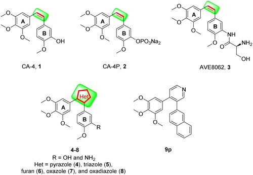
Six-membered heterocycles are broadly used as useful structural fragments in the design of drug molecules. There are as well numerous successful examples of replacing other heterocycles with six-membered heterocycles in drug discovery and development, such as from fluvastatin to pitavastatinCitation20 and from fluconazole to voriconazoleCitation21. It is reasonable to anticipate that the six-membered heterocycles play a comparable role to that of other heterocycles in CA-4 analogs. However, very few researches have been reported on it.
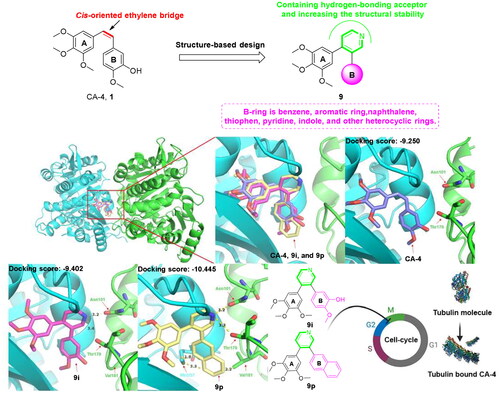
To search for potential six-membered heterocycles to replace other heterocycles of CA-4 analogs and find promising tubulin polymerisation inhibitors, molecular docking calculations were performed for diverse structural analogs of CA-4 with six-membered heterocycles to the CA-4 binding site of tubulin. To evaluate the molecular docking properties, the co-crystallized ligand CA-4 (PDB code: 5LYJ) was first docked and its binding mode was studied. The results of the docking of pyridine derivatives 9i and 9p were drawn to our attention. As shown in , CA-4 (purple), 9i (plum), and 9p (yellow) are well-oriented and superimposed with each other. 9i and 9p are bound to the active site via a hydrogen bond between the methoxy group (A-ring) and the amino acid residue Cysβ241, which is similar to CA-4. Another hydrogen bond is also present between Valα181 and the hydroxyl group on the B-ring of 9i and CA-4. It is worth noting that the nitrogen atom of pyridine of 9i and 9p established two critical hydrogen bonds with amino acid residues Asnα101 and Thrα179. Besides, the docking score of 9i (Docking score: −9.402) and 9p (Docking score: −10.445) in the 5LYJ was lower than that of CA-4 (Docking score: −9.250), suggesting that these compounds might have strong antiproliferative activities and the pyridine skeleton might result in the development of a series of novel compounds with apparent inhibitory potency against tubulin. For these reasons, we report the synthesis, biological evaluation, and preliminary structure-activity relationships (SARs) of 3-aryl-4–(3,4,5-trimethoxyphenyl)pyridines as potent tubulin polymerisation inhibitors.
Figure 2. The rational design of target compounds.
Result and discussion
Chemistry
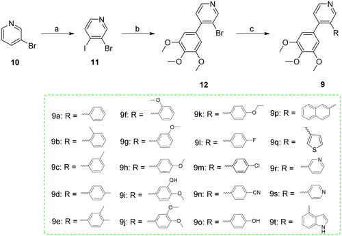
Scheme 1 illustrates the general synthetic method for the preparation of 3-aryl-4–(3,4,5-trimethoxyphenyl)pyridines (9). Firstly, commercially available 3-bromopyridine (10) was used as the starting material and reacted with I2 in presence of LDA in tetrahydrofuran to yield intermediate 3-bromo-4-iodopyridine (11)Citation22,Citation23. Subsequently, the intermediate 11 was further reacted with 3,4,5-trimethoxyphenylboric acid in the presence of tetrakis(triphenylphosphine)palladium and potassium carbonate to give key 3-bromo-4–(3,4,5-trimethoxyphenyl)pyridine (12) at 126 °CCitation24. Lastly, the target compounds 3-aryl-4–(3,4,5-trimethoxyphenyl)pyridines (9) were generated via the Suzuki cross-coupling reaction between 12 and the corresponding arylboronic acidCitation25,Citation26. All of the substrates decorated with electron donating groups (EDG) or electron-withdrawing groups (EWG) on the B-ring of phenylboronic acids were successfully obtained as a wide variety of 3-aryl-4–(3,4,5-trimethoxyphenyl)pyridines. The results showed that most arylboronic acids with EDG on the aromatic ring yielded moderate yields of the target compounds ranging from 70% to 96%, while 4-fluorophenylboronic acid, 4-chlorophenylboronic acid, and 4-cyanophenylboronic acid (EWG) gave efficient access to the corresponding coupling products (9 l, 9 m, and 9n) in 94%, 75%, and 83% yields, respectively. In addition, naphthalen-2-ylboronic acid and (1H-indol-4-yl)boronic acid also exhibited excellent reactivity, despite their increased steric hindrance (9p and 9t).
Scheme 1. Reagents and conditions (a) LDA, THF, -85 °C, then I2/THF, -78 °C, overnight; (b) 3,4,5-trimethoxyphenylboric acid, Pd(PPh3)4, K2CO3, 1,4-dioxane/H2O, N2 atmosphere,125 °C, M.W., 20 min; (c) Substituted phenylboronic acid, Pd(PPh3)4, K2CO3, 1,4-dioxane/H2O, N2 atmosphere,126 °C, M.W., 25 min.
Biological evaluation
In vitro antiproliferative activity
The antiproliferative activities of the newly synthesised compounds 9a-t were evaluated against three representative cancer cell lines (HeLa, MCF-7, and A549) using an accepted MTT assay with CA-4 as the positive control. The majority of compounds exhibited moderate potency against three cancer cell lines with IC50 values in the submicromolar to micromolar range. These results of the MTT assay showed that the introduction of the pyridine moiety as a suitable mimic for the cis-orientation of the olefin bond present in CA-4 could maintain the effective antiproliferative activity.
We first introduced different rigid aromatic groups such as phenyl (9a), naphthyl (9p), thienyl (9q), pyridyl (9r and 9s), and indolyl (9t) into the B-ring to explore the effect of different skeletons on antiproliferative activities against three different cell lines. Amongst these, 9p displayed excellent antiproliferative activity against HeLa cells (IC50 values of 0.047 µM). To gain insight into the SARs, the substitution pattern and position on the benzene ring (B-ring) of target compounds (9a-9o) were varied. As presented in , it is noteworthy that EDG located in para position (9d, 9h, 9k, and 9o) caused a significant promotion in three cancer cell growth inhibitory properties. The introduction of EWG, such as fluorine (9 l) and nitro (9n) in para position of compound 9a reduced the inhibitory activities to varying degrees. The activities of target compounds (9d, 9h, 9k vs 9 l, 9 m, 9n) revealed that the electronic effect of substituents on the benzene ring plays a vital role in antiproliferative potency.
Table 1. In vitro anticancer activity (IC50 in μM) a,b and docking score of the compounds (9a-t).
Effect on tubulin polymerisation
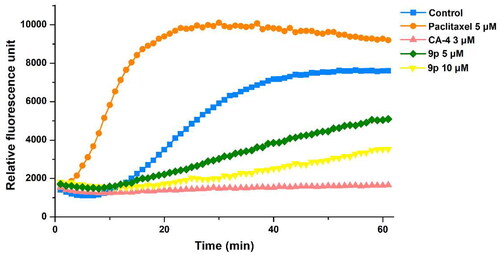
To clarify whether these 3-aryl-4–(3,4,5-trimethoxyphenyl)pyridines target the tubulin-microtubule system, we evaluated the inhibition of tubulin polymerisation by the most active compound 9p and used CA-4 and paclitaxel as positive and negative controls, respectively. As demonstrated in , compared with paclitaxel, both 9p and CA-4 exhibited effective inhibition against tubulin polymerisation. Besides, 9p inhibited tubulin polymerisation in a concentration-dependent manner. These results clearly made clear that 9p inhibited the polymerisation of tubulin in a very similar way to CA-4.
Figure 3. A cell-free tubulin polymerisation assay of 9p with purified tubulin. CA-4 and paclitaxel were used as positive and negative controls, respectively.
Analysis of immunofluorescence staining
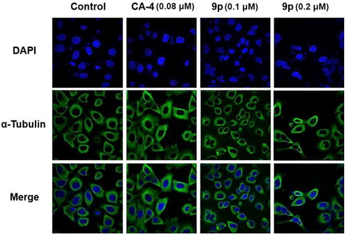
To verify the direct effects of compound 9p on tubulin, we examined whether 9p could destabilise microtubule dynamics in HeLa cells using an immunofluorescence assay, and CA-4 was used as a reference. Additionally, cellular microtubule structures were observed by indirect immunofluorescence. As illustrated in , cells treated with 9p (0.1 µM and 0.2 µM) displayed changes in nuclear shape (blue) and the microtubule network (green) became constricted and disorganised compared to the control group. These results suggested that 9p destroyed the cytoskeleton and suppressed microtubule assembly, similar to CA-4.
Figure 4. Effects of compound 9p (0.1 µM and 0.2 µM) and CA-4 (0.08 µM), on the cellular microtubule network and microtubule reassemble by immunofluorescence.
Cell cycle analysis
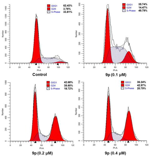
To further explore the biological target, we investigated the effects of the most promising compound 9p on the cell cycle. HeLa cells were treated with 0.1 µM, 0.2 µM, and 0.4 µM of 9p, and the percentage of HeLa cells at different cell cycle stages was analysed by flow cytometry after treatment, respectively.
As demonstrated in , the percentage of HeLa cells stalled in the G2/M phase increased from 14.47% to 42.97% after treatment with the three concentrations of 9p compared to the control (3.76%). Consequently, 9p could concentration-dependently induce G2/M phase arrest in HeLa cells. Cell cycle distribution indicated that 9p could cause HeLa cells to arrest in the G2/M phase, followed by apoptosis.
Figure 5. Effects of compound 9p on cell cycle. HeLa cell lines were treated with compound 9p (0.1 µM, 0.2 µM, and 0.4 µM) for 24 h.
Induction of cell apoptosis
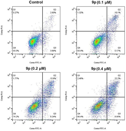
To study whether compound 9p could induce cell apoptosis, we performed an Annexin VFITC/PI assay. As shown in , the total percentage of early (Annexin-V+/PI-) and late (Annexin-V+/PI-) apoptotic cells was only 6.46% after 48 h of treatment in the control group. However, 15.27% of the total number of apoptotic cells was obtained after 48 h treatment with 0.1 µM of 9p. In addition, incubation of HeLa cells with higher concentrations of 9p at 0.2 µM and 0.4 µM increased the percentage of apoptotic cells to 24.04% and 31.15%, respectively. These results demonstrated that 9p effectively induced apoptosis in HeLa cells in a concentration-dependent manner.
Figure 6. Analyses of apoptosis induction in Hela cells. Cells were harvested and stained with Annexin-V/PI for analysis after treatment with different concentrations of compound 9p (0.1 µM, 0.2 µM, and 0.4 µM) and control for 48 h. The diverse cell stages were given as live (Q4), early apoptotic (Q3), late apoptotic (Q2), and necrotic cells (Q1).
Physicochemical properties
In addition, to examine the drug-like properties of 3-aryl-4–(3,4,5-trimethoxyphenyl)pyridines, conventional physicochemical properties of CA-4, 9i, and 9p were predicted using a free online website (http://www.swissadme.ch/index.php) or ChemBioDraw Ultra 14.0 software for their fit with Lipinski’s five rulesCitation27. As summarised in , CA-4, 9i, and 9p fit well with the Lipinski’s five rule. Compared to CA-4, 9i has a lower value of lipid water partition coefficient. This finding suggests that 9i may have better water solubility than CA-4. Furthermore, the presence of five hydrogen bonding receptors in 9i, much more than CA-4, contributes to the lower binding energy of the compound to the site of action.
Table 2. Prediction of physicochemical properties of CA-4, 9i, and 9pTable Footnotea.
Conclusion
Overall, based on the analysis of the X-ray crystal structure of tubulin in complex with DMAM-CA-4, we reported the design and discovery of a new series of 3-aryl-4–(3,4,5-trimethoxyphenyl)pyridines on the cis-orientation of the olefin bond of CA-4 as the novel tubulin polymerisation inhibitors with improved structural stability.
The MTT assay results proved that target compounds 9a-t exhibited moderate antiproliferative potencies against three human tumour cells (HeLa, MCF-7, and A549) in vitro. The replacement of cis-orientation of the olefin bond of CA-4 with the pyridine scaffold led to the comparable antiproliferative activity of the representative compound, 9p, compared with CA-4. According to the analysis of SARs, the introduction of naphthalene moiety was favourable for potency antitumor activity, and the compounds with naphthalene as B-ring were more active than that with substituted phenyl as B-ring. Among all the designed compounds, 9p displayed the most potent antiproliferative activity with an IC50 value of 0.047-0.90 μM as well as with improved structural stability than CA-4. Moreover, 9p was capable of inhibiting tubulin polymerisation in vitro. Preliminary mechanistic investigations showed that 9p evoked G2/M phase cell cycle arrest and apoptosis in HeLa cells. Further mechanistic investigations revealed that 9p could potently destabilise the microtubule network in HeLa cells in a concentration-dependent manner. Lastly, the prediction of physicochemical properties indicated that 9p fits well with five Lipinski’s rule. All of these results indicate 9p as a promising tubulin polymerisation inhibitor for further investigation in anticancer drug development.
Experimental
Chemistry
Materials and methods
All of the reagents and solvents were purchased from chemical companies.1H NMR and 13C NMR spectra were tested in CDCl3 with TMS as the internal reference on a Bruker AVANCE (1H at 500 MHz,13C at 126 MHz). High-resolution mass spectra (HRMS) were recorded by Agilent Accurate-Mass Q-TOF 6530 instrument in ESI mode. The microwave reactions were carried out in a single mode cavity microwave synthesiser (CEM Corporation, NC, USA). TLC analysis was used for determining the extent of reactions under UV light (wavelength: 365 nm and 254 nm).
General synthetic procedure for 3-bromo-4-iodopyridine (11)
The stirred dry tetrahydrofuran was cooled to an internal temperature of −78 °C using a liquid N2/CO2/hexane bath. Diisopropylamine (0.085 mol) was then added by syringe, and after a further 15 min stirring, n-butyllithium (1.6 M in hexanes; 0.10 mol) was syringed into the solution. The resulting lithium diisopropylaminoamine solution was stirred at −78 °C for 1 h to allow for completion of the reaction, and subsequently cooled to −85°С. Neat 3-bromopyridine (10, 0.10 mol) was then syringed into the stirred lithium diisopropylaminoamine solution and stirred for 1 h. The iodine (0.20 mol) in dry tetrahydrofuran was then added dropwise by syringe to the lithiopyridine reaction mixture. The mixture was stirred at −78 °C for a further 5 h, then allowed to warm to ambient temperature with continued stirring overnight. Distilled water (20 ml) was added to quench the reaction and the solvent volume was reduced to approximately 75 ml by distillation under reduced pressure on a water bath. The remaining dark mixture was then partitioned between diethyl ether and excess saturated NaS2O3, and subsequently extracted repeatedly with diethyl ether (4 × 200 ml) due to the poor solubility of the suspended 3-bromo-4-iodopyridine (11). The combined organic extracts were dried (anhydrous Na2SO4), gravity filtered, and concentrated in vacuo. The crude material was purified by column chromatography over silica gel using n-hexane/dichloromethane (5:1) as a solvent.
General synthetic procedure for 3-bromo-4–(3,4,5-trimethoxyphenyl)pyridine (12)
The intermediate 11 (0.5 mmol), 3,4,5-trimethoxyphenylboric acid (0.6 mmol), K2CO3 (2 mmol), and Pd(PPh3)4 (0.05 mmol) were dissolved in 1,4-dioxane (10 ml) and H2O (3 ml). Then the mixture was stirred at irradiated in a microwave reactor for 20 min at 125 °C. When the reaction was completed, the mixture was extracted with ethyl acetate (15 ml × 3). The combined organic layers were washed with brine, dried over Na2SO4, and purified by silica gel column chromatography or recrystallized to afford 3-bromo-4–(3,4,5-trimethoxyphenyl)pyridine (12).
General synthetic procedure for 3-aryl-4–(3,4,5-trimethoxyphenyl)pyridines (9a-t)
A mixture of 3-bromo-4–(3,4,5-trimethoxyphenyl)pyridine (12, 0.10 mmol), Pd(PPh3)4 (0.005 mmol), and K2CO3 (0.40 mmol), and substituted phenylboronic acid (0.11 mmol) in 1,4-dioxane/H2O (5 ml, 3:1) was degassed and purged with N2 for about three times. After stirring at irradiated in a microwave reactor for 25 min at 126 °C (indicated by TLC) under N2 atmosphere, H2O (50 ml) was added to the reaction mixture and extracted with ethyl acetate. The combined organics were washed with brine, dried over anhydrous Na2SO4, filtered, and concentrated under vacuum to give a residue, which was purified by column 400 chromatography using a mixture of n-hexane/ethyl acetate (4:1) as an eluent to provide the target compounds 3-aryl-4–(3,4,5-trimethoxyphenyl)pyridines (9a-t).
3-phenyl-4–(3,4,5-trimethoxyphenyl)pyridine (9a)
White solid; yield: 92%; 1H NMR (500 MHz, CDCl3) δ 8.64 (s, 2H), 7.38 (d, J = 5.0 Hz, 1H), 7.32 − 7.27 (m, 3H), 7.18 (dd, J = 7.7, 1.7 Hz, 2H), 6.35 (s, 2H), 3.83 (s, 3H), 3.61 (s, 6H); 13C NMR (126 MHz, CDCl3) δ 152.92 (2 C), 150.97, 148.79, 147.43, 137.98, 137.82, 135.80, 133.59, 129.68 (2 C), 128.36 (2 C), 127.31, 124.02, 106.85 (2 C), 60.91, 55.93 (2 C); HRMS calcd for C20H19NO3 [M + H]+ 322.1443, found 322.1442.
3-(o-tolyl)-4–(3,4,5-trimethoxyphenyl)pyridine (9b)
White solid; yield: 90%; 1H NMR (500 MHz, CDCl3) δ 8.66 (d, J = 4.0 Hz, 1H), 8.54 (s, 1H), 7.43 (d, J = 5.1 Hz, 1H), 7.24 − 7.20 (m, 2H), 7.20 − 7.16 (m, 1H), 7.15 − 7.11 (m, 1H), 6.35 (s, 2H), 3.82 (s, 3H), 3.59 (s, 6H), 1.86 (s, 3H); 13C NMR (126 MHz, CDCl3) δ 152.82 (2 C), 151.08, 148.75, 147.76, 137.94, 137.67, 136.41, 135.48, 133.48, 130.38, 130.17, 127.95, 125.86, 123.42, 106.33 (2 C), 60.89, 55.90 (2 C), 19.88; HRMS calcd for C21H22NO3 [M + H]+ 336.1600, found 336.1598.
3-(m-tolyl)-4–(3,4,5-trimethoxyphenyl)pyridine (9c)
White solid; yield: 91%;1H NMR (500 MHz, CDCl3) δ 8.63 (s, 2H), 7.37 (d, J = 4.7 Hz, 1H), 7.18 (t, J = 7.6 Hz, 1H), 7.09 (d, J = 7.6 Hz, 1H), 7.01 (s, 1H), 6.95 (d, J = 7.6 Hz, 1H), 6.36 (s, 2H), 3.83 (s, 3H), 3.62 (s, 6H), 2.28 (s, 3H); 13C NMR (126 MHz, CDCl3) δ 152.86 (2 C), 150.93, 148.67, 147.36, 137.96, 137.89, 137.84, 135.93, 133.68, 130.27, 128.22, 127.99, 126.84, 123.95, 106.88 (2 C), 60.92, 55.95 (2 C), 21.32; HRMS calcd for C21H22NO3 [M + H]+ 336.1600, found 336.1598.
3-(p-tolyl)-4–(3,4,5-trimethoxyphenyl)pyridine (9d)
White solid; yield: 95%; 1H NMR (500 MHz, CDCl3) δ 8.62 − 8.59 (m, 2H), 7.36 (d, J = 5.0 Hz, 1H), 7.11 (d, J = 7.9 Hz, 2H), 7.06 (d, J = 8.1 Hz, 2H), 6.35 (s, 2H), 3.84 (s, 3H), 3.62 (s, 6H), 2.33 (s, 3H); 13C NMR (126 MHz, CDCl3) δ 152.88 (2 C), 150.99, 148.55, 147.40, 137.76, 137.07, 135.77, 134.95, 133.78, 129.54 (2 C), 129.02 (2 C), 123.98, 106.82 (2 C), 60.92, 55.91 (2 C), 21.08; HRMS calcd for C21H22NO3 [M + H]+ 336.1600, found 336.1599.
3-(3,4-dimethylphenyl)-4–(3,4,5-trimethoxyphenyl)pyridine (9e)
White solid; yield: 80%; 1H NMR (500 MHz, CDCl3) δ 8.62 (s, 1H), 8.59 (d, J = 5.0 Hz, 1H), 7.35 (d, J = 5.0 Hz, 1H), 7.04 (d, J = 7.7 Hz, 1H), 6.96 (s, 1H), 6.87 (d, J = 9.0 Hz, 1H), 6.37 (s, 2H), 3.84 (s, 3H), 3.62 (s, 6H), 2.24 (s, 3H), 2.19 (s, 3H); 13C NMR (126 MHz, CDCl3) δ 152.83 (2 C), 150.96, 148.44, 147.37, 137.78, 136.48, 135.88, 135.68, 135.36, 133.86, 130.74, 129.55, 127.13, 123.91, 106.87 (2 C), 60.92, 55.93 (2 C), 19.65, 19.37; HRMS calcd for C22H24NO3 [M + H]+ 350.1756, found 350.1759.
3-(2-methoxyphenyl)-4-(3,4,5-trimethoxyphenyl)pyridine (9f)
White solid; yield: 90%; 1H NMR (500 MHz, CDCl3) δ 8.62 (d, J = 4.4 Hz, 1H), 8.58 (s, 1H), 7.38 (d, J = 5.0 Hz, 1H), 7.33 − 7.27 (m, 1H), 7.20 (dd, J = 7.4, 1.7 Hz, 1H), 6.99 (t, J = 7.4 Hz, 1H), 6.77 (d, J = 8.2 Hz, 1H), 6.37 (s, 2H), 3.81 (s, 3H), 3.61 (s, 6H), 3.39 (s, 3H); 13C NMR (126 MHz, CDCl3) δ 156.56, 152.67 (2 C), 151.35, 148.82, 148.38, 137.60, 134.66, 132.67, 131.25, 129.43, 127.22, 123.28, 120.79, 110.88, 105.72 (2 C), 60.88, 55.90 (2 C), 55.04; HRMS calcd for C21H22NO4 [M + H]+ 352.1549, found 352.1546.
3-(3-methoxyphenyl)-4-(3,4,5-trimethoxyphenyl)pyridine (9 g)
White solid; yield: 82%; 1H NMR (500 MHz, CDCl3) δ 8.64 − 8.62 (m, 2H), 7.37 (d, J = 4.9 Hz, 1H), 7.24 − 7.19 (m, 1H), 6.83 (dd, J = 8.3, 2.5 Hz, 1H), 6.79 (d, J = 7.6 Hz, 1H), 6.71 − 6.67 (m, 1H), 6.38 (s, 2H), 3.83 (s, 3H), 3.67 (s, 6H), 3.64 (s, 3H); 13C NMR (126 MHz, CDCl3) δ 159.49, 152.95 (2 C), 150.82, 148.82, 147.48, 139.26, 137.89, 135.64, 133.64, 129.40, 124.00, 122.12, 115.18, 113.13, 106.77 (2 C), 60.91, 56.00 (2 C), 55.22; HRMS calcd for C21H22NO4 [M + H]+ 352.1549, found 352.1552.
3-(4-methoxyphenyl)-4–(3,4,5-trimethoxyphenyl)pyridine (9h)
White solid; yield: 96%; 1H NMR (500 MHz, CDCl3) δ 8.61 (s, 2H), 7.35 (d, J = 4.0 Hz, 1H), 7.09 (d, J = 8.7 Hz, 2H), 6.84 (d, J = 8.7 Hz, 2H), 6.36 (s, 2H), 3.84 (s, 3H), 3.79 (s, 3H), 3.64 (s, 6H); 13C NMR (126 MHz, CDCl3) δ 159.01, 152.95 (2 C), 150.90, 148.30, 147.41, 137.76, 133.88, 130.78 (2 C), 130.11, 124.14, 124.12, 113.85 (2 C), 106.75 (2 C), 60.92, 55.97 (2 C), 55.30; HRMS calcd for C21H22NO4 [M + H]+ 352.1549, found 352.1552.
2-methoxy-5-(4–(3,4,5-trimethoxyphenyl)pyridin-3-yl)phenol (9i)
White solid; yield: 70%; 1H NMR (500 MHz, CDCl3) δ 8.61 (s, 1H), 8.55 (d, J = 5.0 Hz, 1H), 7.34 (d, J = 5.0 Hz, 1H), 7.00 (s, 1H), 6.85 (d, J = 2.1 Hz, 1H), 6.75 (d, J = 8.3 Hz, 1H), 6.58 (dd, J = 8.3, 2.0 Hz, 1H), 6.39 (s, 2H), 3.86 (s, 3H), 3.84 (s, 6H), 3.65 (s, 3H); 13C NMR (126 MHz, CDCl3) δ 152.94 (2 C), 150.71, 148.06, 147.66, 146.48, 145.98, 137.84, 135.66, 133.79, 130.85, 124.20, 121.47, 116.09, 110.75, 106.69 (2 C), 60.91, 56.01 (2 C), 55.97; HRMS calcd for C21H22NO5 [M + H]+ 368.1498, found 368.1496.
3–(3,4-dimethoxyphenyl)-4–(3,4,5-trimethoxyphenyl)pyridine (9j)
White solid; yield: 93%; 1H NMR (500 MHz, CDCl3) δ 8.64 (s, 1H), 8.58 (d, J = 4.9 Hz, 1H), 7.53 (td, J = 7.3, 1.4 Hz, 1H), 7.34 (d, J = 5.0 Hz, 1H), 6.80 (dd, J = 8.2, 1.8 Hz, 1H), 6.59 (d, J = 1.8 Hz, 1H), 6.38 (s, 2H), 3.86 (s, 3H), 3.82 (s, 3H), 3.64 (s, 6H), 3.63 (s, 3H); 13C NMR (126 MHz, CDCl3) δ 153.03 (2 C), 150.80, 148.65, 148.49, 148.46, 147.40, 137.82, 135.41, 132.96, 130.31, 124.07, 121.93, 113.17, 111.11, 106.69 (2 C), 60.92, 56.06 (2 C), 55.93, 55.78; HRMS calcd for C22H24NO5 [M + H]+ 382.1654, found 382.1655.
3-(4-ethoxyphenyl)-4–(3,4,5-trimethoxyphenyl)pyridine (9k)
White solid; yield: 96%; 1H NMR (500 MHz, CDCl3) δ 8.64 − 8.58 (m, 2H), 7.34 (d, J = 4.9 Hz, 1H), 7.07 (d, J = 8.8 Hz, 2H), 6.82 (d, J = 8.7 Hz, 2H), 6.36 (s, 2H), 4.01 (q, J = 7.0 Hz, 2H), 3.84 (s, 3H), 3.64 (s, 6H), 1.40 (t, J = 7.0 Hz, 3H); 13C NMR (126 MHz, CDCl3) δ 158.35, 152.94 (2 C), 150.97, 148.33, 147.36, 137.73, 135.47, 133.92, 130.76 (2 C), 129.97, 124.06, 114.42 (2 C), 106.75 (2 C), 63.48, 60.92, 55.97 (2 C), 14.76; HRMS calcd for C22H24NO4 [M + H]+ 366.1705, found 366.1709.
3-(4-fluorophenyl)-4–(3,4,5-trimethoxyphenyl)pyridine (9 l)
White solid; yield: 94%; 1H NMR (500 MHz, CDCl3) δ 8.62 (s, 2H), 7.38 (d, J = 5.0 Hz, 1H), 7.14 (dd, J = 8.6, 5.4 Hz, 2H), 7.01 (t, J = 8.7 Hz, 2H), 6.33 (s, 2H), 3.84 (s, 3H), 3.64 (s, 6H); 13C NMR (126 MHz, CDCl3) δ 162.24, 153.05 (2 C), 150.56, 148.63, 147.78, 137.98, 134.84, 133.82, 133.37, 131.30 (2 C), 124.24, 115.40 (2 C), 106.75 (2 C), 60.94, 55.98 (2 C); HRMS calcd for C20H19FNO3 [M + H]+ 340.1349, found 340.1353.
3-(4-chlorophenyl)-4-(3,4,5-trimethoxyphenyl)pyridine (9 m)
White solid; yield: 75%; 1H NMR (500 MHz, CDCl3) δ 8.64 − 8.61 (m, 2H), 7.38 (d, J = 5.0 Hz, 1H), 7.29 (d, J = 8.5 Hz, 2H), 7.11 (d, J = 8.5 Hz, 2H), 6.33 (s, 2H), 3.85 (s, 3H), 3.65 (s, 6H); 13C NMR (126 MHz, CDCl3) δ 153.06 (2 C), 150.59, 148.95, 147.66, 136.36, 133.27, 130.94 (2 C), 130.23, 129.02, 128.59 (2 C), 128.17, 124.21, 106.76 (2 C), 60.95, 55.98 (2 C); HRMS calcd for C20H19ClNO3 [M + H]+ 356.1053, found 356.1054.
4-(4–(3,4,5-trimethoxyphenyl)pyridin-3-yl)benzonitrile (9n)
White solid; yield: 83%; 1H NMR (500 MHz, CDCl3) δ 8.67 (d, J = 4.9 Hz, 1H), 8.61 (s, 1H), 7.61 (d, J = 8.4 Hz, 2H), 7.40 (d, J = 5.0 Hz, 1H), 7.31 (d, J = 8.3 Hz, 2H), 6.29 (s, 2H), 3.84 (s, 3H), 3.64 (s, 6H); 13C NMR (126 MHz, CDCl3) δ 153.21 (2 C), 150.49, 149.80, 147.76, 142.96, 138.27, 133.93, 132.82, 132.09 (2 C), 130.39 (2 C), 124.35, 118.45, 111.23, 106.73 (2 C), 60.98, 56.02 (2 C); HRMS calcd for C21H19N2O3 [M + H]+ 347.1396, found 347.1404.
4-(4–(3,4,5-trimethoxyphenyl)pyridin-3-yl)phenol (9o)
White solid; yield: 90%; 1H NMR (500 MHz, CDCl3) δ 9.59 (s, 1H), 8.60 (s, 1H), 8.55 (d, J = 4.9 Hz, 1H), 7.38 (d, J = 5.1 Hz, 1H), 6.99 (d, J = 8.5 Hz, 2H), 6.85 (d, J = 8.5 Hz, 2H), 6.39 (s, 2H), 3.84 (s, 3H), 3.64 (s, 6H); 13C NMR (126 MHz, CDCl3) δ 157.11, 152.94 (2 C), 150.40, 147.86, 147.45, 137.79, 133.78, 131.47, 130.79 (2 C), 128.45, 124.35, 115.69 (2 C), 106.76 (2 C), 60.93, 55.99 (2 C); HRMS calcd for C20H19NO4 [M + H]+ 338.1392, found 338.1396.
3-(Naphthalen-2-yl)-4–(3,4,5-trimethoxyphenyl)pyridine (9p)
White solid; yield: 66%; 1H NMR (500 MHz, CDCl3) δ 8.75 (s, 1H), 8.66 (d, J = 4.9 Hz, 1H), 7.85 − 7.77 (m, 3H), 7.69 (d, J = 8.5 Hz, 1H), 7.52 − 7.45 (m, 2H), 7.42 (d, J = 5.1 Hz, 1H), 7.14 (dd, J = 8.4, 1.5 Hz, 1H), 6.39 (s, 2H), 3.81 (s, 3H), 3.50 (s, 6H); 13C NMR (126 MHz, CDCl3) δ 152.96 (2 C), 151.22, 148.90, 147.56, 137.91, 135.68, 135.57, 133.59, 133.30, 132.29, 128.29, 127.92, 127.75, 127.68, 127.59, 126.34, 126.32, 124.06, 106.91 (2 C), 60.90, 55.89 (2 C); HRMS calcd for C24H22NO3 [M + H]+ 372.1600, found 372.1601.
3-(Thiophen-3-yl)-4–(3,4,5-trimethoxyphenyl)pyridine (9q)
Light yellow solid; yield: 68%; 1H NMR (500 MHz, CDCl3) δ 8.72 (s, 1H), 8.62 (s, 1H), 7.36 (d, J = 5.0 Hz, 1H), 7.22 (dd, J = 4.9, 3.0 Hz, 1H), 7.19 (dd, J = 2.9, 1.3 Hz, 1H), 6.73 (d, J = 6.1 Hz, 1H), 6.41 (s, 2H), 3.86 (s, 3H), 3.68 (s, 6H); 13C NMR (126 MHz, CDCl3) δ 153.07 (2 C), 150.12, 148.22, 147.76, 133.60, 131.94, 130.95, 128.73, 125.56, 124.79, 124.23, 123.70, 106.38 (2 C), 60.96, 56.01 (2 C); HRMS calcd for C18H18NO3S [M + H]+ 328.1007, found 328.1008.
4–(3,4,5-trimethoxyphenyl)-3,3'-bipyridine (9r)
White solid; yield: 78%; 1H NMR (500 MHz, CDCl3) δ 8.67 (d, J = 4.9 Hz, 1H), 8.63 (s, 1H), 8.54 (d, J = 3.9 Hz, 1H), 8.50 (s, 1H), 7.46 (dt, J = 7.8, 1.9 Hz, 1H), 7.40 (d, J = 5.0 Hz, 1H), 7.23 (dd, J = 7.8, 4.8 Hz, 1H), 6.31 (s, 2H), 3.83 (s, 3H), 3.63 (s, 6H); 13C NMR (126 MHz, CDCl3) δ 153.19 (2 C), 150.71, 150.04, 149.61, 148.57, 148.04, 138.14, 136.90, 133.79, 132.97, 132.17, 124.33, 123.06, 106.82 (2 C), 60.94, 56.03 (2 C); HRMS calcd for C19H19NO3 [M + H]+ 323.1396, found 323.1399.
4-(3,4,5-trimethoxyphenyl)-3,4'-bipyridine (9s)
White solid; yield: 87%; 1H NMR (500 MHz, CDCl3) δ 8.68 − 8.55 (m, 4H), 7.40 (d, J = 5.0 Hz, 1H), 7.13 (d, J = 5.6 Hz, 2H), 6.32 (s, 2H), 3.84 (s, 3H), 3.64 (s, 6H); 13C NMR (126 MHz, CDCl3) δ 153.19 (2 C), 150.39, 149.94, 149.79 (2 C), 147.77, 146.10, 138.30, 133.08, 132.75, 124.40 (2 C), 124.34, 106.71 (2 C), 60.97, 56.03 (2 C); HRMS calcd for C19H19NO3 [M + H]+ 323.1396, found 323.1399.
4–(4–(3,4,5-trimethoxyphenyl)pyridin-3-yl)-1H-indole (9t)
White solid; yield: 78%; 1H NMR (500 MHz, CDCl3) δ 8.75 (s, 1H), 8.71 − 8.56 (m, 2H), 7.33 (d, J = 8.2 Hz, 1H), 7.16 − 7.11 (m, 1H), 7.11 − 7.06 (m, 1H), 6.89 (d, J = 7.2 Hz, 1H), 6.40 (s, 2H), 6.17 (s, 1H), 3.76 (s, 3H), 3.47 (s, 6H); 13C NMR (126 MHz, CDCl3) δ 152.67 (2 C), 151.60, 148.58, 147.92, 137.63, 135.67, 135.07, 133.96, 130.37, 127.50, 124.66, 123.76, 121.91, 121.34, 110.52, 106.32 (2 C), 101.59, 60.81, 55.80 (2 C); HRMS calcd for C22H21N2O3 [M + H]+ 361.1552, found 361.1551.
Biological evaluation
Cell culture
Human cervical carcinoma HeLa cells, human breast cancer MCF-7 cells, and human lung cancer A549 cells were cultured in RPMI-1640 medium containing 10% FBS, 100 U/mL streptomycin, and 100 U/mL penicillin at 37 °C in a humidified atmosphere containing 5% CO2. All cell lines were purchased from the American Type Culture Collection (ATCC, Manassas, VA).
In vitro antiproliferative activity
The in vitro antiproliferative activities of CA-4 and all of the target compounds were determined by MTT assayCitation14. Briefly, HeLa, MCF-7, and A549 cells were seeded into 96-well plates at a density of 2 × 10⁴/well, depending on the growth rate of the cell line. 24 h later, triplicate wells were treated with media and the compounds being tested. After 72 h of incubation at 37 °C in 5% CO2, the drug-containing medium was removed and replaced with 100 μL of fresh medium containing 5 mg/mL MTT solution. After 4 h of incubation, the medium with MTT was removed, and 100 μL of dimethyl sulfoxide was added to each well. The plates were gently agitated until the purple formazan crystals were dissolved, and the OD490 values were determined using a microplate reader. The data were calculated and plotted as the percent viability compared to the control. The IC50 was defined as the drug concentration that resulted in an absorbance of 50% of that of the untreated wells in the MTT assay.
Effect on tubulin polymerisation
The effects of compound 9p and CA-4 on tubulin polymerisation were determined using a fluorescence-based tubulin polymerisation assay kit (Cytoskeleton-Cat.#BK011P) according to the manufacturer’s protocolCitation28. Tubulin was resuspended in ice-cold G-PEM buffer and added to wells of a 96-well plate containing the designated concentrations of the drug or vehicle. The samples were mixed well, and tubulin assembly was monitored at 1 min intervals for 90 min at 37 °C using a plate reader. The IC50 values were calculated after 20 min using SPSS software.
Analysis of immunofluorescence staining
Immunostaining was performed to detect the microtubule-associated tubulin protein after exposure to CA-4 and the investigated compound 9pCitation29,Citation30. HeLa cells were seeded in a 24-well plate at 1 × 10⁴ cells per well and grown for 24 h. The cells were treated with the vehicle, CA-4, or 9p for 24 h. The control and treated cells were fixed with 4% formaldehyde in PBS for 30 min at −20 °C, washed twice with PBS and permeabilized with 0.1% (v/v) Triton X-100 in PBS for 5 min. The cells were then blocked with 5% bovine serum albumin in PBS for 10 min. The primary α-tubulin antibody was diluted (1/100) with 2% bovine serum albumin in PBS, and the plates were incubated overnight at 4 °C. The cells were washed with PBS to remove unbound primary antibody, and the cells were then incubated with FITC-conjugated secondary antibody diluted (1/100) with 2% BSA in PBS for 3 h at 37 °C. The cells were washed with PBS to remove unbound secondary antibody, and the nuclei were stained with DAPI. Then, immunofluorescence was detected using a fluorescence microscope.
Cell cycle analysis
HeLa cells (8 × 10⁴ cells) were incubated with designated concentrations of 9p, CA-4, or 0.05% DMSO for the indicated timesCitation25,Citation31. The cells were collected by centrifugation, washed with PBS, and fixed in ice-cold 70% ethanol. The fixed cells were harvested by centrifugation and resuspended in 500 ml of PBS containing 1 mg/mL RNase. After 30 min of incubation at 37 °C, the cells were stained with 50 mg/mL propidium iodide at 4 °C in the dark for 30 min. The samples were then analysed by FACS can flow cytometry. The experiments were repeated at least three times.
Induction of cell apoptosis
To investigate whether the target compound can induce apoptosis, an Annexin Van -FITC/PI experiment was carried outCitation26. HeLa cells were grown in 6-well plates (3 × 10⁵ cells/well) and incubated with various concentrations of 9p or vehicle control for 48 h. Subsequently, cells were harvested by centrifugation, washed with PBS, and resuspended in binding buffer. Then, 10 μL of PI Staining Solution and 5 μL of Annexin V-FITC were added to the cell suspension for 15 min at room temperature in the dark. Finally, the samples were detected by a CytoFLEX flow cytometer and the percentage of apoptotic cells was calculated using Flowjo 10.8 software.
Molecular docking analysis
The ligands in .sdf format applied in molecular docking were created with ChemBioDraw Ultra and prepared by LigPrep in Schrödinger package (version 2018)Citation27. For protein preparation, the crystal structure of Tubulin (PDB code: 5LYJ) in complex with CA-4, 9i, and 9k was downloaded from RCSB PDB Bank (http://www.rcsb.org/) and prepared by Protein Preparation Wizard in Schrödinger package (version 2018). The ligands were prepared by LigPrep Wizard in Schrödinger package (version 2018). All hydrogen atoms were added to residues, and all bond orders were assigned. Whereafter, the OPLS3 force field was applied to minimise the protein energy and eliminate steric hindrance. During the docking, a 15 Å × 15 Å × 15 Å grid box was generated around the active site of protein. The docking was performed by Ligand Docking in Schrödinger package (version 2018). The docking results were analysed using PyMOL (https://pymol.org/2/).
Author contributions
Chao Wang: methodology, validation, investigation, writingoriginal draft, writing-review & editing, funding acquisition. Yujing Zhang: methodology, validation, investigation. Shanbo Yang: synthesis of the compound. Lingyu Shi: investigation, visualisation. Yutao Xiu: validation, resources. Yudong Wu: conceptualisation, project administration. Hongfei Jiang: supervision, writing-review and editing.
Supplemental Material
Download PDF (5.6 MB)Disclosure statement
The authors declare that they have no known competing financial interests or personal relationships that could have appeared to influence the work reported in this paper.
Additional information
Funding
References
- Dumontet C, Jordan MA. Microtubule-binding agents: a dynamic field of cancer therapeutics. Nat. Rev. Drug Discov. 2010;9:790–803.
- Steinmetz MO, Prota AE. Microtubule-targeting agents: strategies to hijack the cytoskeleton. Trends Cell Biol. 2018;28:776–792.
- Kaul R, Risinger AL, Mooberry SL. Microtubule-targeting drugs: more than antimitotics. J. Nat. Prod. 2019;82:680–685.
- Lawrence EJ, Zanic M. Rescuing microtubules from the brink of catastrophe: CLASPs lead the way. Curr Opin Cell Biol. 2019;56:94–101.
- Cermak V, Dostal V, Jelínek M, Libusova L, Kovar J, Rosel D, Brabek J. Microtubule-targeting agents and their impact on cancer treatment. Eur. J. Cell Biol. 2020;99:151075.
- Pettit G, Singh S, Hamel E, Lin CM, Alberts D, Garcia-Kendal D. Isolation and structure of the strong cell growth and tubulin inhibitor combretastatin A-4. Experientia. 1989;45:209–211.
- Pettit GR, Singh SB, Boyd MR, Hamel E, Pettit RK, Schmidt JM, Hogan F. Antineoplastic agents. 291. Isolation and synthesis of combretastatins A-4, A-5, and A-6. J. Med. Chem. 1995;38:1666–1672.
- Tron GC, Pirali T, Sorba G, Pagliai F, Busacca S, Genazzani AA. Medicinal chemistry of combretastatin A 4: present and future directions. J. Med. Chem. 2006;49:3033–3044.
- Li Q, Sham HL. Discovery and development of antimitotic agents that inhibit tubulin polymerisation for the treatment of cancer, Expert. Opin. Ther. Pat. 2002;12(11):1663–1702.
- Mustafa M, Anwar S, Elgamal F, Ahmed ER, Aly OM. Potent combretastatin A-4 analogs containing 1,2,4-triazole: synthesis, antiproliferative, anti-tubulin activity, and docking study. Eur J Med Chem. 2019;183:111697.
- Perez-Perez MJ, Priego EM, Bueno O, Martins MS, Canela MD, Liekens S. Blocking blood flow to solid tumors by destabilizing tubulin: an approach to targeting tumor growth. J Med Chem. 2016;59:8685–8711.
- Von Pawel J, Gorbounova V, Reck M, Kowalski DM, Allard A, Chadjaa M, Rey A, Bennouna J, Grossi F. DISRUPT: a randomised phase 2 trial of ombrabulin (AVE8062) plus a taxane-platinum regimen as first-line therapy for metastatic non-small cell lung cancer. Lung Cancer. 2014;85:224–229.
- Metzler M, Neumann H. Epoxidation of the stilbene double bond, a major pathway in aminostilbene metabolism. Xenobiotica. 1977;7:117–132.
- Wang C, Yang S, Du J, Ni J, Wu Y, Wang J, Guan Q, Zuo D, Bao K, Wu Y, et al. Synthesis and bioevaluation of diarylpyrazoles as antiproliferative agents. Eur. J. Med. Chem. 2019;171:1–10.
- Odlo K, Fournier-Dit-Chabert J, Ducki S, Gani OA, Sylte I, Hansen TV. 1, 2, 3-Triazole analogs of combretastatin A-4 as potential microtubule-binding agents. Bioorg. Med. Chem. 2010;18:6874–6885.
- Theeramunkong S, Caldarelli A, Massarotti A, Aprile S, Caprioglio D, Zaninetti R, Teruggi A, Pirali T, Grosa G, Tron GC, et al. Regioselective Suzuki coupling of dihaloheteroaromatic compounds as a rapid strategy to synthesize potent rigid combretastatin analogues. J Med Chem. 2011;54(14):4977–4986.
- Zhang M, Liang YR, Li H, Liu MM, Wang Y. Design, synthesis, and biological evaluation of hydantoin bridged analogues of combretastatin A-4 as potential anticancer agents. Bioorg. Med. Chem. 2017;25:6623–6634.
- Kumar D, Patel G, Chavers AK, Chang KH, Shah K. Synthesis of novel 1, 2, 4-oxadiazoles and analogues as potential anticancer agents. Eur. J. Med. Chem. 2011;46:3085–3092.
- Kamal A, Dastagiri D, Ramaiah MJ, Bharathi EV, Reddy JS, Balakishan G, Sarma P, Pushpavalli SNCVL, Pal-Bhadra M, Juvekar A, et al. Synthesis, anticancer activity and mitochondrial mediated apoptosis inducing ability of 2, 5-diaryloxadiazoleepyrrolobenzodiazepine conjugates. Bioorg Med Chem. 2010;18(18):6666–6677.,
- Sorbera LA, Leeson PA, Castaner J. AMP-579: treatment of acute myocardial infarction adenosine A1/A(2A) agonist, Drug. Future. 1998;23:847–859.
- Shi A, Zhang L, Wan H, Wang S, Yang M, Guan Q, Bao K, Zhang W. Design, synthesis and bioevaluation of 2-mercapto-6-phenylpyrimidine-4-carboxylic acid derivatives as potent xanthine oxidase inhibitors. Eur. J. Med. Chem. 2018;155:590–595.
- Baxter PN. Synthesis and fluorescence ion-sensory properties of the first dehydropyridoannulene-type cyclophane with enforced exotopic metal ion binding sites. Chemistry. 2003;9(11):2531–2541.
- Imahori T, Uchiyama M, Sakamoto T, Kondo Y. Regiocontrolled deprotonative-zincation of bromopyridines using aminozincates. Chem. Commun (Camb). 2001;23:2450–2451.
- Liu R, Huang M, Zhang S, Li L, Li M, Sun J, Wu L, Guan Q, Zhang W. Design, synthesis and bioevaluation of 6-aryl-1-(3,4,5-trimethoxyphenyl)-1H-benzo[d]imidazoles as tubulin polymerization inhibitors. Eur. J. Med. Chem. 2021;226:113826.
- Shi L, Yang S, Chang J, Zhang Y, Liu W, Zeng J, Meng J, Zhang R, Wang C, Xing D. Design, synthesis and biological evaluation of 9-aryl-5H-pyrido[4,3-b]indole derivatives as potential tubulin polymerization inhibitors. Front. Chem. 2022;10:1004835.
- Yang S, Wang C, Shi L, Chang J, Zhang Y, Meng J, Liu W, Zeng J, Zhang R, Shao Y, et al. Design, synthesis and biological evaluation of novel diarylpyridine derivatives as tubulin polymerisation inhibitors. J. Enzyme. Inhib. Med. Chem. 2022;37(1):2755–2764.
- Wang C, Li Y, Liu Z, Wang Z, Liu Z, Man S, Zhang Y, Bao K, Wu Y, Guan Q, et al. Design, synthesis and biological evaluation of 1-Aryl-5-(4-arylpiperazine-1-carbonyl)-1H-tetrazols as novel microtubule destabilizers. J. Enzyme. Inhib. Med. Chem. 2021;36(1):549–560.
- Yang XC, Cheng BB, Xiao Y, Xue MM, Liu T, Cao H, Chen JJ. Discovery of novel CA-4 analogs as dual inhibitors of tubulin polymerization and PD-1/PD-L1 interaction for cancer treatment. Eur. J. Med. Chem. 2021;213:113058.
- Deng B, Sun Z, Wang Y, Mai R, Yang Z, Ren Y, Liu J, Huang Ma Z, Chen T, Zeng, C, et al. Design, synthesis, and bioevaluation of imidazo [1,2-a] pyrazine derivatives as tubulin polymerization inhibitors with potent anticancer activities. Bioorg Med Chem. 2022;76:117098.
- Sana S, Reddy VG, Srinivasa Reddy T, Tokala R, Kumar R, Bhargava SK, Shankaraiah N. Cinnamide derived pyrimidine-benzimidazole hybrids as tubulin inhibitors: Synthesis, in silico and cell growth inhibition studies. Bioorg Chem. 2021;110:104765.
- Parupalli R, Akunuri R, Spandana A, Phanindranath R, Pyreddy S, Bazaz MR, Vadakattu M, Joshi SV, Bujji S, Gorre B, et al. Synthesis and biological evaluation of 1-phenyl-4,6-dihydrobenzo[b]pyrazolo[3,4-d]azepin-5(1H)-one/thiones as anticancer agents. Bioorg Chem. 2023;135:106478.