Sirs,
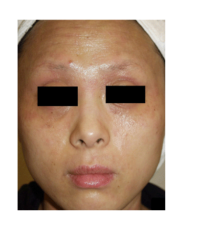
A 31‐year‐old Korean female patient with Fitzpatrick type IV skin presented with brownish pigmented patches, coarse and fine wrinkles, and alopecic patches on both eyebrows. She was diagnosed with atopic dermatitis (AD) and had been treated with systemic antihistamines, topical glucocorticosteroids and tacrolimus ointment for over 10 years. On her first visit, she still presented active lesions of chronic AD on the face (). Although the patient treated her eyebrows with tattooing 3 years ago, its effect had not lasted for more than a year.
Figure 1. Active lesions of chronic AD, brownish pigmented patches, coarse and fine wrinkles, and alopecic patches on both eyebrows are noted.
We treated the patient with three sessions of tattooing at 2‐month intervals using dark brown and turboblack pigments (permanent make‐up color numbers 2010 and 5120, respectively; MLW®, Intermed, Berlin, German). Also, for the treatment of post‐inflammatory hyperpigmentation (PIH) and wrinkles, five sessions of 1064‐nm Q‐switched Nd:YAG (QSNY) laser treatment using the MedLite C6™ (HoyaConBio Inc., Fremont, CA, USA) with a low fluence at 2‐week intervals and one session of fractional photothermolysis system (FPS) therapy were performed. The 1064‐nm QSNY treatment was delivered to the entire face, including the PIH, with the settings of 2.5 J/cm2, a 6‐mm spot size, and two to three passes with appropriate overlapping. Additional treatment on the PIH was followed with the settings of 4 J/cm2, a 4‐mm spot size, and two to three passes with appropriate overlapping.
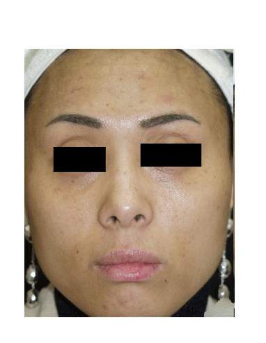
A 1550‐nm wavelength FPS was performed using the Sellas II™ laser (Dinona Corporation, Seoul, Korea). The treatment settings of 12.5 mJ for the pulse energy and 169 pulses per area (PPA) for spot density were delivered twice on the face with the moving operating mode. Additional treatment, 10–12.5 mJ for the pulse energy and 100 PPA for the spot density, was followed on the face with the stamp operating mode; in this setting, three passes were delivered with an appropriate overlap. After each laser procedure, the patient was recommended to apply Physiogel® cream (Stiefel‐Korea, Seoul, Korea) once every night without any topical or systemic glucocorticosteroids. She did not present any major complications or side effects, such as aggravation of AD, post‐therapy erythema, or pigmentation abnormalities. After treatment, distinct improvement was observed () and the patient was satisfi ed.
Figure 2. Clinical improvement with five sessions of 1064‐nm Q‐switched Nd:YAG (QSNY) laser treatment with low fluence at 2‐week intervals and one session of fractional photothermolysis system (FPS) therapy.
Patients with chronic AD characteristically present lichenified, dry, and lackluster skin. Although AD often subsides with the patient's age, many patients over their twenties continue to suffer from chronic AD with frequent aggravation and complications, including PIH, wrinkles, textural abnormalities, loss of eyebrows, and looking older than their present age. Since AD is associated with a marked decrease in skin barrier function and chronic reoccurrence (Citation1), many patients as well as dermatologists are reluctant to use cosmetic therapeutic methods, such as chemical p eeling or laser modalities.
The 1064‐nm QSNY is a non‐ablative, selective photothermolysis system; therefore, if there are no specific light‐absorbing factors on the epidermal architecture, such as the epidermal barrier, light can project through skin without a pronounced effect on its barrier function. A 1064‐nm QSNY with low fluence therapy is easily applicable for various pigmentary disorders, such as melasma, PIH, café‐au‐lait macules, and acquired bilateral nevus of Ota‐like macules (Citation2). Using this method, most of the patients experience minimal downtime without post‐therapy bleeding or crust formation. Posttherapy erythema tends to be spontaneously resolved within a few hours.
A 1550‐nm erbium‐doped FPS has already proved to provide effective clinical outcomes in various dermatologic conditions with a relatively low complication rate (Citation3). Because the stratum corneum remains intact and each wound is surrounded by healthy epidermal architecture, epidermal barrier function is preserved and recovery time is rapid (Citation3,Citation4).
Although the efficacy of the pulsed‐dye laser in the treatment of eczematous dermatoses, including AD, has been reported previously (Citation5), our report is insufficient in determining whether the lasers affect the superficial dermal microvasculature or inflammatory cells, eventually leading to the improvement of AD. However, we observed that the AD lesions in our patient were not aggravated during the laser treatment periods. We suggest that therapeutic modalities without barrier function disturbance, such as the 1064‐nm QSNY with low fluence and the 1550‐nm erbium‐doped FPS, can be utilized for the treatment of some chronic AD complications, especially in cases where potential emotional distress and cosmetic purposes are involved.
References
- Cork MJRobinson DAVasilopoulos YFerguson AMoustafa MMacGowan A, et al. New perspectives on epidermal barrier dysfunction in atopic dermatitis: gene‐environment interactions. J Allergy Clin Immunol. 2006;118:3–21.
- Kim JSKim MJCho SB. Treatment of segmental café‐au‐lait macules using 1,064‐nm Q‐switched Nd:YAG laser with low pulse energy. Clin Exp Dermatol (in press).
- Graber EMTanzi ELAlster TS. Side effects and complications of fractional laser photothermolysis: Experience with 961 treatments. Dermatol Surg. 2008;34:301–5.
- Manstein DHerron GSSink RKTanner HAnderson RR. Fractional photothermolysis: a new concept for cutaneous remodeling using microscopic patterns of thermal injury. Lasers Surg Med. 2004;34:426–38.
- Syed SWeibel LKennedy HHarper JI. A pilot study showing pulsed‐dye laser treatment improves localized areas of chronic atopic dermatitis. Clin Exp Dermatol. 2008;33:243–8.