
Abstract
Objectives
Recent advances in Spatial Temporal Image Correlation (STIC) 4 D fetal echocardiography include the application of eSTIC based on electronic probe image acquisition. We aimed to directly compare the performance of conventional STIC versus eSTIC technique (B-Mode and color Doppler imaging) during off-line reconstruction of STIC/eSTIC fetal heart volume pairs.
Methods
Pairs of B-Mode and Color Doppler STIC volumes were acquired sequentially by firstly conventional (STIC) followed by electronic (eSTIC) probes during 33 consecutive obstetric scans at median 23 (range 13–31) gestational weeks. The resulting 66 fetal heart volume pairs were assessed blindly off-line by a fetal cardiologist who documented feasibility of reconstruction, presence of motion artifacts, subjective image quality on a 4-level scale: 1-best to 4-non-diagnostic and morphological diagnosis, to enable a paired comparison of STIC and eSTIC in the same fetus under similar scanning conditions.
Results
eSTIC volumes had higher temporal resolution (37 vs. 24 frames per second, p < .001), less motion during acquisition (12 vs. 20 cases, O.R. 7.0, p = .002) and better average image quality (1.9 vs. 2.2, p = .006) compared to STIC volumes. More diagnostic reconstructions were achieved by eSTIC (n = 55, 86%) than STIC (n = 52, 78.8%), p = .001), in a comparable analysis time (mean 4.96 vs. 4.94 min). During a comparison of image quality of the original acquisition (A) and reconstructed planes (B and C planes) e STIC was superior in 22 (33%), 39 (59%) and 21 (38%) volumes, respectively, with the remaining cases being of similar quality (<10% in each plane in favor of STIC). Imaging mode and gestational age had a similar impact on both eSTIC and STIC performance: diagnostically acceptable studies in 49 (75.8%) vs. 48 (72.2%) by B-Mode, 60 (90.9%) vs. 56 (84.8%) by Color Doppler Mode, 8 (62.5%) vs. 10 (50%) in early scans, 38 (95%) vs. 38 (95%) in mid-gestation scans, and 7 (70%) vs. 6 (60%) in third trimester scans. Eight obstetric scans identified a fetus with a cardiac variant or structural abnormality. Diagnostic concordance of the two STIC approaches was comparable (40/48 concordant interpretations, kappa 0.657) all confirmed by fetal and/or postnatal echocardiography.
Conclusions
eSTIC was associated with more effective 4 D fetal heart reconstruction due to reduced motion artifacts and superior image quality in all planes, when compared to STIC. Early gestation reconstructions were not generally successful using either technology. Further study is needed to define the cost-effectiveness and diagnostic impact of eSTIC over conventional STIC and their role over, or in addition to, screening 2 D fetal echocardiography by appropriately trained sonographers.
Introduction
Since its first description as an off-line 4 D fetal echocardiography technique [Citation1], Spatial Temporal Image Correlation (STIC) analysis has been validated as a useful clinical tool [Citation2], offering remote evaluation of digitally stored fetal heart volumes by experts [Citation3], improved imaging of complex vascular malformations [Citation4], unique views of the fetal heart valves and interventricular septum [Citation2,Citation5,Citation6]. It also represents a promising research tool, allowing for fetal heart volumetry [Citation7], biometry [Citation8] and assessment of systolic ventricular function [Citation9,Citation10], while it enhances fetal echocardiography teaching [Citation11] and morphological interpretation especially when assisted by designated software applications [Citation12,Citation13]. Recent modification of electronic probes to enable electronic STIC (eSTIC) may address the current limitations due to faster acquisition [Citation1,Citation14]
The main aim of this study is to compare the feasibility of acquisition and diagnostic concordance in fetal heart volumes obtained consecutively using STIC and eSTIC during routine obstetric sonography. Secondary aims include the impact of imaging mode (2 D, color Doppler) and of gestational age (early, mid-, late gestation) on the performance of each STIC approach.
Methods
The study received Institutional Research Board approval (717/19) including permission for retrospective analysis of anonymized sonograms. Following informed consent, 33 pregnant women presenting consecutively for routine obstetric sonographic studies were included in the study.
All sonographic studies were performed for clinical indications by a single certified obstetric sonographer, with prior expertise in STIC volume acquisition, using a GE Voluson E10 ultrasound system, equipped with a standard mechanical 4 D probe (RMC 6 C) and an electronic 4 D probe (eM6C), capable of conventional and eSTIC volume acquisition, respectively. After the completion of a 2 D sonographic study, a pair (one B-Mode and one color Doppler) of STIC fetal heart volumes was acquired using the standard and electronic 4 D probes consecutively under identical image acquisition settings, scanning depth and almost identical fetal heart projection relative to the ultrasound beam. The angle of each STIC sweep was chosen to include transverse views from the upper abdomen through the fetal heart (4chamber view and outflows) including the mediastinum (3 vessel-trachea view). Four STIC volumes were available: two volume pairs (B-Mode STIC-eSTIC) and two color Doppler volume pairs (col.D STIC-eSTIC). Following each sonographic study, an anonymized folder with a unique ID (acquisition order-fetal gestational week) was created to store the 4 anonymized STIC volume (vol) files, one B-Mode and one col.D pair (eSTIC-STIC). A total of 33 folders including 132 STIC vol. files (33 B-Mode and 33 col.D eSTIC-STIC pairs) were available for retrospective off-line analysis by a single pediatric cardiologist with expertise in fetal cardiology including STIC volume analysis using 4 D View software (Version 14, GE Healthcare Austria GmbH & Co OG). Each STIC volume was examined to grade image quality, reconstruction feasibility and diagnostic interpretation.
Specifically, the fetal heart projection (angle between intraventricular septum and ultrasound beam 0–360 degrees, plane A), the presence of fetal motion artifacts and subjective image quality assessment (4-level scale, 1 best to 4 non-diagnostic) was documented for each plane (A,B,C) of a given volume file including an image quality comparison between eSTIC and STIC pairs in the 2 D and color volume pairs (STIC superior, e STIC superior, both equal), for each of the three planes. (, Supplementary Video 1(a,b))
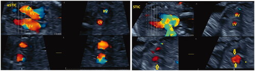
Figure 1. Paired comparison of color Doppler eSTIC vs STIC – plane B. Color Doppler e STIC (left) and STIC (right), B-plane (upper right and lower frames) reconstructed images obtained in a rapid sequence during the same study, corresponding to short axis (sagittal) views of fetal heart ventricles (LV: left ventricle; RV: right ventricle). Obvious distortion of ventricular contour in B-plane reconstruction (arrows right) in STIC frames resulting in inferior image quality compared to corresponding eSTIC B-plane frames.
The observer documented whether specific recommended fetal heart anatomy details could be successfully demonstrated (yes/no) and appeared normal (yes/no). In the A plane structures sought included abdominal situs (the relationship of aorta and inferior caval vein to the spine and stomach position in the transverse view), 4 chamber view details (atrial and ventricular size symmetry, AV valve offsetting, 2 pulmonary veins, foramen ovale flap motion, intact intraventricular septum), left and right ventricular outflow details (concordant ventriculoarterial connections with identifiable crossover and of similar size), and 3 vessel and trachea view (transverse aortic and ductal arches of similar sizes passing to the left of the trachea with a right-sided superior caval vein and identifiable brachiocephalic vein and normal sized thymus just cephalad) [Citation15]. The reconstructed planes B and C were evaluated to assess the short axis views of the aortic, mitral and tricuspid valve morphology and sagittal views of the arterial duct (AD) and aortic arch (AoA) [Citation16]. In addition to the anatomical details, flow patterns through the valves (mitral, tricuspid, aortic, pulmonary), foramen ovale, arterial duct, aortic isthmus and in the 3 vessel view flows were evaluated from the color Doppler volumes. The observer made a final diagnosis of normal, possibly abnormal, abnormal, impossible to interpret and the off-line analysis time for each volume. The off-line STIC diagnoses were compared to their corresponding 2 D anomaly scan diagnoses, and abnormal fetal heart findings to the confirmatory diagnosis including fetal and/or neonatal echocardiography (which followed only in the presence of abnormal findings in routine sonographic fetal heart imaging)
Statistical analysis
The overall as well as the gestational age- and imaging mode-based success rate of STIC and eSTIC analysis were compared. Appropriate tests (Cross tabs Chi square test, Odds ratio, Mc Nemar test, paired t-test) were used to compare STIC vs eSTIC performance (SPSS 18.0). Kappa value was estimated to show the level of agreement (diagnostic concordance). A p-value <.05 was considered as level of statistical significance
Results
The 33 obstetric sonograms were performed at median age 23 (range 13–31) gestational weeks (GW). Eight (24%) were early (<18th GW), twenty (61%) were mid gestation (18–26thGW) and five (15%) late gestation (>27thGW) sonographic studies. Seven fetuses had abnormal sonographic findings, 6 of which were confirmed by fetal and/or neonatal echocardiography. Details are presented in .
Table 1. Diagnostic concordance between STIC, e STIC and anomaly scan/confirmatory fetal echocardiography.
The eSTIC volume files were of larger size (63 vs. 37 MB) and of higher temporal resolution (37 vs. 24 frames per cycle) compared to their corresponding STIC volume pairs (p < .001).
The original acquisition plane (A) heart axis angle was almost identical (189 vs. 200 degrees, p = .183) in eSTIC and STIC acquisition. The spine was down in 15/33 (45%) studies with only 7 (21%) having a favorable fetal heart projection for STIC acquisition (spine between 5-7 o clock). [Citation17] Motion during acquisition was less frequently observed in eSTIC vs STIC acquisition (12/66 vs. 20/66 cases, O.R: 7.0, 95% CI: 1.7–27.2, p = .002).
Subjective image quality assessment was documented as superior in eSTIC compared to STIC analysis, in the original acquisition plane A (1.45 vs. 1.67, p = .022) as well as the reconstructed planes B (2.44 vs. 2.79, p = .008) and C (3.56 vs. 3.77, p = .015). The average reconstruction image quality (average of individual plane image quality scores) was also superior in e STIC compared to STIC (1.94 vs. 2.27, p = .006)
eSTIC was associated with superior image quality in a direct comparison of volume pairs in 22 (33%), 39 (59%) and 21 (32%) volumes, in planes A, B and C, respectively, with the remaining cases being classified either as of similar performance or in favor of STIC (<10% in each plane) as presented in . The success of reconstruction of the fetal echocardiography views and recognition of morphological details is presented in . eSTIC was particularly effective in the reconstruction of fetal echocardiography views obtained normally from sagittal and oblique image planes such as ductal and aortic arch views. Overall, eSTIC analysis provided diagnostic reconstructions more frequently compared to STIC (55/66 (83.3%) vs. 52/66 (78.8%) cases, p = .001). The imaging mode and gestational age affected eSTIC and STIC performance similarly: overall, B Mode-based analysis was less successful [n = 49 (75.8%)/n = 48 (72.2%)] than Color Doppler [n = 60 (90.9%)/n = 56 (84.8%)], by both eSTIC/STIC, respectively. Mid-gestation scans were more often diagnostic [n = 38 (95%)/n = 38 (95%)] compared to early scans [n = 8 (62.5%)/n = 10 (50%)] and late-gestation scans [n = 7 (70%)/n = 6 (60%)], by both eSTIC/STIC. summarizes the feasibility of each approach, according to gestational age and imaging mode groups.
Table 2. Comparison of subjective image quality between eSTIC and STIC pairs, during off-line analysis.
Table 3. Paired comparison between STIC and eSTIC off-line analysis to demonstrate fetal cardiac morphology.
Table 4. Comparison of successful STIC volume reconstruction between STIC-eSTIC.
Average off-line analysis time was similar for both approaches (4.94 min, range 2–12 vs. 4.96 min, range 2.2–9.3 min, for STIC and eSTIC, respectively, p = .896).
The studies were defined as normal or abnormal (including definitive abnormal and probable abnormal). There were 40 concordant interpretations (normal: 24, abnormal: 16) compared with 8 discordant (4 abnormal by each approach), indicating relatively good agreement (kappa = 0.657, p < .001).
presents the original 2 D anomaly scan diagnoses along with corresponding eSTIC/STIC diagnoses (B Mode and color Doppler imaging), and the confirmatory diagnosis (fetal and/or neonatal echocardiogram) for each case. In all 6 cases of fetal heart variants/abnormalities, both STIC and eSTIC also documented the presence of abnormal findings: 4 cases had concordant diagnoses and in 2 cases STIC analysis provided additional minor (presence of retroesophageal course of arterial duct and aberrant left subclavian artery from right aortic arch) and major (presence of atrioventricular septal defect) information, compared with the initial routine anomaly scan diagnoses (cases 25, 26 of ).
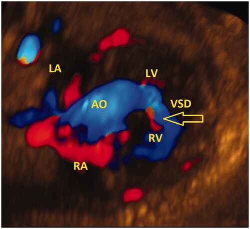
Discordant anomaly scan (normal) and STIC/eSTIC (abnormal) diagnoses included mainly small ventricular septal defects, detected only during off-line STIC (color Doppler) volume reconstruction (, Supplementary Video 2).
Figure 2. Color Doppler eSTIC off-line analysis: Muscular ventricular septal defect (arrow), off-line color Doppler eSTIC analysis. LA: left atrium; RA: right atrium; LV: left ventricle; RV: right ventricle; VSD: ventricular septal defect.
Discussion
In this direct paired comparison of consecutive eSTIC and STIC 4 D fetal echocardiograms, of the same subjects under almost identical image settings and their off-line analysis by a single expert we have demonstrated the expected superiority of eSTIC over STIC. The technical factors contributing to this include fundamental differences in 4 D volume acquisition design of the two techniques (electronic artefact-free sub-volume sweep in eSTIC compared with the mechanical artefact-prone total volume sweep in STIC) [Citation2]. In our study we found eSTIC analysis provided significantly more diagnostically acceptable reconstructions compared to STIC, especially in the more difficult early and late scans, and was associated with less motion and superior image reconstruction in the original and in both reconstructed planes. Gestational age (mid gestational imaging advantage) and imaging mode (in favor of color Doppler), both had an impact on the diagnostic feasibility of both approaches.
We designed this study to minimize variability in scanning conditions to enable a true comparison of eSTIC and STIC performance [Citation17]. Feasibility and diagnostic information is known to be affected by fetal projection, motion [Citation1–5], gestational age [Citation18], imaging mode (B-mode, color-, power-Doppler Mode) [Citation4], personnel experience both during STIC acquisition, off-line reconstruction and its interpretation [Citation19]. Additional supporting diagnostic software may also affect performance [Citation12,Citation13].
Our study concurs with the results of a previous study by Guasina et al. [Citation14], who reported superior eSTIC with STIC performance (94 vs. 76%) in different, though randomized, subjects scanned at mid-gestation with normal cardiac anatomy. However, when we limited our analysis to only the mid-gestational scans, we found similar diagnostic performance at 90% for both eSTIC and STIC when compared within the same subjects under almost identical imaging conditions. The superiority of eSTIC in terms of diagnostic feasibility in early and later gestational ages is likely to be attributed to reduced motion artifacts during a more rapid acquisition combined with improved reconstruction of imaging planes other than the original acquisition plane, while most failures of eSTIC reconstruction could be attributed to unfavorable fetal position [Citation2,Citation14].
We assessed the impact of gestational age and color Doppler imaging, in the abnormal cardiac studies. Both approaches diagnosed all fetal cardiac malformations correctly. However, eSTIC resulted in improved off-line reconstruction of the outflow tracts and of great arteries (transverse and sagittal views). These views are mandatory in the complete mid-trimester anomaly scan guidelines [Citation15,Citation16], but still represent a technical challenge for many obstetric sonographers resulting in increased scanning time, missed diagnoses of congenital heart disease and considerable investment in continuous medical education. The unique advantage that STIC (and especially e STIC) provides is in off-line reconstruction of the more challenging imaging planes and can provide diagnostic support for both the obstetric sonographer as well as the expert fetal cardiologist.
In the present study, eSTIC resulted in three additional diagnostic reconstructions over the 52 successful STIC analysis, corresponding to a relative improvement of 6%. Although the magnitude of diagnostic improvement offered by eSTIC seems relative small, the expected clinical impact of eSTIC could be more important if it aids earlier and better fetal cardiac imaging [Citation20] and reduces the considerable morbidity and mortality associated with undiagnosed fetal congenital heart disease. [Citation21] The application of color STIC resulted in a relative improvement of at least 10% in both STIC and eSTIC analysis, in accordance with previous studies [Citation4,Citation13].
The limitations of the present study include a relatively small sample size (partially addressed by the paired analysis design), retrospective analysis of STIC volumes, single observer analysis, and incomplete information regarding final outcome of all cases we included.
A further limitation represents the lack of a “gold standard” diagnostic test against which both STIC and STIC diagnostic performance could be compared: although all abnormal routine anomaly scans with associated fetal heart abnormalities received confirmatory diagnostic imaging (fetal and/or neonatal echocardiography) corresponding therefore to “true positive” STIC and eSTIC diagnoses, the possibility and extent of “false positive” STIC diagnoses cannot be assessed in our study. Cases of suspected AVSD in early scans by STIC (case 1) or by e STIC (case 8) analysis, represented either false positive (case 1, with normal following midgestation fetal heart imaging) or not confirmed findings (case 8, pregnancy termination). The diagnosis of AVSD is a challenging during early sonographic fetal evaluation (including STIC), due to lack of atrioventricular valve offsetting in early fetal life. (The increased prevalence of ventricular septal defects in STIC/eSTIC analysis might represent true (though clinically insignificant) findings which were not detected during the routine anomaly scan, or image artifacts associated with volume acquisition. Given the additional diagnostic information that both STIC approaches provided in two suspected fetal cardiac abnormalities, which were confirmed, we consider the discordant findings (mostly of VSDs) to represent probably true though clinical insignificant findings, especially if present in both B-Mode and color Doppler imaging. Finally, a cost effectiveness analysis was not performed due to the small sample size.
We believe that in certain conditions (superior image quality, absence of artifacts, analysis by an expert fetal cardiologist) eSTIC (and STIC) could provide more reliable diagnostic information compared to real time fetal heart sonographic screening by obstetric sonographers; however we must emphasize that despite the large body of available literature supporting the clinical and research applications of STIC, the technique (including eSTIC) should still not be considered as a stand-alone diagnostic tool, replacing 2 D fetal echocardiography performed by experts [Citation22].
Conclusions
eSTIC was associated with more effective 4 D fetal heart reconstruction due to reduced motion artifacts and superior image quality in all planes, when directly compared to STIC. Early gestation reconstructions were not generally successful. Further study is needed to define the cost-effectiveness and diagnostic impact of eSTIC over conventional STIC and their role over, or in addition to, screening 2 D fetal echocardiography by appropriately trained sonographers.
Supplemental data for this article can be accessed here.
Download Microsoft Video (AVI) (14.9 MB)Supplemental Material
Download Microsoft Video (AVI) (9.7 MB)Supplemental Material
Download Microsoft Video (AVI) (53.8 MB)Acknowledgments
We would like to thank Prof. H. Gardiner for her valuable comments regarding the submitted manuscript, School of Medicine University of Crete and Chania Medial Association, for approving and supporting the presented research.
Disclosure statement
No potential conflict of interest was reported by the author(s).
References
- DeVore GR, Falkensammer P, Sklansky MS, et al. Spatio-temporal image correlation (STIC): new technology for evaluation of the fetal heart. Ultrasound Obstet Gynecol. 2003;22(4):380–387.
- DeVore GR, Satou G, Sklansky M. 4D fetal echocardiography-an update. Echocardiography. 2017;34(12):1788–1798.
- Viñals F, Mandujano L, Vargas G, et al. Prenatal diagnosis of congenital heart disease using four-dimensional spatio-temporal image correlation (STIC) telemedicine via an Internet link: a pilot study. Ultrasound Obstet Gynecol. 2005;25(1):25–31.
- Chaoui R, Hoffmann J, Heling KS. Three-dimensional (3D) and 4D color Doppler fetal echocardiography using spatio-temporal image correlation (STIC). Ultrasound Obstet Gynecol. 2004;23(6):535–545.
- Rolo LC, Santana EF, da Silva PH, et al. Fetal cardiac interventricular septum: volume assessment by 3D/4D ultrasound using spatio-temporal image correlation (STIC) and virtual organ computer-aided analysis (VOCAL). J Matern Fetal Neonatal Med. 2015;28(12):1388–1393.
- Adriaanse BM, Uittenbogaard LB, Tromp CH, et al. Prenatal visualization of the pulmonary and aortic valves and leaflets is feasible using 4-dimensional sonography. J Ultrasound Med. 2016;35(3):497–504.
- Araujo Júnior E, Novoa Y Novoa VA, Barros FS, Rocha LA, et al. Reference values for the volumes of foetal heart atrial wall by three-dimensional ultrasound using STIC and VOCAL methods between 20w0d and 33w6d weeks of gestation. J Matern Fetal Neonatal Med. 2016;29(19):3076–3083.
- Tedesco GD, de Souza Bezerra M, Barros FS, et al. Reference ranges of fetal cardiac biometric parameters using three-dimensional ultrasound with spatiotemporal image correlation M mode and their applicability in congenital heart diseases. Pediatr Cardiol. 2017;38(2):271–279.
- Tanis JC, Mohammed N, Bennasar M, et al. Online versus offline spatiotemporal image correlation (STIC) M-mode for the evaluation of cardiac longitudinal annular displacement in fetal growth restriction. J Matern Fetal Neonatal Med. 2018;31(14):1845–1850.
- Molina FS, Faro C, Sotiriadis A, et al. Heart stroke volume and cardiac output by four-dimensional ultrasound in normal fetuses. Ultrasound Obstet Gynecol. 2008;32(2):181–187.
- Avnet H, Mazaaki E, Shen O, et al. Evaluating spatiotemporal image correlation technology as a tool for training nonexpert sonographers to perform examinations of the fetal heart. J Ultrasound Med. 2016;35(1):111–119.
- Veronese P, Bogana G, Cerutti A, et al. A prospective study of the use of Fetal Intelligent Navigation Echocardiography (FINE) to obtain standard fetal echocardiography views. Fetal Diagn Ther. 2017;41(2):89–99.
- Yeo L, Markush D, Romero R. Prenatal diagnosis of tetralogy of Fallot with pulmonary atresia using: Fetal Intelligent Navigation Echocardiography (FINE). J Matern Fetal Neonatal Med. 2019;32(21):3699–3702.
- Guasina F, Bellussi F, Morganelli G, et al. Electronic spatiotemporal image correlation improves four-dimensional fetal echocardiography. Ultrasound Obstet Gynecol. 2018;51(3):357–360.
- Carvalho JS, Allan LD, Chaoui R, International Society of Ultrasound in Obstetrics and Gynecology, et al. S. ISUOG practice guidelines (updated): sonographic screening examination of the fetal heart. Ultrasound Obstet Gynecol. 2013;41(3):348–359.
- Fetal Echocardiography Task Force; American Institute of Ultrasound in Medicine Clinical Standards Committee; American College of Obstetricians and Gynecologists; Society for Maternal-Fetal Medicine. AIUM practice guideline for the performance of fetal echocardiography. J Ultrasound Med. 2013;32:1067–1082.
- Yeo L, Romero R. How to acquire cardiac volumes for sonographic examination of the fetal heart: Part 1. J Ultrasound Med. 2016;35(5):1021–1042.
- Votino C, Cos T, Abu-Rustum R, et al. Use of spatiotemporal image correlation at 11–14 weeks’ gestation. Ultrasound Obstet Gynecol. 2013;42(6):669–678.
- Novaes JY, Zamith MM, Araujo Júnior E, et al. Congenital heart diseases by three-dimensional ultrasound using spatiotemporal image correlation: influence of professional experience. Echocardiography. 2016;33(1):99–104.
- Germanakis I, Sifakis S. The impact of fetal echocardiography on the prevalence of liveborn congenital heart disease. Pediatr Cardiol. 2006; 27(4):465–472.
- Plana MN, Zamora J, Suresh G, et al. Pulse oximetry screening for critical congenital heart defects. Cochrane Database Syst Rev. 2018;3:CD011912.
- Donofrio MT, Moon-Grady AJ, Hornberger LK, American Heart Association Adults With Congenital Heart Disease Joint Committee of the Council on Cardiovascular Disease in the Young and Council on Clinical Cardiology, Council on Cardiovascular Surgery and Anesthesia, and Council on Cardiovascular and Stroke Nursing, et al. Diagnosis and treatment of fetal cardiac disease: a scientific statement from the American Heart Association. Circulation. 2014;129(21):2183–2242.