
ABSTRACT
Background: Neuropathy is a common adverse effect of bortezomib. Isolated central nervous system (CNS) relapse in MM remains exceedingly rare and carries a dismal prognosis. We present an unusual case of bortezomib related neuropathy masking a CNS relapse of MM. Case presentation: A 57-year-old female was diagnosed with standard-risk MM with clinical and cytogenetic features not typically associated with CNS involvement. She was treated with 4 cycles of bortezomib/cyclophosphamide/dexamethasone (VCD) and achieved a VGPR, after which she underwent an autologous stem cell transplant (ASCT) followed by bortezomib maintenance. Six months after ASCT she developed symptoms suggestive of peripheral neuropathy which was attributed to bortezomib. However the symptoms persisted despite discontinuation of bortezomib. Imaging and cerebrospinal fluid analysis subsequently confirmed a CNS relapse. Discussion: CNS involvement in MM (CNS-MM) is uncommon and is considered an aggressive disease. Recently published literature has reported biomarkers with prognostic potential. However, isolated CNS relapse is even less common; an event which carries a very poor prognosis. Given the heterogeneous neurologic manifestations associated with MM, clinical suspicion may be masked by confounding factors such as bortezomib-based therapy. The disease may further remain incognito if the patient does not exhibit any of the high risk features and biomarkers associated with CNS involvement. Conclusion: In the era of proteasome inhibitor (PtdIns)/immunomodulator (IMID)-based therapy for MM which carries neurologic adverse effects, it is prudent to consider CNS relapse early. This case further highlights the need for more robust biomarkers to predict CNS relapse and use of newer novel agents which demonstrate potential for CNS penetration.
Introduction
Multiple myeloma (MM) is a mature B-cell malignancy which accounts for 13% of all hematologic malignancies in whites and 33% in blacks. It is characterized by a clonal expansion of plasma cells, typically within the bone marrow but sometimes also in extramedullary sites.Citation1-3 CNS involvement in MM (CNS-MM) remains rare, accounting for 1% of all MM cases, and exhibits a dismal prognosis with an overall survival (OS) of less than 6 months.Citation4,5 These include CNS-MM cases both at the time of diagnosis as well as relapse. CNS involvement is defined by the presence of monoclonal malignant plasma cells in the CSF during the course of MM disease, with or without radiologic features on Magnetic Resonance Imaging (MRI) suggestive of MM.Citation6,7
Relapse of MM with isolated CNS involvement, after attainment of complete remission (CR) post-ASCT (autologous stem cell transplantation), is even less common and is reported only as case reports or small case series.Citation7,8 Novel agents (NA) in the last decade have improved the outlook for patients with MM.Citation4,9,10 One of the more commonly used NA, the proteasome inhibitor (PI), bortezomib (velcade) is frequently associated with peripheral neuropathy.Citation11 This case underscores the importance of maintaining a high level of suspicion for CNS relapse while patients are on bortezomib-based regimens, even in the absence of biomarkers and clinical parameters commonly associated with CNS involvement, as a missed diagnosis may result in inferior outcomes.
Case presentation
A 57 year-old Chinese female with no other past medical history of significance, presented with recurrent epistaxis and was found to have thrombocytopaenia. Subsequent investigations showed infiltration of the bone marrow with clonal plasma cells with plasmablastic morphology. Although she had mild renal impairment, there was no hypercalcaemia or anaemia. Her skeletal survey was normal; however, there were fluorodeoxyglucose (FDG)-avid bone lesions on PET-CT. She was diagnosed with ISS stage II, IgG kappa multiple myeloma (MM) with normal cytogenetics. Flourescent in situ hybridization (FISH) was not done. Serum M protein was 61.8 g/L at diagnosis.
She underwent 4 cycles of bortezomib, cyclophosphamide and dexamethasone (VCD) and achieved very good partial remission (VGPR). Thereafter she underwent ASCT with a reduced dose of melphalan (140 mg/m2) due to renal impairment. She was subsequently treated with monthly bortezomib maintenance but developed peripheral neuropathy during the fifth month of maintenance after SCT. Her main symptoms were paraesthesia and numbness in the palms and soles which worsened despite symptomatic treatment and discontinuation of bortezomib.
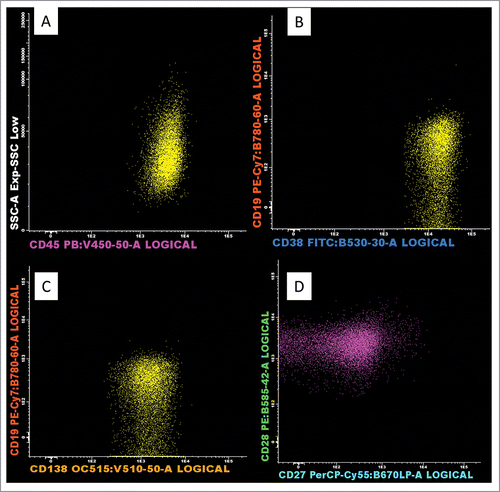
She further developed right upper and lower limb weakness. Her neurologic examination revealed decreased proprioception in a glove and stocking distribution, areflexia, bilateral foot drop and power of 3/5 in the right arm and leg. An MRI brain showed lytic bone lesions with dural-based masses within both occipital lobes. Lesions suspicious of myelomatous involvement were also detected in the pituitary, hypoglossal canal, cavernous sinus and sella turcica. Her MRI spine did not show any myelomatous involvement. Cerebrospinal fluid (CSF) cytology revealed 88% plasma cells which were confirmed by flow cytometry. Her CSF plasma cell morphology and flow cytometric immunophenotyping are shown in respectively. She underwent concurrent cranial irradiation with intra-thecal (IT)-methotrexate/cytarabine and thalidomide-dexamethasone (Thal-Dex) for control of CNS disease. She remained stable for 6 months after relapse, while on lenalidomide and dexamethasone (Len-Dex). She developed progressive disease thereafter, with a rising serum Free Light Chain ratio (sFLC) and multiple FDG-avid PET lesions. She is currently receiving salvage therapy.
Figure 1. Cerebrospinal fluid (CSF) cytological examination showed presence of abnormal plasma cells featuring enlarged and hyperchromatic nuclei, nuclear contour irregularity and occasional cells with binucleation. (Giemsa stain, original magnification, x 400).
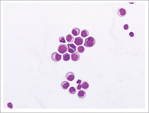
Figure 2. Flow cytometric analysis of the CSF specimen demonstrating plasma cells (PC). The PC are of intermediate side scatter and are CD45 positive (A), CD 38 brightly positive, CD 19 negative and CD 138 positive (B and C). They are also dimly positive for CD 28 and are CD 27 negative (D); this is a feature of aberrant PC. (Van Dongen et al Leukemia 2012).
Discussion
We present a case of MM with isolated CNS relapse and review the data related to outcomes of CNS MM in the era of NAs, biomarkers associated with CNS MM and NAs that show promise for activity in CNS disease.
Given the heterogeneity in neurologic manifestations of MM and incorporation of immunomodulators (IMIDs) at the core of MM treatment in the last decade, an occurrence as rare as MM relapse with isolated CNS involvement exhibits a potential for missed diagnosis.
Some of the more commonly used NAs include immunomodulators (IMIDs) such as thalidomide, lenalidomide, pomalidomide and proteasome inhibitors (PtdIns) such as bortezomib. Bortezomib-induced peripheral neuropathy (BIPN) is a dose-limiting adverse effect and occurs in as many as 75% of patients treated with bortezomib.Citation12,13
Though there is significant paucity of data related to risk factors for CNS MM in the era of novel therapeutics, recently published reports suggest association with high risk features such as raised LDH and β2M, IgG paraprotein, high risk baseline genetic abnormalities and secondary plasma cell leukemia.Citation14-16 In terms of disease outcome, Nazanin M et al recently reported in their retrospective study involving 9 patients with CNS-MM, who were treated with novel agents, to have a median OS of 3.5 months which is not different in comparison with OS in reports published prior to the era of novel drugs.Citation16-19 However, it is of note that commonly used IMID/PI drugs have poor CNS penetration except pomalidomide and marizomib which have shown promise in terms of CNS penetration.Citation20-24. presents treatment and outcomes of CNS myeloma.
Table 1. Treatment and outcomes of CNS-MM in the era of NAs.
It remains a judgment call on the part of the treating physician to suspect, perform a comprehensive neurologic exam and assess the risk for CNS relapse. However, this entity carries a poor prognosis and may define the beginning of a terminal course and is, therefore, worth consideration even when the disease does not carry the biomarkers and genetic features associated with CNS relapse. Further large-scale studies are needed to better understand the heterogeneity of CNS-MM, biomarkers and genetic features that may demonstrate potential for prognostication and outcomes with PtdIns/IMID-based therapy with CNS penetration.
Clinical pearls
Due diligence needs to be paid to CNS involvement in patients with MM, typically in the relapsed setting as that carries a dismal prognosis.
CNS MM has reportedly been associated with poor prognostic features such as high LDH, high B2M and secondary plasma cell leukemia but statistical strength is still lacking.
MM is associated with variable neurologic manifestations and it poses a clinical challenge to differentiate symptoms from disease versus those due to therapy.
CNS relapse must be considered in all patients with MM and appropriate radiologic and CSF investigations be performed should symptoms persist, even in standard risk disease.
Disclosure of potential conflicts of interest
No potential conflicts of interest were disclosed.
Authors' contributions
MBA collected the data, wrote the manuscript and coordinated the project. SDM provided flow cytometry images and assisted with revision of manuscript. MAA drafted the table and contributed to revision of manuscript. TKB provided CSF cytology image. CWJ treated the patient, conceived of the project and assisted with revision of manuscript. All authors read and approved the final manuscript.
References
- Palumbo A, Anderson KC. Multiple myeloma. N Engl J Med 2011; 364:1046-60; PMID:21410373; http://dx.doi.org/10.1056/NEJMra1011442
- Swerdlow SH, Campo E, Harris NL, et al. WHO Classification of Tumours of Haematopoietic and Lymphoid Tissues. 4th ed. Lyon, France: World Health Organization 2008; 200-13.
- Altekruse SF, Kosary CL, Krapcho M, et al. SEER cancer statistics review, 1975–2007. Bethesda, MD: National Cancer Institute.
- Kumar SK, Rajkumar SV, Dispenzieri A, Lacy MQ, Hayman SR, Buadi FK, Zeldenrust SR, Dingli D, Russell SJ, Lust JA, et al. Improved survival in multiple myeloma and the impact of novel therapies. Blood 2008; 111(5):2516-20; PMID:17975015; http://dx.doi.org/10.1182/blood-2007-10-116129
- Abdallah AO, Atrash S, Shahid Z, Jameel M, Grazziutti M, Apewokin S, Kumar NS, Restrepo A, Waheed S, Van Rhee F, et al. Patterns of central nervous system involvement in relapsed and refractory multiple myeloma. Clin Lymphoma Myeloma Leuk 2014; 14(3):211-4; PMID:24373936; http://dx.doi.org/10.1016/j.clml.2013.11.004
- Chen CI, Masih-Khan E, Jiang H, Rabea A, Cserti-Gazdewich C, Jimenez-Zepeda VH, Chu CM, Kukreti V, Trudel S, Tiedemann R, et al. Central nervous system involvement with multiple myeloma: Long term survival can be achieved with radiation, intrathecal chemotherapy, and immunomodulatory agents. Br J Haematol 2013; 162:483-8; PMID:23772701; http://dx.doi.org/10.1111/bjh.12414
- Petersen S, Wagner A, Gimsing P. Cerebral and meningeal multiple myeloma after autologous stem cell transplantation. A case report and review of the literature. Am. J. Hematol 1999; 62:228-33; PMID:10589078; http://dx.doi.org/10.1002/(SICI)1096-8652(199912)62:4%3c228::AID-AJH5%3e3.0.CO;2-3
- Veinstein A, Brizard A, Randriamalala E, Babin P, Preud'homme JL, Guilhot F. Central nervous system relapses after autologous stem cell transplantation for myeloma. Report of two cases. Hematol. Cell Ther 1997; 39:327-30; PMID:9497892; http://dx.doi.org/10.1007/s00282-997-0327-6
- Ria R, Reale A, Vacca A. Novel agents and new therapeutic approaches for treatment of multiple myeloma. World J Methodol 2014; 4(2):73-90; PMID:25332907; http://dx.doi.org/10.5662/wjm.v4.i2.73
- Brenner H, Gondos A, Pulte D. Recent major improvement in long-term survival of younger patients with multiple myeloma. Blood 2008; 111: 2521-26; PMID:17901246; http://dx.doi.org/10.1182/blood-2007-08-104984
- Richardson PG, Delforge M, Beksac M, Wen P, Jongen JL, Sezer O, Terpos E, Munshi N, Palumbo A, Rajkumar SV, et al. Management of treatment-emergent peripheral neuropathy in multiple myeloma. Leukemia 2012; 26(4):595-608; PMID:22193964; http://dx.doi.org/10.1038/leu.2011.346
- Cavaletti G, Jakubowiak AJ. Peripheral neuropathy during bortezomib treatment of multiple myeloma: a review of recent studies. Leuk Lymphoma 2010; 51(7):1178-87; PMID:20497001; http://dx.doi.org/10.3109/10428194.2010.483303
- Corso A, Mangiacavalli S, Varettoni M, Pascutto C, Zappasodi P, Lazzarino M. Bortezomib-induced peripheral neuropathy in multiple myeloma: A comparison between previously treated and untreated patients. Leuk Res 2010; 34(4):471-4; PMID:19674790; http://dx.doi.org/10.1016/j.leukres.2009.07.022
- Fassas AB, Ward S, Muwalla F, Van Hemert R, Schluterman K, Harik S, Tricot G. Myeloma of the central nervous system: strong association with unfavourable chromosomal abnormalities and other high-risk disease features. Leuk Lymphoma 2004; 45:291-300; PMID:15101714; http://dx.doi.org/10.1080/10428190310001597964
- Alsobhi ES, Hashim IA, Abdelaai MA, Aljifri AM, Alshamy AM. Elevated cerebrospinal fluid B2 microglobulin as a tumor marker in a patient with multiple myeloma of the central nervous system. Saudi Med J 2007; 28:128-30; PMID:17206305
- Fassas A, Muwalla F, Berryman T, Benramdane R, Joseph L, Anaissie E, Sethi R, Desikan R, Siegel D, Badros A, et al. Myeloma of the central nervous system: association with high-risk chromosomal abnormalities, plasmablastic morphology and extramedullary manifestations. Br J Haematol 2002; 117:103-8; PMID:11918539; http://dx.doi.org/10.1046/j.1365-2141.2002.03401.x
- Nazanin M, Wei X, Demopoulos A, Hormigo A, Chari A. Characterization of central nervous system multiple myeloma in the era of novel therapies. Leuk Lymphoma 2016; 4:1-5; PMID:26727654
- Katodritou E, Terpos E, Kastritis E, Delimpasis S, Symeonidis AS, Repousis P, Kyrtsonis MC, Vadikolia C, Michalis E, Polychronidou G, et al. Lack of survival improvement with novel anti-myeloma agents for patients with multiple myeloma and central nervous system involvement: the Greek Myeloma Study Group experience. Ann Hematol 2015; 94:2033-42; PMID:26420061; http://dx.doi.org/10.1007/s00277-015-2484-y
- Nieuwenhuizen L, Biesma DH. Central nervous system myelomatosis: review of the literature. Eur J Haematol 2007; 80:1-9; PMID:17961180
- Ocio EM, Richardson PG, Rajkumar SV, Palumbo A, Mateos MV, Orlowski R, Kumar S, Usmani S, Roodman D, Niesvizky R, et al. New drugs and novel mechanisms of action in multiple myeloma in 2013: a report from the International Myeloma Working Group (IMWG). Leukemia 2014; 28(3):525-42; PMID:24253022; http://dx.doi.org/10.1038/leu.2013.350
- Mussetti A, Dalto S, Montefusco V. Effective treatment of pomalidomide in central nervous system myelomatosis. Leuk Lymphoma 2013; 54:864-6; PMID:22880953; http://dx.doi.org/10.3109/10428194.2012.718343
- Potts BC, Albitar MX, Anderson KC, Baritaki S, Berkers C, Bonavida B, Chandra J, Chauhan D, Cusack JC Jr, Fenical W, et al. Marizomib, a proteasome inhibitor for all seasons: preclinical profile and a framework for clinical trials. Curr Cancer Drug Targets 2011; 11: 254-84; PMID:21247382; http://dx.doi.org/10.2174/156800911794519716
- Short KD, Rajkumar SV, Larson D, Buadi F, Hayman S, Dispenzieri A, Gertz M, Kumar S, Mikhael J, Roy V, et al. Incidence of extramedullary disease in patients with multiple myeloma in the era of novel therapy, and the activity of pomalidomide on extramedullary myeloma. Leukemia 2011; 25: 906-08; PMID:21350560; http://dx.doi.org/10.1038/leu.2011.29
- Li Z, Qiu Y, Personett D, Huang P, Edenfield B, Katz J, Babusis D, Tang Y, Shirely MA, Moghaddam MF, et al. Pomalidomide shows significant therapeutic activity against CNS lymphoma with a major impact on the tumor microenvironment in murine models. PLoS One 2013; 8(8):e71754; PMID:23940785; http://dx.doi.org/10.1371/journal.pone.0071754
- Paludo J, Painuly U, Kumar S, Buadi FK, Hayman SR, Lacy MQ, Dispenzieri A, Lust JA, Dingli D, McCurdy A, et al. Myelomatous involvement of the central nervous system: Mayo Clinic experience. ASH Annual Meeting Abstracts 2013, 122: abstr. 3119.
- Gangatharan SA, Carney DA, Prince HM, Wolf MM, Januszewicz EH, Ritchie DS, Harrison SJ. Emergence of central nervous system myeloma in the era of novel agents. Hematol Oncol 2012; 30(4):170-4; PMID:22144117; http://dx.doi.org/10.1002/hon.1021
- Lee D, Kalff A, Low M, Gangatharan S, Ho P, Bajel A, Ritchie D, Grigg A, Spencer A. Central nervous system multiple myeloma–potential roles for intrathecal therapy and measurement of cerebrospinal fluid light chains. Br J Haematol 2013; 162(3): 371-5; PMID:23718539; http://dx.doi.org/10.1111/bjh.12404