
ABSTRACT
Inflammatory myofibroblastic tumor (IMT) is currently recognized as an intermediate mesenchymal neoplasm. It can arise anywhere in the body, but it is particularly common in the lungs. Gastric IMT is very rare in adults. In this study, we report a case of a 68-year-old woman with IMT in the gastric cardia, with invasion into the spleen and diaphragm. Because of its location and aggressive clinical features, it was first mistaken for gastric cancer. However, pathology and immunohistochemistry were used to finally confirm the diagnosis of IMT after total resection of the tumor and spleen and partial resection of the diaphragm. In order to provide better understanding of this rare tumor, targeted next-generation sequencing (NGS) and IHC were performed to assess genetic and protein abnormalities of the tumor. Both IHC and NGS were found to be negative for ALK or other gene fusions. However, double amplification of CDK4 and MDM2 were found by NGS, and IHC also found CDK4 and MDM2 to be positive. To the best of our knowledge, this is the first gastric IMT report to show double invasion of the spleen and the diaphragm, and double amplification of CDK4 and MDM2 in IMT are also reported for the first time. This genomic aberration with protein overexpression is the most likely tumorigenic driver of this rare and aggressive tumor.
Introduction
Inflammatory myofibroblastic tumor (IMT) is an uncommon mesenchymal neoplasm composed of spindle cells, myofibroblasts, lymphocytes, plasma cells, and eosinophils.Citation1 IMT was once considered a benign inflammatory process,Citation2 while recent studies have declaimed that IMT has an intermediate malignant potential of a tendency for local recurrence and invasion but rarely metastasis. IMT can occur across the entire age range but has a higher incidence in children and young adults.Citation3,Citation4 The lung is the most common affected site of IMT, whereas various extrapulmonary sites have also been reported, including the mesentery, omentum, liver, retropertomeum, pelvis, and central nervous system.Citation5,Citation6 Gastric IMT is very rare in adults. To the best of our knowledge, only 39 cases of IMT of the stomach have been reported in the English-language literature.Citation6–Citation9 Gastric IMT with local invasion to multiple organs is even rarer.Citation9 Previous studies have reported IMT to be genetically heterogeneous. Approximately 50 % of IMT cases display clonal rearrangements of the anaplastic lymphoma kinase (ALK) gene.Citation10 In contrast, ALK-negative IMTs may be more aggressive than ALK-positive IMTs.Citation3 Recent studies have shown ALK-negative IMT might harbor other kinase fusions, such as receptor tyrosine kinase c-ros oncogene 1 (ROS1),Citation11 which initiated genome-level research into potential tumorigenic drivers in ALK-negative subsets of IMTs.
Figure 1. Endoscopy and CT scan. (a) Endoscopy feature capture showing the protruding lesion on the posterior wall of the gastric cardia, with an ulcer in the middle. (b) CT scan showing a non-homogeneously enhanced irregular mass in the gastric cardia, with suspected invasion to the spleen.
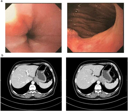
Figure 2. Copy number (CN) scatter diagram. The 520 oncogenic genes of the panel all have their own positions on the chromosome (horizontal axis). Every gene position corresponds to one capture domain, and every capture domain is cut into several continuous bins. The copy number (vertical axis) of every bin is examined and recorded as every spot in the diagram. The copy-number spots of key genes are highlighted in color. Each gene’s final copy number is calculated as the average of all its bin’s copy numbers.
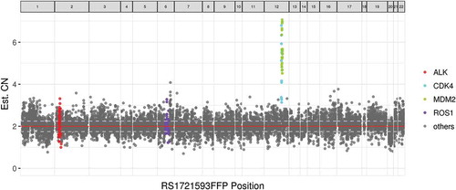
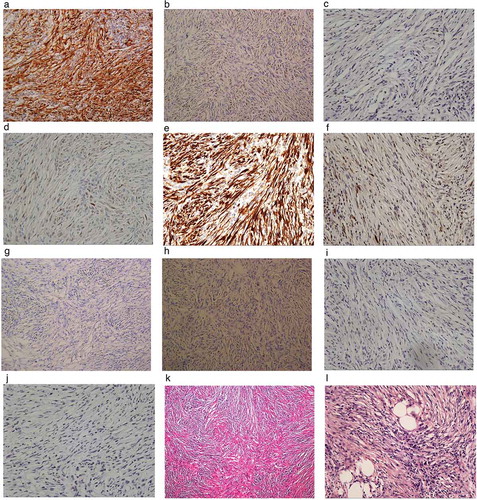
Figure 3. Histopathological and immunohistochemical appearance. (a) Positive immunostaining with monoclonal rabbit anti-human CDK4 (EP189, Zhongshan). (b) Positive immunostaining with monoclonal mouse anti-human MDM2 (SMP14, Thermo Fisher). (c) Negative immunostaining with monoclonal rabbit anti-human CDK2 (E304, Abcam). (d) Sporadically positive immunostaining with monoclonal cyclin D1 (EP12, Roche). (e) Diffusely positive immunostaining with p16ink4a (E6H4, Roche). (f) Sporadically positive immunostaining with polyclonal rabbit anti-human p19ink4d (Abcam). (g) Negative immunostaining with monoclonal mouse anti-human ALK (OTI1A4, Zhongshan). (h) Negative immunostaining with monoclonal mouse anti-human p53 (D0-7, Leica). (i) Negative immunostaining with monoclonal rabbit anti-human cyclin E1 (SP146, Abcam). (j) p21 WAF1 (DCS-60.2, Zhongshan Golden Bridge). (k) Low-power view showing the tumor to be mainly composed of spindle cells, collagen fibers, and inflammatory infiltration consisting of an increasing number of plasma cells and lymphocytes (H&E staining, × 10). (l) High-power view shows very little atypia and low-level mitosis (H&E staining, × 400).
In this study, we present a rare case of gastric IMT mimicking cancer, with invasion of the spleen and diaphragm. After complete surgical resection, when the tumor was pathologically diagnosed as IMT, its rare behavior, specifically its local aggressiveness, prompted us to perform immunohistochemical examination of various markers and oncogene screening based on targeted next-generation sequencing (NGS). Both IHC and NGS showed the tumor to be negative for ALK, and neither ROS1 nor any other genetic fusion was detected using NGS. However, double amplifications of cyclin-dependent kinase 4 (CDK4) and the human homolog of the murine double minute type 2 (MDM2) were identified and found to be characteristic and prognostic factors of dedifferentiated liposarcoma.Citation12 This double amplification was first reported in gastric IMT, which might be a potential tumorigenic driver of this subset of IMTs.
Materials and methods
Case report
A 68-year-old female with a two-year history of dysphagia, presented at a GI clinic. CT scan showed mildly enhanced irregular soft tissue mass (4.5 × 3.1 cm) at gastric cardia with a susceptible invasion to the spleen (Fig. 1b). Esophagogastroduodenoscopy (EGD) () and endoscopic ultrasound (EUS) showed an ulcerated, asymmetrical hypoechoic lesion at the back wall of the cardia with absence of the local gastric parietal natural structure, and a diagnosis of cancer was suspected. However, ultrasound-guided FNAC at pylorus, gastric angle, and cardia suggested chronic mucosal inflammation with mild intestinal metaplasia. Because malignancy was still strongly suspected, left thoracotomy was performed. During the surgery, a solid mass was found at the junction of the esophagus and stomach with invasion into the spleen and the diaphragm. The tumor was completely resected with splenectomy and partial diaphragmatic resection. All specimens were sent for pathological and immunohistochemical examination for diagnosis.
Pathological examination showed the tumor originated from the gastro-muscular layer and sub-mucosal layer of the cardia with invasion to the spleen and partially striated muscle (diaphragm). It was composed of spindle cells with inflammatory background consisting of a large number of plasma cells and lymphocytes. High-power vision showed very little atypia and low-level mitosis, only 2 per 10 high power fields (HPF). Immunohistochemistry (IHC) reported that the spindle cells were diffusely positive for vimentin and smooth muscle actin (SMA); focally positive for myogenic desmin; and negative for calponin, calretinin, and GIST-specific markers such as CD-117, DOG-1, and S-100. The approximate Ki-67 label index was 10 %. A diagnosis of gastric inflammatory myofibroblastic tumor (IMT) can be concluded from the results above.
The patient was discharged from the hospital on the 11th day after surgery. She had no postoperative complications. No subsequent therapy was prescribed. The patient has now been followed up for 8 months and she has experienced no weight loss and consumes a good and regular diet. The most recent abdominopelvic CT scan showed no sign of tumor recurrence or metastasis.
Next-generation sequencing
DNA was extracted from FFPE samples. Hematoxylin – and eosin-stained slides were reviewed to evaluate tumor content and viability. DNA concentration and genomic DNA quality were measured using a Qubit dsDNA assay and 260 nm/280 nm absorption ratio, respectively. Sequencing was performed on a Nextseq500 sequencer (Illumina, Inc., US) for 520 cancer-related genes, among which, whole exons of 312 genes and critical exons, introns, and promoter regions of the remaining 208 genes were captured. Resultant sequences were analyzed for base substitutions, insertions, deletions, copy number alterations, and select gene fusions.
Immunohistochemical examination for additional markers
Immunohistochemical analysis was performed to detect the protein products of known oncogenes, including ALK (anaplastic lymphoma kinase) (OTI1A4, Zhongshan; working solution), MDM2 (murine double minute 2) (SMP14, Thermo Fisher; 1:100), CDK4 (cyclin-dependent kinase 4) (EP180, Zhongshan; Working solution), and P53 (D0-7, Leica; working solution). Positive reactivity was defined as the labeling of > 10 % of the tumor cells for each antibody.
After the results of NGS and IHC came back, we performed further immunohistochemical analysis to derive better understanding of the protein biomarker levels, including p21 WAF1 (DCS-60.2, Zhongshan Golden Bridge; working solution), p16 (E6H4, Roche; working solution), p19 (Polyclone, Abcam; 1:50), cyclin D1 (EP12, Roche; working solution), cyclin E1 (SP146, Abcam; 1:10), and CDK2 (E304, Abcam; 1:100).
Results
Targeted next-generation sequencing was negative for gene fusions in the panel, including those of ALK and ROS1, and also negative for mutations of other common oncogenes. However, copy number alterations of double amplifications of CDK4 and MDM2 were detected. During the NGS process, every gene position corresponded to one capture domain. Every capture domain was cut into several continuous bins, and the copy number of every bin was examined and recorded for every spot in the diagram (). Finally, each gene’s copy number was calculated as the average of all its bin’s copy numbers. Copy number alterations were analyzed here based on the following algorithm. First, the copy number baseline of every gene was established using tests of massive numbers of negative samples of the normal population. Then copy numbers of examined genes were compared to the baseline. For one examined gene to be judged to have undergone a copy number alteration, more than 60 % of its bin’s copy numbers were required to be significantly higher than the baseline (P-value < 0.005 in z test), and the examined gene’s copy number over its whole capture domain must have been significantly higher than the baseline (P-value < 0.005 in z test). The copy number of CDK4 was 4.46 and that of MDM2 was 5.63 ().
IHC showed positive results for CDK4 and MDM2 and confirmed the overexpression of the corresponding proteins, which coincided with amplifications detected at the genetic level. IHC also showed negative results for ALK and p53. During the second-batch IHC analysis, the tumor was diffusely stained with anti-P16, sporadically stained with anti-P19 and anti-cyclin D1, and negative for CDK2, cyclin E, and p21 WAF1 ().
Discussion
IMT is a rare tumor originally described in the lung and later reported in other sites throughout the body.Citation5 The stomach is regarded as one of the most uncommon locations of IMT.Citation7 Currently, IMT is classified as an intermediate mesenchymal neoplasm.Citation13 Adjacent organ infiltration involving the peritoneal cavity or spleenCitation8,Citation14 indicated the malignant potential of gastric IMT. The 39 gastric IMTs previously reported showed only 5 locally aggressive cases with 3 cases of invasion to the spleen, 1 to the liver, and 1 to the pancreas.Citation6–Citation9 To the best of our knowledge, our study is the first report of gastric IMT invading both the spleen and diaphragm. Its unusual local aggressive behavior explains the difficulty of clinical diagnosis. Pathologic diagnosis, however, was clear. Immunohistochemical markers and histological patterns of the resected tissue help to distinguish IMT from other types of gastric tumors, such as gastrointestinal stromal tumor (GIST) and inflammatory fibroid polyps (IFP). IMT cells are immunopositive for vimentin, desmin, and SMA but negative for IFP-specific CD34,Citation15 GIST-specific CD117, S-100, and DOG1.Citation16
The ALK gene plays a very important role in the tumorigenesis of IMT. Approximately 50 %–70 % of the tumors have an ALK gene rearrangement.Citation3 More than 10 different ALK fusion partner genes have been identified in IMT, including TPM3/4, RANBP2, TFG, CARS, ATIC LMNA, PRKAR1A, CLTC, FN1, SEC31A, and EML4.Citation10,Citation17,Citation18 Recent studies have also reported additional kinase fusion genes in IMT, such as ROS1 and PDGFRB.Citation17 These new findings establish the existence of tumorigenic drivers in the IMT other than the ALK fusion. Kinase fusion genes were detected in up to 85 % of IMT by targeted NGS, and they are all therapeutically targetable with existing FDA-approved TKIs.Citation17
Only 15 of the 39 gastric IMT cases previously reported were evaluated using ALK testing by IHC, and 8 of them were positive. None of these patients underwent genetic testing.Citation6–Citation9 In our study, neither IHC nor NGS indicated any gene rearrangement, overexpression, or amplification of ALK. NGS also showed negative results for ROS1 and other kinase fusions. The patient had no known history of surgery, trauma or steroid use, and her HIV and EBV tests were negative. The finding of double amplification of CDK4 and MDM2 genes by NGS, reported in IMT for the first time in the present work, is the strongest tumorigenic driver of this locally aggressive gastric IMT. There has been only one report of an IMT case in which CDK4 and MDM2 were both positive (an oral case), but only immunohistochemical analysis was conducted with no examination at the genetic level.Citation19
MDM2 and CDK4 are both located in the chromosome 12q13-15 region and are both recognized as important genes in oncogenesis of a variety of malignant human neoplasms, including both carcinomas and sarcomas.Citation12,Citation20 The main function of MDM2 is to inactivate tumor suppressor gene p53 by targeting it for proteasomal degradation and blocking transactivation domain, which causes stabilization of the p53 protein.Citation21 The p53-independent tumor-driving activities of MDM2 have also been described.Citation22 CDK4, however, plays a role in cell cycle progression by forming a complex with cyclin D1, which causes phosphorylation of the RB protein and inactivates its cell-growth-suppressive effect.Citation23 Cyclin D1 was also detected by IHC in this case, which might also indicate that CDK4 plays a role in the tumorigenesis of this IMT. CDK4 and the cyclin D1 complex are inhibited by INK4 proteins, which include p16ink4a, p17ink4b, p18ink4c, and p19ink4d.Citation23,Citation24 Under strong oncogenic stress, p16ink4a overexpression ultimately regulates RB protein to suppress growth and cell cycle progression, which promotes oncogene-induced senescenc.Citation23,Citation24 In certain pre-malignant lesions, high levels of p16ink4a are observed and are believed to contribute to arresting the lesions’ progression.Citation24 In our study, p16ink4a and p19ink4d were both found positive by IHC, which might be a sign of the strong oncogenic stress of this tumor.
CDK2 is from the CDK family and assembles with cyclin E to form an active kinase complex, which contributes to further phosphorylation of RB and the initiation of DNA replication downstream of CDK4. The CDK2-cyclin E complex can be bound and inhibited by p21.Citation23 IHC analysis was negative for CDK2, cyclin E, and p21, which suggested that the stimulus for tumorigenesis might not come from this pathway.
Double amplification of CDK4 and MDM2 has been reported in different types of sarcomas and also in carcinomas. The amplification levels of the two genes were recently reported to have close relationships with the prognosis of dedifferentiated liposarcoma, and a significant synergistic effect was observed between them.Citation12 MDM2 and CDK4 interfere with the cell cycle through different mechanisms. It is here speculated that the loss of function through one pathway may be counterbalanced by retention of function through the other. The blockade of both pathways by double amplification of the two genes may have synergistic effects on the dysregulation of the cell cycle.Citation12,Citation20 A recent study also reported that the combined drug-targeting of MDM2 and CDK4 have synergistic effects on dedifferentiated liposarcomas,Citation23,Citation25 which might have therapeutic potential for tumors caused by these two genes.
In our study, NGS showed that double amplification of MDM2 and CDK4 and IHC confirmed protein level overexpression of the two genes. This may explain the tumorigenesis of this locally aggressive ALK-negative IMT. NGS showed no mutations in p53 or other oncogenic genes, and IHC was also negative for p53, which may indicate MDM2 might work via a p53-independent mechanism in this tumor. The amplification levels of MDM2 and CDK4 are not very high in this tumor, and it has been reported that copy numbers of the two genes may be correlated with the level of malignancy of the tumor.Citation11 Meanwhile, a high level of p16ink4a, which was detected by IHC, suppresses the oncogenic stimulus. This may explain the pathologic findings of little cell atypia and low-level mitosis of this tumor, and although it invaded multiple adjacent organs, which is very rare for IMT, it did not grow into a higher-level malignancy, such as myofibroblastic sarcoma. Until the last follow-up 8 months after the operation, the patient showed no signs of recurrence. However, long-term, close follow-up is still necessary.
To conclude, IMT is a rare intermediate tumor with several types of biological potential. We investigated a gastric IMT with invasions of the spleen and diaphragm and observed overexpression and double amplification of MDM2 and CDK4, which may be an additional potential tumorigenic driver of IMT and the cause of the tumor’s malignant behavior. Further investigations into the relationship between the two genes and IMT are needed, and the therapeutic potential of targeting therapy against these two genes may also be of interest for future studies.
Acknowledgments
The authors thank Burning Rock Biotech for providing excellent NGS technical support. We are also grateful to the MDT team of the Peking Union Medical College Hospital for their support.
Additional information
Funding
References
- Albayrak F, Dursun H, Albayrak Y, Altas S, Uyanik A, Yildirm R. Inflammatory myofibroblastic tumor of the stomach in an adult woman: a rare intermittent cause of gastric outlet obstruction. Tumori. 2010;96(3):492–495.
- Goldin SB, Osborne D, Paidas C, Iannello J, Gilbert-Barness E, Karl R, Wilsey MJ Jr. Inflammatory myofibroblastic tumor of the midesophagus. Fetal Pediatr Pathol. 2007;26(5–6):243–254. doi:10.1080/15513810801893421.
- Coffin CM, Hornick JL, Fletcher CD. Inflammatory myofibroblastic tumor: comparison of clinicopathologic, histologic, and immunohistochemical features including ALK expression in atypical and aggressive cases. Am J Surg Pathol. 2007;31(4):509–520. doi:10.1097/01.pas.0000213393.57322.c7.
- Coindre JM. [Histologic classification of soft tissue tumors (WHO, 1994)]. Ann Pathol. 1994;14(6):426–427.
- Coffin CM, Humphrey PA, Dehner LP. Extrapulmonary inflammatory myofibroblastic tumor: a clinical and pathological survey. Semin Diagn Pathol. 1998;15(2):85–101.
- Qiu JF, Shi YJ, Fang L, Wang HF, Zhang MC. High fever as an initial symptom of primary gastric inflammatory myofibroblastic tumor in an adult woman. Int J Clin Exp Med. 2014;7(5):1468–1473.
- Jadhav M, Harvi R, Patil R, Kittur S. Inflammatory Myofibroblastic Tumor of the Stomach Presenting as an Exophytic Mass - A Diagnostic Dilemma. Turk Patoloji Derg. 2017;01388. [E-pub ahead of print.]
- Shi H, Wei L, Sun L, Guo A. Primary gastric inflammatory myofibroblastic tumor: a clinicopathologic and immunohistochemical study of 5 cases. Pathol Res Pract. 2010;206(5):287–291. doi:10.1016/j.prp.2009.09.002.
- Chen WC, Jiang ZY, Zhou F, Wu ZR, Jiang GX, Zhang BY, Cao LP. A large inflammatory myofibroblastic tumor involving both stomach and spleen: A case report and review of the literature. Oncol Lett. 2015;9(2):811–815. doi:10.3892/ol.2014.2761.
- Zhao Z, Verma V, Zhang M. Anaplastic lymphoma kinase: role in cancer and therapy perspective. Cancer Biol Ther. 2015;16(12):1691–1701. doi:10.1080/15384047.2015.1095407.
- Hornick JL, Sholl LM, Dal Cin P, Childress MA, Lovly CM. Expression of ROS1 predicts ROS1 gene rearrangement in inflammatory myofibroblastic tumors. Mod Pathol. 2015;28(5):732–739. doi:10.1038/modpathol.2014.165.
- Ricciotti RW, Baraff AJ, Jour G, Kyriss M, Wu Y, Liu Y, Li SC, Hoch B, Liu YJ. High amplification levels of MDM2 and CDK4 correlate with poor outcome in patients with dedifferentiated liposarcoma: A cytogenomic microarray analysis of 47 cases. Cancer Genet. 2017; 218-219: 69–80. doi:10.1016/j.cancergen.2017.09.005.
- Jo VY, Fletcher CD. WHO classification of soft tissue tumours: an update based on the 2013 (4th) edition. Pathol. 2014;46(2):95–104. doi:10.1097/PAT.0000000000000050.
- Kim KA, Park CM, Lee JH, Cha SH, Park SW, Hong SJ, Seol HY, Cha IH, Mok YJ, Kim YS. Inflammatory myofibroblastic tumor of the stomach with peritoneal dissemination in a young adult: imaging findings. Abdom Imaging. 2004;29(1):9–11. doi:10.1007/s00261-003-0085-z.
- Makhlouf HR, Sobin LH. Inflammatory myofibroblastic tumors (inflammatory pseudotumors) of the gastrointestinal tract: how closely are they related to inflammatory fibroid polyps? Hum Pathol. 2002;33(3):307–315.
- Miettinen M, Sobin LH, Sarlomo-Rikala M. Immunohistochemical spectrum of GISTs at different sites and their differential diagnosis with a reference to CD117 (KIT). Mod Pathol. 2000;13(10):1134–1142. doi:10.1038/modpathol.3880210.
- Lovly CM, Gupta A, Lipson D, Otto G, Brennan T, Chung CT, Borinstein SC, Ross JS, Stephens PJ, Miller VA, et al. Inflammatory myofibroblastic tumors harbor multiple potentially actionable kinase fusions. Cancer Discov. 2014;4(9):889–895. doi:10.1158/2159-8290.CD-14-0377.
- Debelenko LV, Arthur DC, Pack SD, Helman LJ, Schrump DS, Tsokos M. Identification of CARS-ALK fusion in primary and metastatic lesions of an inflammatory myofibroblastic tumor. Lab Invest. 2003; (9): 1255–1265. doi:10.1097/01.LAB.0000088856.49388.EA.
- Brooks JK, Nikitakis NG, Frankel BF, Papadimitriou JC, Sauk JJ. Oral inflammatory myofibroblastic tumor demonstrating ALK, p53, MDM2, CDK4, pRb, and Ki-6 immunoreactivity in an elderly patient. Oral Surg Oral Med Oral Pathol Oral Radiol Endod. 2005;99(6):716–726. doi:10.1016/j.tripleo.2004.11.023.
- Nikitakis NG, Drachenberg CB, Papadimitriou JC. MDM2 and CDK4 Expression in Carcinosarcoma of the Esophagus: comparison with Squamous Cell Carcinoma and Review of the Literature. Exp Mol Pathol. 2002;73(3):198–208. doi:10.1006/exmp.2002.2465.
- Ashcroft M, Vousden KH. Regulation of p53 stability. Oncogene. 1999;18(53):7637–7643. doi:10.1038/sj.onc.1203012.
- Lundgren K, Montes De Oca Luna R, McNeill YB, Emerick EP, Spencer B, Barfield CR, Lozano G, Rosenberg MP, Ca F. Targeted expression of MDM2 uncouples S phase from mitosis and inhibits mammary gland development independent of p53. Genes Dev. 1997;11(6):714–725.
- Asghar U, Witkiewicz AK, Turner NC, Knudsen ES. The history and future of targeting cyclin-dependent kinases in cancer therapy. Nat Rev Drug Discov. 2015;14(2):130–146. doi:10.1038/nrd4504.
- Witkiewicz AK, Knudsen KE, Dicker AP, Knudsen ES. The mining of p16ink4a expression in tumors. Cell Cycle. 2011;10(15):2497–2503. doi:10.4161/cc.10.17.17068.
- Laroche-Clary A, Chaire V, Algeo MP, Derieppe MA, Loarer FL, Italiano A. Combined atargeting of MDM2 and CDK4 is synergistic in dedifferentiated liposarcomas. J Hematol Oncol. 2017;10(1):123. doi:10.1186/s13045-017-0482-3.