
ABSTRACT
The co-regulation of DNA replication and gene transcription is still poorly understood. To gain a better understanding of this important control mechanism, we examined the DNA replication and transcription using the Dbf4 origin-promoter and Dbf4 pseudogene models. We found that origin firing and Dbf4 transcription activity were inversely regulated in a cell cycle-dependent manner. We also found that proteins critical for the regulation of replication (ORC, MCM), transcription (SP1, TFIIB), and cohesin (Smc1, Smc3) and Mediator functions (Med1, Med12) interact with specific sites within and the surrounding regions of the Dbf4 locus in a cell cycle-dependent manner. As expected, replication initiation occurred within a nucleosome-depleted region, and nucleosomes flanked the 2 replication initiation zones. Further, the histone H3 in this region was distinctly acetylated or trimethylated on lysine 9 in a cell cycle-dependent fluctuation pattern: H3K9ac was most prevalent when the Dbf4 transcription level was highest whereas the H3K9me3 level was greatest during and just after replication. The KDM4A histone demethylase, which is responsible for the H3K9me3 modification, was enriched at the Dbf4 origin in a manner coinciding with H3K9me3. Finally, HP1γ, a protein known to interact with H3K9me3 in the heterochromatin was also found enriched at the origin during DNA replication, indicating that H3K9me3 may be required for the regulation of replication at both heterochromatin and euchromatin regions. Taken together, our data show that mammalian cells employ an extremely sophisticated and multilayered co-regulation mechanism for replication and transcription in a highly coordinated manner.
Introduction
DNA replication is semiconservative, such that each strand of the parent double helix serves as a template for and eventually becomes the complement of the newly synthesized daughter strand.Citation1 The replication process begins by binding of a 6-member origin recognition complex (ORC) at an origin of DNA replication around late M-early G1 phase, followed by the formation of a pre-replication complex (pre-RC) at the ORC-bound locus in a step-wise manner.Citation1 In mammals, origins tend to cluster to the transcription promoter regions.Citation2 It is therefore not surprising to find that transcription and replication are often co-regulated,Citation3,4 and high transcription activities may restrict replication initiation.Citation2,4
Recent studies show that cohesin, a protein known for its function in connecting 2 sister chromatids, also plays a role in positioning origins.Citation5 The down-regulation of cohesin causes a slow progression of S phase as a result of reduced origin firing frequency and larger chromatin loop formation in G1.Citation5 In the promoter regions in particular, the cohesion complex interacts with the Mediator complex to form DNA loops by bring in 2 distant chromatin regions together.Citation6-8 For example, an enhancer located at a distance is brought close to the promoter region by the cohesin complex. This topological change of chromatin structure results in the formation of a DNA loop which, in turn, helps to facilitate the binding of transcription factors and eventually transcription activity.Citation9-13 However, it is currently unclear how this control mechanism contributes to the regulation of DNA replication at origins, especially those localized at the promoter region.
Epigenetic features are also known to be involved in determining origin functions in at least 2 different ways: (i) through post-translational modifications on specific DNA sequences such as CpG islands and G-quadruplexes; and (ii) through 3-dimensional organization of the nucleus, histone modifications and nucleosome positioning.Citation13-16 These modifications may be directly relevant to the opening of chromatin structure and thus transcription activities. This notion is consistent with the observation that the transcriptionally active euchromatin region is replicated during early S phase while heterochromatin is replicated later.Citation13,17 Aside from the general hindrance to access to rate-limiting replication proteins induced by chromatin compaction,Citation18 the highly dynamic post-translational modifications of the nucleosome also play a critical role in regulating chromatin transactions as described below.Citation19,20
There are more than 60 unique histone amino acid residues that can be post-translationally modified (PTM) by acetylation, methylation, phosphorylation, and other modifications.Citation21-23 The majority of histone post-translational modifications described thus far are associated with transcription and DNA repair activities.Citation21,24 However, nucleosomes also influence replication activities in 2 important ways: (i) by their genomic positioning, and (ii) through the PTM of the amino-terminal tails of the constituent histone proteins.Citation25 Histone modifications affecting replication tend to be repressive, such as trimethylation of histone H3 at lysine 9 (H3K9me3) by the Suppressor of Variegation 3–9 Homolog 1 (SUV39H1). H3K9me3 is associated with heterochromatin formation, heterochromatin protein 1 (HP1) binding, and late replication initiation.Citation26 As well, H3K9me3 may play a role in preventing re-replication in heterochromatin in concert with HP1γ and the demethylase lysine (K)-specific demethylase 4A (KDM4A).Citation27,28 In contrast, acetylation has been identified as a modification implicated in promoting DNA replication initiation, transcription, DNA repair, and mitotic chromatin condensation.Citation21 In general, histone acetylation on lysine residues is an activator of chromatin transactions because it relaxes DNA by making histone-DNA interactions less favorable.Citation29,30 Histone H3 acetylation on lysine 9 (H3K9ac) is a well-studied transcription-related modification; and H3K9ac is enriched at active gene promoter regionsCitation31 and is positively correlated with the high level of gene expression.Citation32
Dbf4 is the regulatory subunit of Cdc7, a serine/threonine kinase essential for the activation of individual origins through the phosphorylation of the MCM proteins.Citation33 The entire DNA replication machinery including Dbf4 must be tightly regulated in time and space to ensure that each origin fires only once per cell cycle, which is critical for maintaining genetic stability.
The level of Dbf4 mRNA is low during G1 and the post-metaphase/anaphase transition but high during S and G2.Citation34-37 Interestingly, however, we have recently found that human Dbf4 protein levels are not dramatically changed during the cell cycle, with only a slight decrease in the Dbf4 protein level during a brief period after prometaphase.Citation38 Our data thus showed that the levels of Dbf4 transcription and proteins are not necessarily tightly coupled, raising the possibility that the cell cycle regulation of Dbf4 transcription activity may have an unknown function.Citation38
We previously identified a strong early-firing origin in the human Dbf4 gene promoter locus (Fig. S1).Citation39-41 In the current study, we used the human Dbf4 origin-promoter model to examine the mechanism how transcription and DNA replication are co-regulated. We found that gene transcription activity at the Dbf4 locus was quite dramatically down-regulated during origin firing. Our data also demonstrate that many proteins that are critical for the regulation of DNA replication, transcription, cohesion and the Mediator complex are associated with and dissociated from specific sites within and the surrounding region of the Dbf4 locus in a cell cycle-dependent manner. Similarly, histone H3 modifications at the Dbf4 locus also fluctuated in a cell cycle-dependent manner. Together, our data are consistent with the notion that the dynamic and well-coordinated multi-layered control mechanisms co-regulate replication and transcription at the human Dbf4 locus.
Results
Dbf4 transcription is downregulated when origin activity is high
To gain insights into the co-regulation of DNA replication and transcription at the Dbf4 locus, we determined the replication initiation timing in the context of transcription activity in HeLa S3 cells synchronized at the G1/S boundary by double thymidine treatment (DT). Upon release from the G1/S arrest, cells synchronously progressed through S phase and reached to G2/M by 6 h post-DT (). The origin activity, determined by a nascent single strand abundance assay,Citation39,42 rapidly increased as cells entered the cell cycle, and reached peak at 1 h post-G1/S, followed by rapid decline to an undetectable level by 2 h post-G1/S (). In contrast, the level of mRNA was the lowest at 1 h post-release, and remained low until 4 h, followed by a dramatic increase by 5 h post-G1/S (). To more accurately measure the transcription initiation activity as the function of cell cycle, we examined the levels of pre-mRNA. We found that the pattern of pre-mRNA levels was similar to that of mRNA; however, the lowest level of pre-mRNA was observed at 0.5 h post-G1/S, followed by a gradual increase starting from 1 h post-G1/S (). We see a similar dichotomy between the low level of Dbf4 transcript and the high level of replication activity when we synchronize cells in prometaphase with nocodazole and release into the next cell cycle (Fig. S2). Thus, the increase in Dbf4 transcription coincides with the decrease in replication activity at the human Dbf4 origin-promoter locus ().
Figure 1. The levels of transcription and DNA replication activities at the Dbf4 locus showed an inverse relation. (A). A representative cell cycle profile of HeLa S3 cells synchronized using a double thymidine (DT) arrest-and-release method. “AS” denotes asynchronous cells. (B) The levels of transcription and DNA replication activities at the human Dbf4 locus showed an inverse relation. Nascent strand abundance assays were performed using short, single-stranded (1–2 kb) DNA isolated at the indicated time points post-G1/S, and drawn in a schematic from (for detail, see in the cited reference). Citation39,40 Quantification data for mRNA abundance for 2 independent experiments were plotted against Dbf4 origin activity to show relative abundance of transcript versus replication activity. (C) Northern blotting of Dbf4 mRNA isolated from HeLa S3 cells synchronized at G1/S-and-released is shown. Total 18S rRNA was used as a loading control. (D) The level of Dbf4 pre-mRNA and Dbf4 origin activity are regulated in a cell-cycle dependent manner. HeLa S3 cells were synchronized at the G1/S border by DT treatment (0 h), followed by release into cell cycle for the indicated duration (0.5 h to 6 h). Total RNA was extracted from each sample and reverse transcription was performed using a primer specific for the first intron of the Dbf4 pre-mRNA. PCR reactions were carried out using a primer pair mapped within the first exon of the Dbf4 mRNA. NTC, no template control; -RT, control reactions in the absence of reverse transcriptase. To confirm that the amplification was within a semi-quantifiable range, PCR reactions were performed using serial dilutions of cDNA prepared from asynchronous HeLa S3 cells. The mid-point of this dilution range (0.1) was used for amplification of the subsequent reactions. (E) The level of Dbf4 pre-mRNA was dramatically decreased as the origin activity increased. Quantification data for pre-mRNA abundance was plotted against Dbf4 origin activity to show relative abundance of transcript vs. replication activity.
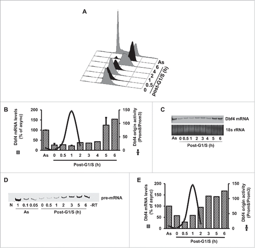
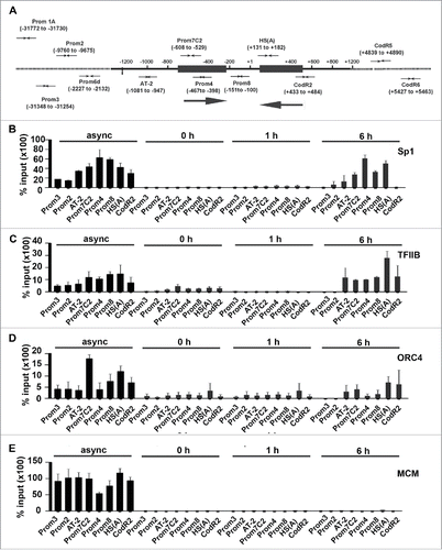
Figure 2. Transcription and DNA replication proteins localize to the Dbf4 locus in a cell cycle-dependent manner. (A) The physical map of the Dbf4 locus and primer locations within the region are shown. Numbers in parenthesis correspond to relative positions with respect to the “A” of the Dbf4 translation start codon, which is taken as +1. Replication initiation zones I and II are represented by black boxes, and directions of replication initiations are indicated by large black arrows. Primer sequences are shown in Table S1. (B, C) Transcription factors Sp1 and TFIIB are absent from the Dbf4 locus during DNA replication. ChIP analysis was performed using an antibody specific for Sp1 or TFIIB on extracts isolated from asynchronous HeLa S3 cells and cells synchronized at the G1/S border (0 h)-and-released for the indicated duration (1 h and 6 h). Resultant chromatin pull-down by anti-Sp1 or anti-TFIIB antibody was assayed by qPCR using indicated primer sets. Values represent mean ± SD, n>2. (D) Orc4 enrichment is diminished during S phase at the Dbf4 locus. ChIP was performed with an antibody specific for Orc4 as described above. Values represent mean ± SD, n > 2. (E) MCM is absent from the Dbf4 locus during G1-S phase. Lysates collected as above were analyzed using ChIP with antibodies mix of MCM proteins (Mcm 2/3/6/7). Values represent mean ± SD, n > 2.
DNA replication and transcription factors are bound to the Dbf4 origin-promoter locus in a cell cycle-dependent manner
In light of the observation that the levels of origin firing and transcription activities at the Dbf4 locus have an inverse relation, we determined the association of proteins involved in the regulation of DNA replication and gene transcription with chromatin at the locus. The protein-chromatin interactions of asynchronous and G1/S arrest-and-released cells were determined by chromatin immunoprecipitation (ChIP) and subsequently quantitative PCR (qPCR) with primer sets representing the sequence throughout the entire region ( and Table S1). Data from this experiment are summarized as follows: (i) The Sp1 transcription factor was strongly associated with the Dbf4 promoter region in asynchronous cells, as represented by Prom4, Prom7C2 and Prom8 (). However, data from synchronized cell population showed that Sp1 does not associate with the Dbf4 promoter region in the beginning of S phase (0–1 h post-G1/S, ). By 6 h post-G1/S, which coincides with cells' entry into G2, the association of Sp1 with the Dbf4 promoter locus was strong, particularly the Prom4 (replication zone I) and HS(A) (replication zone II) loci. (ii) A similar binding pattern was observed with the TFIIB transcription factor (). A notable difference between the binding patterns of Sp1 and TFIIB is that the latter showed slightly higher chromatin association in cells arrested at the G1/S boundary (0 h). In addition, the binding of TFIIB was extended to the down-stream of the translational start codon (+1 in ). In fact, its binding to the region represented by HS(A) primer was the highest within the entire Dbf4 locus by 6 h post-G1/S. This coincides with high Dbf4 transcription activity (). The lack of Sp1 and TFIIB at the Dbf4 promoter region in the beginning of S phase coincides with low transcription activity at the same time point.
We next examined the cell cycle-dependent enrichment of replication proteins associated with pre-RC at the chromatin. We focused on Orc4 and the MCM complex as we had previously shown them to be associated with the origin region.Citation39 We found that Orc4 enriched strongly at the chromatin flanking the promoter region (Prom7C2 and HS(A)) in asynchronous cells (). These regions were previously defined as replication zones I and II, respectively ().Citation39 The Orc4 enrichment at HS(A) was reduced by half in cells arrested at G1/S, and by ∼90% at Prom7C2, and remained low as the DNA in the region was replicated (). The association remained low until 6 h post-G1/S, when Orc4 levels began to increase, but did not return to the level observed in asynchronous cells. The difference may reflect the time it takes for Orc4 to associate with newly replicated DNA, as the ORC associates with origins at late M and early G1.
We next found that MCM was highly enriched throughout most of the Dbf4 promoter and non-promoter regions in asynchronous cells, with a marked reduction in the Sp1 binding site (). However, when we examined the association of MCM in the synchronous population, we did not see the enrichment of MCM at the Dbf4 locus during G1/S to G2 (0–6 h post-G1/S). The absence of MCM from the origin during S phase likely indicates that the helicase does not associate with origin or other specific sequence during this time (which is anticipated); however, the MCM complex would associate with origin (region) when a new pre-RC is formed in G1. Our data indicates that replication proteins, like transcription factors, associate with origin region in a cell-cycle dependent manner. Thus, the association and dissociation of replication and transcription factors with/from the Dbf4 locus are well-correlated with the replication and transcription activities shown in .
Cohesin and Mediator flank the Dbf4 promoter region in a cell cycle-dependent manner
To determine the regulatory mechanism behind the origin firing and gene transcrition dichotomy at the Dbf4 locus, we further characterized the chromatin landscape surrounding the Dbf4 origin-promoter region. Using a ChIP assay, we first determined the localization of 2 subunits of the cohesion complex, Smc1 and Smc3 (Structural Maintenance of Chromosomes 1 and 3).Citation43 For cells arrested at the G1/S border, Smc1 was strongly associated with both replication zones I and II (Prom4, HS(A)), the flanking region (CodR5), and upstream putative enhancer region represented by AT-2 primer.Citation39,40 Smc3 also showed strong association with origin and the flanking region at 0 h post-G1/S. By 1 h post-G1/S, both Smc1 and Smc3 were dissociated from replication zone I, although Smc1 still showed strong association with replication zone II (; summarized in Fig. S3).
Figure 3. Chromatin modifying proteins flank Dbf4 origin in a cell cycle-dependent manner. (A, B) Cohesin subunits, Smc1 and Smc3, are present at the Dbf4 locus in a cell cycle-dependent manner. ChIP assays were carried out with antibodies specific for Smc1 and Smc3 from cell extracts prepared from asynchronous HeLa S3 cells or those sampled at indicated time points post-G1/S. Chromatin was analyzed using qPCR with primer sets indicated. (C, D) The association of the Med1 and Med12 with the Dbf4 origin is cell cycle-dependent. ChIP assays were carried out with antibodies specific for Med1 or Med12 from extracts isolated from asynchronous HeLa S3 cells or those sampled at indicated time points post-G1/S. DNA bound by the protein was analyzed using qPCR with indicated primer sets. In all cases, percent input was determined by the amount of DNA enriched, less background, recovered by rabbit IgG (normal rabbit IgG), and then expressed the result relative to 6 ng input DNA sample. Values represent mean ± SD, n = 6. The entire data set is summarized in Figure S3.
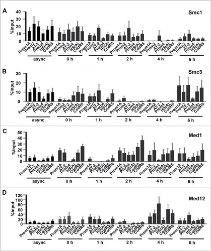
Unlike Smc1, Smc3 was not strongly associated with the enhancer (represented by AT-2) at the G1/S boundary. Smc3, like Smc1, showed strong association with the enhancer region at 1 h post-G1/S when replication initiation was very active (). The association of both Smc1 and Smc3 with CodR5 (far-downstream of the origin-promoter locus) was relatively strong until 1 h (Smc3) and 2 h (Smc1) post-G1/S. By 6 h post-G1/S (i.e., most cells are in G2), Smc3 was strongly associated with enhancer and replication zone II (HS(A)) and the downstream CodR5 region, but not with replication zone I.
We next examined the binding of the Mediator complex to the Dbf4 origin locus using 2 subunits, Med1 and Med12. In cells arrested at G1/S, Med1 proteins were strongly associated with enhancer (AT-2) and far-downstream from origin (i.e., CodR5 and 6), but not with the origin (; summarized in Fig. S3). By 1 h post-G1/S, however, the binding of Med1 to the entire region was minimal. By 2 h post-G1/S, its binding pattern became similar to that at G1/S, followed by more dynamic association patterns. Nevertheless, its binding to origin was always much lower than the flanking regions. This set of data is consistent with the notion that the formation of a loop by bringing the upstream (Prom1A & AT-2) and downstream (CodR5 & CodR6) regions together may negatively regulate origin activity but positively regulate transcription activity. The level of Med12 at the Dbf4 locus was generally low during early S phase (). However, high level of Med12 was found at replication zone I (Prom4) at 4–6 h post-G1/S. Since replication zone I overlaps the Dbf4 transcription initiation sites,Citation39,40 the strong presence of Med12 at the region represented by Prom4 during late S phase coincides with the strong activity of Dbf4 transcription and continuous suppression of replication ().
The Dbf4 origin is within a nucleosome-depleted area
To gain further insight into the co-regulation of DNA replication and gene transcription at the Dbf4 origin locus, we examined the nucleosome positions in the region. Replication origins tend to be located in nucleosome-poor regions as it is in the case of transcription promoters.Citation44 In fact, origins and promoters are often co-localized to the nucleosome-depleted regions (NDRs) within or around the 5′ end of genes.Citation4,45 As the existence of an NDR has already been identified within the Dbf4 origin-promoter locus,Citation39 we sought to further resolve the region with more primer sets. At the on-set of this experiment, asynchronous HeLa S3 cells were exposed to gradually increasing concentrations of DNase I, followed by isolation and quantification of DNA (8 ng from each sample) by qPCR (). The Prom3 primer set was derived from sequence in the eighth intron of the SLC25A40 gene (i.e., 31 kb upstream of Dbf4 origin) and served as a reference control. As expected, the amount of recovered DNA represented by Prom3 was changed relatively little between the minimum and maximum dose of enzyme used (0–50 U), indicating the intron locus includes normal nucleosome configuration. In contrast, 2 regions within the Dbf4 origin locus showed DNase I hypersensitivity: the regions represented by Prom4 and AT-2. The most dramatic decrease in DNA recovery was at the Prom4 region, which showed 96% reduction (i.e., 9.0 vs 0.4 ng) of the control by 50 units of DNase1 treatment. This region coincides with replication zone I and with the auxiliary promoter region, which is just downstream of the major RNA Pol II binding site (Fig. S1). The hypersensitive site was approximately 400 bp spanning from PromC7UC to Prom4C, and the sensitivity decreased in magnitude from Prom4 outward. This region covers almost the entire replication zone I. The other DNase 1 hypersensitive site (92% reduction at 50 units of DNase1) exists at AT-2, which lies at the most downstream portion of the Dbf4 enhancer.
Figure 4. The human Dbf4 origin-promoter locus contains a nucleosome-depleted region. (A) The region represented by prom4 is the most hypersensitive to DNase I digestion within the Dbf4 gene locus. HeLa S3 cells were treated with different concentrations of DNase I. An aliquot of 8 ng of chromatin isolates was quantitated by qPCR with various primer pairs indicated (all sequences were derived from chromosome 7). The non-origin sequence amplified by the Prom3 primer set represents a negative control.Citation39,40 Values represent mean ± standard error of the mean (SEM), n = 2–5. Values are presented on a logarithmic scale. (B) Histone H3 binding locations at the human Dbf4 locus. A ChIP assay with sonicated chromatin was carried out with asynchronous HeLa S3 cells using an antibody specific for histone H3. The resultant randomly fragmented DNA was subjected to quantification by qPCR. Percent input values was determined by subtracting output value recovered by normal (pre-immune) rabbit immunoglobulin G from H3 output value, then expressed the result relative to 6 ng input DNA sample. Values represent mean ± SD, n = 2. *P < 0.05; **P < 0.01 relative to Prom4 percent input. (C) Histone H3 binds to the non-origin control region. ChIP was performed as above for regions represented by Prom3, Prom4 and HS(A) primer sets. Values represent mean ± SD, n = 2. **P < 0.01. (D) Summary of results shown in . Putative nucleosome locations identified are indicated by gray hexagons.
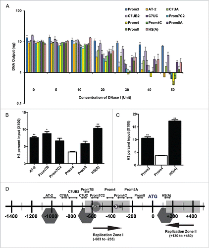
The two hypersensitive regions are separated by a region moderately resistant to DNase I digestion. The region represented by C7UB2 showed 77% reduction in DNA recovery at 50 units of DNase I, when compared to untreated control. Prom8A, located on the downstream edge of initiation zone I and encompassing the major transcription start site, is a region of local resistance to DNase I relative to its 2 nearest primer sets, Prom4C and Prom8. Primer HS(A) that represent the upstream border of initiation zone II exhibited a similar resistance to DNase I to that of Prom8A ().
To confirm our finding of NDRs by the DNase1 hypersensitivity assay, we determined the nucleosome positions using a ChIP assay. Asynchronous HeLa S3 cells were cross-linked with formaldehyde and processed for ChIP (). Relative to Prom4, histone H3 was significantly enriched at Prom7B (P < 0.05) and HS (A) (P < 0.01) as the latter being the greatest output for histone H3 with a 3.0-fold enrichment over Prom4. The regions represented by AT-2 (downstream boundary of enhancer) and Prom7C2 (< 25 bp downstream of Prom7B) also showed enrichment of histone H3, compared to Prom4. Similarly, Prom8 showed 1.7-fold enrichment of histone H3 over Prom4. To gain further insight, we determined if histone H3 is enriched at the region represented by Prom3, a non-origin region that is resistant to DNase I in a similar extent to the Prom7B region (, p < 0.05). In comparison, HS(A) is 50% higher than Prom3, but both regions were enriched above the level at Prom4, which we considered as background (, p < 0.05). Based on the data from the DNase I hypersensitivity and histone H3 ChIP assays, we concluded that 2 nucleosomes exist within the 1.2 kb origin locus, one each at the immediately upstream of initiation zones I and II (). In addition, one nucleosome exists at the downstream boundary of Dbf4 enhancer.
ψDbf410 does not contain an active origin
To gain a better understanding about the role of epigenetics in the regulation of origin activities, we looked for a suitable reference locus that could serve as a negative replication baseline. To that end we reasoned that a Dbf4 pseudogene locus could serve in this capacity as a certain pseudogene shows substantial nucleotide sequence identity without having an equivalent function. We, therefore, retrieved 3 pseudogene sequences by searching a pseudogene database (www.pseudogene.org) using the human Dbf4 Gene ID (ENSG00000006634) as query.Citation46 One Dbf4 pseudogene is mapped to chromosome 10 and 2 others are located on chromosome 2, which are termed here ψDbf410, ψDbf42A and ψDbf42B, respectively (Table S2). The ψDbf410 pseudogene includes all of the Dbf4 exons except exon 11, and showed the highest nucleotide sequence identity (98.2%) to that of the Dbf4 coding region (Table S2 and ). In addition, the nucleotide sequence of the 5′ non-coding region showed substantial identity between the Dbf4 promoter and the ψDbf410 upstream “non-coding” region. Based on this observation, we chose to use ψDbf410 as a baseline control.
Figure 5. ψDbf410 does not contain an origin of DNA replication. (A) Shown are physical maps of the human Dbf4 gene on chromosome 7 and a Dbf4 pseudogene on the chromosome 10 (ψDbf410). Filled or hatched boxes (E1 to E12) denote exons as determined from the nucleotide sequences reported previously (Accession #AF160249). The empty square box denotes the basal Dbf4 promoter.Citation41 Primer sets used for qPCR are shown as pairs of converging arrows. Numbers in brackets denote the positions of 5′ ends of primers relative to the Dbf4 translational start-codon (“A” of ATG is taken as +1). Single arrows at the left-top of the figure (labeled I, II and III) are primers used for standard PCR. Primer sequences are shown in Table S1. (B) The presence of ψDbf410 in the HeLa S3, HEK293 and 293T cell lines was confirmed by PCR-based mapping. Reactions were carried out using genomic DNA isolated from each cell line and the primers indicated (I to III). PCR products were separated by 1% agarose-gel electrophoresis. “L” and “NTC” denote DNA size ladder and no-template-control, respectively. PCR templates were isolated from cell lines indicated. HL denotes HeLa S3 cells. (C) Analysis of replication initiation at the ψDbf410. Nascent strand abundance assays were carried out with 1 SYMBOL 0150 /* MERGEFORMAT 2 kb newly synthesized single-stranded DNA templates isolated from asynchronous HeLa S3 cells and qPCR primer pairs shown (primer positions are shown in ). Relative sequence abundance was calculated using Prom3 sequence on chromosome 7 as reference.Citation39 The DNA segments amplified by Prom7B and HS(A) primer sets represent, respectively, replication initiation zones I and II at the Dbf4 origin locus (which is missing in ψDbf410),Citation39 and the region amplified by the primer sets Chr10 UPS is unique to ψDbf410. * denotes that enrichment of nascent strands at both Prom7B and HS(A) are significantly higher than the Chr10UPS, p < 0.05 for both (*** p < 0.001 for Prom7B) according to Dunnett's test with Chr10 UPS as control. (1-way ANOVA, p = 0.0003, F = 6.521, df = 26).
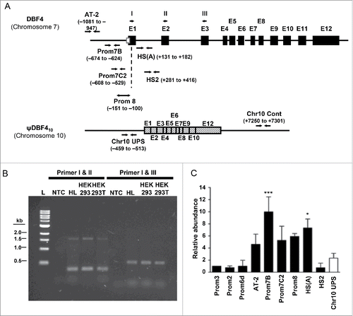
We first confirmed experimentally the existence of ψDbf410 in the human genome by PCR-based analysis using primers I-III ( and Table S2). The genomic DNA templates used in this experiment were prepared from HeLa S3, HEK293 and HEK293T cells. It should be noted that the nucleotide sequences of primers I-III, which were derived from exons 1–3, respectively, are identical between Dbf4 and ψDbf410. As shown in , the combination of primers I and II amplified ∼330 bp and ∼1.6 kb (lanes 3–4). The ∼1.6-kb PCR product was presumably amplified from the 1,603-bp segment spanning from the 5′ end of primer I located at −151 (i.e., 151 nucleotides upstream from the ATG translational start-codon) and the 5′ end of primer II located at +1,451 (i.e., 1,451 nucleotides downstream) from the Dbf4 template on chromosome 7 ( and Table S2). Likewise, the ∼330-bp PCR product was likely amplified from the 329-bp DNA segment between primers I and II on the ψDbf410 template. The difference in DNA size is because ψDbf410 does not contain the 1,274-bp intron 1 ().
The PCR reaction with the combination of primers I and III resulted in the amplification of a single DNA fragment of ∼530 bp (), which is close to the expected size of 529 bp amplified from the ψDbf410 locus. (Note that ψDbf410 does not contain 1,274-bp intron 1 and 6,753-bp intron 2) (). This primer set did not amplify the 8,556-bp DNA segment from the Dbf4 locus on chromosome 7, which contains introns 1 and 2 (). This result is not surprising since the PCR conditions used in this experiment would not efficiently amplify such a long DNA segment. From these data, we concluded the bona fide existence of ψDbf410 in the genomes of HeLa S3, HEK293, and HEK293T cell lines.
To determine whether an active origin exists at the ψDbf410 locus, a nascent strand abundance assay was carried out using a qPCR approach.Citation39,40 Consistent with our previous report,Citation39 the Dbf4 promoter region (represented by primers Prom7B, Prom7C2, Prom8 and HS(A)) contained an active origin (). In contrast, origin activity was not observed with the primer set representing the chromosome 10 upstream promoter sequence (Chr10 UPS) (). It should be noted that the primer sets Prom7C2 and Chr10 UPS represent similar positions relative to the ATG translational start-codon of Dbf4 and ψDbf410, respectively (). Taken together, our data demonstrates that no active origin is present at the ψDbf410 locus.
Both H3K9ac and H3K9me3 are present in nucleosomes at the Dbf4 origin
Since ψDbf410 does not contain an origin, we used it as a reference control against the Dbf4 origin in studying the regulation of origin in the context of chromatin structure and modifications. To compare protein binding patterns between the Dbf4 origin and ψDbf4 loci, a set of primers for the pseudogene locus was chosen to roughly correspond to those at the Dbf4 origin in terms of their location relative to the translation start site (). Of the 4 corresponding pairs of primers, only the Prom8-C10P8 pair, which is located within the first exon of the Dbf4 gene, exhibits any sequence similarity between chromosome 7 and chromosome 10; nonetheless, enough sequence dissimilarities exist between the 2 pairs that they are mutually exclusive in amplifying DNA by PCR.
Figure 6. Nucleosomes within the human Dbf4 origin region, but not those at corresponding locations relative to the Dbf4 pseudogene on chromosome 10 (ψDbf4), are subject to both acetylation and trimethylation on lysine 9 of histone H3 (H3K9ac and H3K9me3, respectively). (A) A schematic representation of corresponding primer locations at the human Dbf4 origin-promoter locus on chromosome 7 and the upstream of ψDbf4 ATG site on chromosome 10. For chromosome 7, shaded gray boxes represent replication initiation zones I and II, spotted rectangle represents Dbf4 exon 1, and black rectangle Dbf4 intron 1. For chromosome 10, shaded gray rectangles represent the first 3 Dbf4 exons located within the ψDbf4 region. The diagram shows from position 87,504,848 to 87,506,448 of chromosome 7, and from position 65,599,419 to 65,601,019 of chromosome 10. (B) The presence of histone H3 at Dbf4 origin and corresponding regions relative to ATG position at ψDbf4. ChIP was carried out in asynchronous HeLa S3 cells with an antibody specific for histone H3. The resultant pull-down DNA was subjected to quantification by qPCR, and normalized to total H3 ChIP values. (C) The presence of histone H3 post-translational modifications (left panel: H3K9ac; right panel: H3K9me3) at the Dbf4 origin (black) and at corresponding regions relative to ATG position at ψDbf4 (white). ChIP was carried out on asynchronous HeLa S3 cells with antibodies specific for H3K9ac or H3K9me3; pull-down DNA was subjected to quantification by qPCR, and normalized to total H3 ChIP values. Values represent mean ± SEM, n = 2. *P < 0.05; **P < 0.01 relative to corresponding primer set at Dbf4 pseudogene.
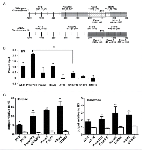
We first determined the nucleosome location in the pseudogene region to compare it with that in the Dbf4 origin locus. We found that the highest enrichment of histone H3 for both loci was found at a similar distance from the ATG, represented by Prom7C2 and C10UPS in the Dbf4 gene and pseudogene region, respectively (). However, the chromatin enrichment was 4-fold higher from the Dbf4 gene region versus the pseudogene region (p < 0.05, ). This could be due to differences between chromatin structures preventing the full accessibility of the H3 antibody, since the Dbf4 gene and ψDbf4 pseudogene are within euchromatin and heterochromatin regions, respectively.
A ChIP assay with sonicated chromatin was carried out with asynchronous HeLa S3 cells using antibodies specific for H3K9ac and H3K9me3. The outputs obtained from qPCR for H3K9ac and H3K9me3 are expressed as relative to the amount of histone H3 (). The Dbf4 origin on chromosome 7 was much more highly acetylated in an asynchronous cell population than was the corresponding region at ψDbf4 on chromosome 10. When the percent inputs at the 4 loci were compared, significant enrichment was observed at all Dbf4 primer sets, AT-2 (p<0.05), Prom8 (p < 0.01), Prom7C2 (p < 0.01), and HS(A) (p < 0.01) relative to their corresponding ψDbf4 primer sets (). Significant enrichment of H3K9me3 was also shown with Prom7C2, Prom8, and HS(A) over their corresponding regions on chromosome 10 (p < 0.01, p < 0.05, p < 0.05, respectively) (). The only exception was AT-2, which also showed enrichment over AT-10, but was not statistically significantly. These data indicate that both acetylation and trimethylation on H3 occur in the promoter region of Dbf4 at a higher level than the pseudogene locus.
Levels of H3K9ac and H3K9me3 fluctuate throughout the cell cycle
Because nucleosomes in the Dbf4 origin locus were both acetylated and trimethylated on lysine 9 of histone H3 in asynchronous cells, a time-course ChIP assay was performed to determine whether an evident switch from one modification to the other could be identified. Samples from G1/S synchronized cells were collected at scheduled time points. MNase, which digests inter-nucleosomal DNA, was used to reduce noisy background signals (which are often observed with sonicated DNA). Three primer sets were used for this experiment: HS(A) was chosen because it is both within the Dbf4 origin and presumed proximal to a nucleosome; Prom4 is also within the Dbf4 origin but not in proximity to a nucleosome and thus would represent the experimental background; finally, Prom3 was chosen as a nucleosome-containing, non-origin control.
When the background (Prom4)-subtracted values of outputs at HS(A) and Prom3 were compared, very little histone H3 acetylation was detected at the control location (i.e., Prom3) in asynchronous cells (). In contrast, highly significant enrichment of H3K9ac was observed at origin represented by Primer HS(A) (). Interestingly, the difference in the enrichment of histone acetylation between HS(A) and Prom3 was relatively insignificant at G1/S transition (0 h in ). However, a significant enrichment of histone acetylation was observed at the Dbf4 origin by 1 h post-G1/S, of which pattern persisted at least until 6 h (G2).
Figure 7. Post-translational modification of histone H3 on lysine 9 is cell cycle-regulated at the Dbf4 origin locus. (A) Shown is the presence of the H3K9ac histone H3 post-translational modifications at Dbf4 origin and non-origin loci in the function of cell cycle positions. Net output values were determined by subtracting output value recovered by an anti-H3K9ac antibody at a non-nucleosomal locus (i.e., Prom4 primer set) from nucleosome-containing primer sets, HS(A) (black bar) and Prom3 (white bar) representing Dbf4 origin and non-origin, respectively. (B) The presence of the H3K9me3 histone H3 post-translational modifications at Dbf4 origin and non-origin loci in the function of cell cycle positions. Values are calculated as in A. Values represent mean ± SD, n = 3. *P < 0.05; **P < 0.01 relative to Prom3 at each time point.
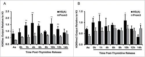
In asynchronous cells, there was significantly more histone H3 trimethylation detected at the origin than the non-origin control (). When following cells through the cell cycle by G1/S synchronization-and-release, the only significant enrichment of H3K9me3 at the origin relative to the histone H3 control occurred immediately following release from the double thymidine block (). Taken together, our data indicate that H3K9me3 is present during replication and H3K9 acetylation occurs when transcription is active.
The binding of KDM4A and HP1γ to Dbf4 origin fluctuates during cell cycle
The presence of H3K9me3 and its cell cycle-dependent fluctuation at the Dbf4 origin-promoter region were unexpected, since this histone modification is usually found in the heterochromatin region. To make sure this observation is not an artifact, we determined if the Dbf4 origin locus is enriched with KDM4A, a H3K9me3 specific de-trimethylase, and with H3K9me3-associated HP1 proteins.Citation47 We found that KDM4A was enriched at the origin compared to background in asynchronous HeLa S3 cells (). When examined cells synchronized at G1/S-and-release, KDM4A strongly enriched at the origin in G1/S-arrested cells (0 h). By 2 h post-G1/S, there was a substantial reduction in enrichment. There appeared a short burst of its localization at 4 h post-release, which was reduced again by 6 h post-G1/S (), at the time when most cells reached G2.
Figure 8. KDM4A and HP1γ are cell cycle-regulated at the Dbf4 origin-promoter locus. (A) KDM4A was within the Dbf4 origin region at G1/S and mid-S. ChIP was carried out with HeLa S3 cell with an antibody specific for KDM4A. The resultant pull-down DNA was then quantified by qPCR. Net output was determined by amount of DNA enriched, less background. Values represent mean ± SD, n = 2. (B) HP1γ is enriched at the Dbf4 origin locus for a short period after replication. Note that HS(A) and Prom2 represent origin and non-origin, respectively. Values were obtained as in panel A. Net output was determined by the amount of DNA enriched, less background. Values represent mean ± SD, n = 3 (* = p < 0.05).
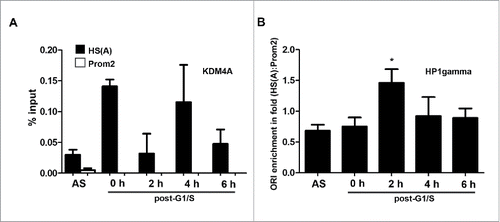
Finally, we examined the localization of the heterochromatin-associated HP1γ at the origin locus, as it was recently reported to associate also with the regulation of DNA replication.Citation27,28 We found that HP1γ was enriched at both Dbf4 origin and non-origin regions in asynchronous cells (). However, the enrichment at non-origin was approximately 1.4-fold higher in asynchronous cells (1.0 vs 0.7). In contrast, HP1γ was approximately 1.4-fold higher at origin than non-origin at 2 h post-G1/S (1.0 vs 1.4).
Discussion
It is generally understood that chromatin structure and chromatin-associated proteins regulate the timing of origin firing.Citation13-16 However, it is poorly understood how replication and transcription timing are co-regulated in origins localized at a promoter region. In the present study, we examined the relationship between replication and transcription during cell cycle at the Dbf4 model using high-resolution ChIP in a synchronized cell population. Data from this study indicate that the chromatin structure within and surrounding the Dbf4 origin-promoter locus plays an important regulatory role in determining the activation and suppression timing of origin and transcription. In particular, the post-translational modifications of the histone H3 on lysine 9 residues are bimodally regulated in a pattern coincident with replication and transcription activities: it is trimethylated early in S phase during DNA replication and acetylated during the high level of Dbf4 gene transcription later in S phase. This data is consistent with the notion that the underlying chromatin structure is primarily in a closed conformation in early S phase when vigorous DNA replication occurs. As acetylation increases post-DNA replication, the chromatin at this locus becomes more open conformation. This bimodal pattern is also seen with proteins associated with the same chromatin region, as it is largely unoccupied by major transcription factors such as Sp1 and TFIIB during DNA replication in early S phase, and then becomes associated with them later in S phase when transcription activity is high ().
The cohesin Smc1 protein was strongly associated with both enhancer and replication zone II during the 0–1 h post-G1/S. The binding pattern of Smc3 was somewhat different from that of Smc1: it showed strong association with replication zone II and CodR5 (i.e., far-downstream of origin) at G1/S; and associated with enhancer and CodR5, but not with origin, at 2 h post-G1/S (). Thus, both Smc1 and Smc3 are strongly associated with enhancer, but not with replication zone I when vigorous replication occurred. Interestingly, a substantial level of Smc1, but not Smc3, was observed at replication zone II at 1 h post-G1/S. At 2 h post-G1/S, both the cohesin proteins were absent from either replication zone. We previously showed that the initiation of DNA replication at the Dbf4 locus starts from replication zone I and proceeds in the “sense” direction (i.e., the direction of transcription) ().Citation32 The replication on the antisense direction initiated from zone II is delayed until the replication of sense strand reaches/passes through the zone II, of which phenomenon is termed asymmetric bidirectional replication (ABR).Citation39,40 The time-dependent dynamic binding pattern of cohesin proteins to replication zones I and II is consistent with the notion that cohesin proteins, especially Smc1, may be involved in the regulation of ABR.Citation39,40
We found that the MCM complex was largely absent from the origin during S phase (). This result is consistent with the previous finding that the MCM licensing factor is loaded onto origins at late M-early G1 phase and functions as a helicase during S phase, at which time it moves away from the origin on the leading edge of the replication complex.Citation48-50 Rather surprisingly, we found that the level of Orc4 was substantially reduced at the origin during DNA replication, with the thought that the ORC always remains at the origin. However, our result may not be necessarily unexpected. In Xenopus, the binding efficiency of the ORC is reduced upon licensing in a “licensing dependent origin inactivation” mechanism,Citation51 which reduces the association of the ORC with chromatin after origin licensing. In mammalian cells, Orc1 is removed from the origin after licensing but not Orc2.Citation52 It is possible from our data that Orc4 is also not as tightly bound to the chromatin upon licensing in a mechanism reminiscent of the “licensing dependent origin inactivation” seen in Xenopus. This interpretation is bolstered by the observation that the association of Orc4 with Orc2 and Orc3 are enhanced in the presence of Orc1, and diminished when Orc1 is broken down.Citation53 It is thus likely that the loosening of ORC after replication initiation leads to a reduced association of Orc4 with the chromatin.
The Med1 subunit of the Mediator complex was strongly associated with enhancer and far-downstream of the origin locus (CodR5 and CodR6) at 0 h and 2 h post-G1/S, but not when DNA replication is at the highest (1 h). Between 2–6 h post-G1/S, the presence of Med1 is very strong at far-downstream of origin. Interestingly, the presence of Med12 is the highest at replication zone I during 4–6 h post-G1/S. These binding patterns of Med1 and Med12 at different time post-G1/S are correlated to the ups and downs of DNA replication and Dbf4 transcription activities. In particular, the absence and presence of Med1 at the regions represented by AT-2 and CodR5/6 are directly correlated with the activation and suppression of DNA replication, respectively. Further, the binding pattern of Med12 to replication zone I is correlated with the suppression of replication initiation and simultaneous activation of transcription activities. The absence and presence of the Mediator complex at the enhancer, replication zone I and far-downstream region (CodR5/6) at different time post-G1/S may be directly relevant to the formation of dynamic chromatin loop structure, which is involved in the co-regulation of origin and transcription activities. Our data are also consistent with the model that vigorous Dbf4 gene transcription cannot start until DNA replication is completed, perhaps through at least the following 2 mechanisms: first, the presence of trimethylated H3K9 prevents the activating acetylation from occurring, which helps to maintain a closed chromatin and, second, through the absence of a loop to initiate the activation of transcription machinery via the Mediator complex.
Initially, it seemed paradoxical to have trimethylated H3K9 present at the Dbf4 origin, as it is generally found enriched at late-firing origins and heterochromatin.Citation26,54 However, genome-wide studies found that between 22–34% of early firing origins in HeLa S3 cells were enriched for H3K9me3 and at least 75% of late firing origins contained the modification,Citation54 suggesting that trimethylated H3K9 may play a role in regulating DNA replication beyond replication timing in a macro scale. In fact, recent evidence suggests that H3K9me3, KDM4A and HP1γ play a role in preventing DNA re-replication in heterochromatin as HP1 blocks re-replication while KDM4A promotes re-replication.Citation27,28 The multi-functional protein HP1 binds to H3K9me3;Citation47 this interaction is required for heterochromatin formation and gene silencing in many different organisms.Citation55-57 Our data also show that HP1 γ binds to Dbf4 origin in early S phase, suggesting that HP1γ may specifically regulate DNA replication at individual origins. Intriguingly, the localization of HP1γ to the origin region was the opposite to that of KDM4A as HP1γ was the highest when KDM4A was lowest (2 h post-G1/S in ). Therefore, our data indicate that the functional relationship of KDM4A and HP1γ can be extended to euchromatin. It is known that ORC also associates with H3K9me3 in heterochromatin,Citation58,59 and HP1 helps to recruit ORC to the heterochromatin.Citation60 Therefore, the enrichment of H3K9me at the Dbf4 origin-promoter locus may help facilitate the rebuilding of the ORC on the nascent strand while at the same time recruiting HP1γ to temporarily “heterochromatize” the origin region. This will directly or indirectly prevent other proteins, such as transcription factors, the reassembled cohesin complex and DNA polymerase from re-associating with the origin and acting as steric hindrance. Once the ORC has been rebuilt, histone H3K9 is demethylated and then acetylated to allow for the reformation of the cohesin complex and the Mediator complex. These complexes, in turn help facilitate Dbf4 gene transcription. Thus, our data is consistent with the notion that H3K9me3, KDM4A and HP1γ play a role in regulating chromatin architecture and DNA replication at euchromatin origins as they do at heterochromatin regions.Citation27,28
The activity of KDM4A at the Dbf4 locus also provides a possible mechanism for the cell cycle regulation of Dbf4 transcrition. We observed that Dbf4 transcription remains low during M-G1-the beginning of S phase ( and S2), which coincides with the formation and licensing of pre-RC. Thus, the pre-RC may occupy the Dbf4 origin-promoter locus, preventing transcription factors from associating with the chromatin at the locus.
It should be noted that it is not possible to determine the replication event of the entire cell population with perfect synchrony. Therefore, the inverse relation between DNA replication and transcription activities observed from the Dbf4 locus is a “collective” of numerous cells at slightly different stages of cell cycle positions. Therefore, if we can capture the perfect moment of replication and transcription events in a single cell, the 2 events are likely mutually exclusive. This notion is strengthened by the fact that DNA replication from zone II occurs in the opposite direction of Dbf4 transcription.Citation39,40 Our interpretation is also consistent with the postulation that the replication at early origins is “temporally separated” from transcription.Citation61
Taken together, our data from Dbf4 origin-promoter model show that the replication and transcription is co-regulated by several layers of different regulatory mechanisms that are involved in post-translational modifications of histone proteins, nucleosome localization/depletion, and the association and dissociation of replication and transcription factors with/from specific sites within and the surrounding region of the origin locus. This complicated but elegant regulation is also coordinated with the formation of chromatin loops in a space and cell-cycle dependent manner. Thus, although very complex and dynamic, a cell employs a very sophisticated and extremely well-coordinated mechanism to seamlessly co-regulate replication and transcription at the origin/promoter locus.
Materials and methods
Cell technique
HeLa S3 cells were maintained and synchronized as described previously.Citation39 Cell cycle distribution was determined by flow cytometry using a Beckman Coulter FC500 flow cytometer as described previously.Citation62
Chromatin Immunoprecipitation (ChIP) and quantitative polymerase chain reaction (qPCR)
ChIP was carried out as described previously,Citation39 with the following modifications. For micrococcal nuclease (MNase) treated ChIP samples, nuclei were digested with MNase prior to sonication. Antibodies used were: histone H3 (Abcam rabbit polyclonal, ab1791, or Santa Cruz rabbit polyclonal, sc-10809), normal rabbit IgG (Santa Cruz, sc-2027), H3K9ac (Cell Signaling rabbit monoclonal, 9671), H3K9me3 (Cell Signaling rabbit monoclonal, 9754), Smc3 (Abcam rabbit polyclonal, ab9263), Smc1 (Abcam rabbit polyclonal, ab 9262), Med1 (Bethyl Labs, rabbit polyclonal A300-793A) Med12 ((Bethyl Labs, rabbit polyclonal, A300-774A), JMJD2A/KDM4A (Santa Cruz, mouse monoclonal, sc-81302), HP1γ (rabbit polyclonal, ab10480) and GFP (Santa Cruz, mouse monoclonal, sc-9996)
Deoxyribonuclease (DNase) I hypersensitivity assay
The sensitivity to DNase I digestion was determined as previously described,Citation39 with some modifications. Briefly, controlled DNase I digestion was carried out in DNase I Reaction Buffer (10 mM Tris pH 7.6, 2.5 mM magnesium chloride, 0.5 mM calcium chloride) as follows: 100 µL of the suspension was added to tubes containing from 0 to 50 units (U) DNase I. The tubes were vortexed briefly, and then incubated for 10 min in a water bath at 37°C. The reaction was stopped by adding 125 µL DNase Stop Solution (20 mM Tris pH 8.0, 10 mM EG1/SA pH 8.0, 600 mM sodium chloride, 1% w/v SDS). Each DNA sample was diluted to 8 ng/6 µL concentration, and specific DNA segment was quantified by qPCR.
Quantitative polymerase chain reaction (qPCR), PCR-based amplification of genomic DNA, and nascent strand abundance assay
qPCR was carried out using an ABI Prism 7900 HT sequence detection system as described previously.Citation39 Primer sequences and genomic locations are shown in Table S1. All primer pairs were designed using the Primer Express software (Applied Biosystems). Dissociation curve analysis was performed after each PCR reaction to confirm the presence of a single amplification product. Amplicon size was also verified by gel electrophoresis. Genomic DNA was isolated using a rapid salt-extraction protocol described in ref. Citation63. Newly synthesized, short, single-stranded DNA was isolated by alkaline gel electrophoresis as described previously.Citation39,42,64 An equal volume of nascent DNA preparation was used for subsequent qPCR-based analyses as described previously.Citation39,42
Abbreviations
| ABR | = | asymmetric bidirectional replication |
| ChIP | = | chromatin immunoprecipitation |
| Chr | = | chromosome |
| DBF4 | = | dumbbell former 4 |
| ψDBF410 | = | DBF4 pseudogene on chromosome 10 |
| ψDBF42A and ψDBF42B | = | DBF4 pseudogenes on chromosome 2 |
| DNase | = | deoxyribonuclease |
| H3K9ac | = | histone H3 acetyl lysine 9 |
| H3K9me3 | = | histone H3 trimethyl lysine 9 |
| HP1 | = | heterochromatin protein 1 |
| KDM4A | = | Lysine (K)-Specific Demethylase 4A |
| MCB | = | Mlu 1-cell cycle box |
| Med | = | mediator |
| NDR | = | nucleosome-depleted region |
| origin | = | origin of DNA replication |
| ORC | = | Origin Recognition Complex |
| PTM | = | post-translational modification |
| qPCR | = | quantitative-polymerase chain reaction |
| SMC | = | Structural Maintenance of Chromosomes |
| SUV39H1 | = | Suppressor Of Variegation 3–9 Homolog 1 |
| TFIIB | = | Transcription Factor II B |
| UPS | = | upstream promoter sequence |
Disclosure of potential conflicts of interest
No potential conflicts of interest were disclosed.
Supplemental Files
Download PDF (197.8 KB)Funding
This work was supported by grants from the Canadian Institutes of Health Research (CIHR #79473) and Natural Sciences and Engineering Council of Canada (NSERC #203528) to HL. JR and JK were supported in part by the Cancer Care Ontario Peter Crossgrove postdoctoral fellowship. IKSL is a recipient of an Ontario Trillium Scholarship.
References
- Sclafani RA, Holzen TM. Cell cycle regulation of DNA replication. Annu Rev Genet 2007; 41:237-80; PMID:17630848; http://dx.doi.org/10.1146/annurev.genet.41.110306.130308
- Martin MM, Ryan M, Kim R, Zakas AL, Fu H, Lin CM, Reinhold WC, Davis SR, Bilke S, Liu H, et al. Genome-wide depletion of replication initiation events in highly transcribed regions. Genome Res 2011; 21:1822-32; PMID:21813623; http://dx.doi.org/10.1101/gr.124644.111
- Hiratani I, Takebayashi S, Lu J, Gilbert DM. Replication timing and transcriptional control: beyond cause and effect–part II. Curr Opin Genet Dev 2009; 19:142-9; PMID:19345088; http://dx.doi.org/10.1016/j.gde.2009.02.002
- Mechali M. Eukaryotic DNA replication origins: many choices for appropriate answers. Nat Rev Mol Cell Biol 2010; 11:728-38; PMID:20861881; http://dx.doi.org/10.1038/nrm2976
- Guillou E, Ibarra A, Coulon V, Casado-Vela J, Rico D, Casal I, Schwob E, Losada A, Mendez J. Cohesin organizes chromatin loops at DNA replication factories. Genes Dev 2010; 24:2812-22; PMID:21159821; http://dx.doi.org/10.1101/gad.608210
- Jiang H, Peterlin BM. Differential chromatin looping regulates CD4 expression in immature thymocytes. Mol Cell Biol 2008; 28:907-12; PMID:18039856; http://dx.doi.org/10.1128/MCB.00909-07
- Miele A, Dekker J. Long-range chromosomal interactions and gene regulation. Mol Biosyst 2008; 4:1046-57; PMID:18931780; http://dx.doi.org/10.1039/b803580f
- Vakoc CR, Letting DL, Gheldof N, Sawado T, Bender MA, Groudine M, Weiss MJ, Dekker J, Blobel GA. Proximity among distant regulatory elements at the beta-globin locus requires GATA-1 and FOG-1. Mol Cell 2005; 17:453-62; PMID:15694345; http://dx.doi.org/10.1016/j.molcel.2004.12.028
- Conaway RC, Sato S, Tomomori-Sato C, Yao T, Conaway JW. The mammalian Mediator complex and its role in transcriptional regulation. Trends Biochem Sci 2005; 30:250-5; PMID:15896743; http://dx.doi.org/10.1016/j.tibs.2005.03.002
- Kornberg RD. Mediator and the mechanism of transcriptional activation. Trends Biochem Sci 2005; 30:235-9; PMID:15896740; http://dx.doi.org/10.1016/j.tibs.2005.03.011
- Malik S, Roeder RG. Dynamic regulation of pol II transcription by the mammalian Mediator complex. Trends Biochem Sci 2005; 30:256-63; PMID:15896744; http://dx.doi.org/10.1016/j.tibs.2005.03.009
- Taatjes DJ. The human Mediator complex: a versatile, genome-wide regulator of transcription. Trends Biochem Sci 2010; 35:315-22; PMID:20299225; http://dx.doi.org/10.1016/j.tibs.2010.02.004
- Evertts AG, Coller HA. Back to the origin: reconsidering replication, transcription, epigenetics, and cell cycle control. Genes cancer 2012; 3:678-96; PMID:23634256; http://dx.doi.org/10.1177/1947601912474891
- Mechali M, Yoshida K, Coulombe P, Pasero P. Genetic and epigenetic determinants of DNA replication origins, position and activation. Curr Opin Genet Dev 2013; 23:124-31; PMID:23541525; http://dx.doi.org/10.1016/j.gde.2013.02.010
- Sherstyuk VV, Shevchenko AI, Zakian SM. Epigenetic landscape for initiation of DNA replication. Chromosoma 2014; 123:183-99; PMID:24337246; http://dx.doi.org/10.1007/s00412-013-0448-3
- Hyrien O. Peaks cloaked in the mist: the landscape of mammalian replication origins. J Cell Biol 2015; 208:147-60; PMID:25601401; http://dx.doi.org/10.1083/jcb.201407004
- Lima-de-Faria A, Jaworska H. Late DNA synthesis in heterochromatin. Nature 1968; 217:138-42; PMID:18300398; http://dx.doi.org/10.1038/217138a0
- Tanaka S, Nakato R, Katou Y, Shirahige K, Araki H. Origin association of Sld3, Sld7, and Cdc45 proteins is a key step for determination of origin-firing timing. Curr Biol 2011; 21:2055-63; PMID:22169533; http://dx.doi.org/10.1016/j.cub.2011.11.038
- Ehrenhofer-Murray AE. Chromatin dynamics at DNA replication, transcription and repair. Eur J Biochem 2004; 271:2335-49; PMID:15182349; http://dx.doi.org/10.1111/j.1432-1033.2004.04162.x
- Rice JC, Allis CD. Histone methylation versus histone acetylation: new insights into epigenetic regulation. Curr Opin Cell Biol 2001; 13:263-73; PMID:11343896; http://dx.doi.org/10.1016/S0955-0674(00)00208-8
- Kouzarides T. Chromatin modifications and their function. Cell 2007; 128:693-705; PMID:17320507; http://dx.doi.org/10.1016/j.cell.2007.02.005
- Li Q, Zhang Z. Linking DNA replication to heterochromatin silencing and epigenetic inheritance. Acta Biochim Biophys Sin (Shanghai) 2012; 44:3-13; PMID:22194009; http://dx.doi.org/10.1093/abbs/gmr107
- Strahl BD, Allis CD. The language of covalent histone modifications. Nature 2000; 403:41-5; PMID:10638745; http://dx.doi.org/10.1038/47412
- Bannister AJ, Kouzarides T. Regulation of chromatin by histone modifications. Cell Res 2011; 21:381-95; PMID:21321607; http://dx.doi.org/10.1038/cr.2011.22
- Ding Q, MacAlpine DM. Defining the replication program through the chromatin landscape. Crit Rev Biochem Mol Biol 2011; 46:165-79; PMID:21417598; http://dx.doi.org/10.3109/10409238.2011.560139
- Barski A, Cuddapah S, Cui K, Roh TY, Schones DE, Wang Z, Wei G, Chepelev I, Zhao K. High-resolution profiling of histone methylations in the human genome. Cell 2007; 129:823-37; PMID:17512414; http://dx.doi.org/10.1016/j.cell.2007.05.009
- Black JC, Allen A, Van RC, Forbes E, Longworth M, Tschop K, Rinehart C, Quiton J, Walsh R, Smallwood A, et al. Conserved antagonism between JMJD2A/KDM4A and HP1gamma during cell cycle progression. Mol Cell 2010; 40:736-48; PMID:21145482; http://dx.doi.org/10.1016/j.molcel.2010.11.008
- Black JC, Manning AL, Van RC, Kim J, Ladd B, Cho J, Pineda CM, Murphy N, Daniels DL, Montagna C, et al. KDM4A lysine demethylase induces site-specific copy gain and rereplication of regions amplified in tumors. Cell 2013; 154:541-55; PMID:23871696; http://dx.doi.org/10.1016/j.cell.2013.06.051
- Struhl K. Histone acetylation and transcriptional regulatory mechanisms. Genes Dev 1998; 12:599-606; PMID:9499396; http://dx.doi.org/10.1101/gad.12.5.599
- Wolffe AP, Hayes JJ. Chromatin disruption and modification. Nucleic Acids Res 1999; 27:711-20; PMID:9889264; http://dx.doi.org/10.1093/nar/27.3.711
- Wang Z, Schones DE, Zhao K. Characterization of human epigenomes. Curr Opin Genet Dev 2009; 19:127-34; PMID:19299119; http://dx.doi.org/10.1016/j.gde.2009.02.001
- Negre N, Brown CD, Ma L, Bristow CA, Miller SW, Wagner U, Kheradpour P, Eaton ML, Loriaux P, Sealfon R, et al. A cis-regulatory map of the Drosophila genome. Nature 2011; 471:527-31; PMID:21430782; http://dx.doi.org/10.1038/nature09990
- Labib K. How do Cdc7 and cyclin-dependent kinases trigger the initiation of chromosome replication in eukaryotic cells? Genes Dev 2010; 24:1208-19; PMID:20551170; http://dx.doi.org/10.1101/gad.1933010
- Cheng L, Collyer T, Hardy CF. Cell cycle regulation of DNA replication initiator factor Dbf4p. MolCell Biol 1999; 19:4270-8; PMID:10330168
- Ferreira MF, Santocanale C, Drury LS, Diffley JF. Dbf4p, an essential S phase-promoting factor, is targeted for degradation by the anaphase-promoting complex. MolCell Biol 2000; 20:242-8; PMID:10594027
- Oshiro G, Owens JC, Shellman Y, Sclafani RA, Li JJ. Cell cycle control of Cdc7p kinase activity through regulation of Dbf4p stability. MolCell Biol 1999; 19:4888-96; PMID:10373538
- Weinreich M, Stillman B. Cdc7p-Dbf4p kinase binds to chromatin during S phase and is regulated by both the APC and the RAD53 checkpoint pathway. EMBO J 1999; 18:5334-46; PMID:10508166; http://dx.doi.org/10.1093/emboj/18.19.5334
- Knockleby J, Kim BJ, Mehta A, Lee H. Cdk1-mediated phosphorylation of Cdc7 suppresses DNA re-replication. Cell Cycle 2016; 15:1494-505; PMID:27105124; http://dx.doi.org/10.1080/15384101.2016.1176658
- Romero J, Lee H. Asymmetric bidirectional replication at the human DBF4 origin. Nat Struct Mol Biol 2008; 15:722-9; PMID:18536724; http://dx.doi.org/10.1038/nsmb.1439
- Lee H, Romero J. Origin of DNA replication at the human lamin B2 locus: OBR or ABR? Cell Cycle 2012; 11:4281-3; PMID:22983127; http://dx.doi.org/10.4161/cc.21992
- Wu X, Lee H. Human Dbf4/ASK promoter is activated through the Sp1 and MluI cell-cycle box (MCB) transcription elements. Oncogene 2002; 21:7786-96; PMID:12420215; http://dx.doi.org/10.1038/sj.onc.1205914
- Romero J, Lee H. One-way PCR-based mapping of a replication initiation point (RIP). Nat Protoc 2008; 3:1729-35; PMID:18927558; http://dx.doi.org/10.1038/nprot.2008.173
- Peters JM, Tedeschi A, Schmitz J. The cohesin complex and its roles in chromosome biology. Genes Dev 2008; 22:3089-114; PMID:19056890; http://dx.doi.org/10.1101/gad.1724308
- Dorn ES, Cook JG. Nucleosomes in the neighborhood: new roles for chromatin modifications in replication origin control. Epigenetics 2011; 6:552-9; PMID:21364325; http://dx.doi.org/10.4161/epi.6.5.15082
- Valenzuela MS, Chen Y, Davis S, Yang F, Walker RL, Bilke S, Lueders J, Martin MM, Aladjem MI, Massion PP, et al. Preferential localization of human origins of DNA replication at the 5′-ends of expressed genes and at evolutionarily conserved DNA sequences. PLoS One 2011; 6:e17308; PMID:21602917; http://dx.doi.org/10.1371/journal.pone.0017308
- Zhang Z, Harrison PM, Liu Y, Gerstein M. Millions of years of evolution preserved: a comprehensive catalog of the processed pseudogenes in the human genome. Genome Res 2003; 13:2541-58; PMID:14656962; http://dx.doi.org/10.1101/gr.1429003
- Maison C, Almouzni G. HP1 and the dynamics of heterochromatin maintenance. Nat Rev Mol Cell Biol 2004; 5:296-304; PMID:15071554; http://dx.doi.org/10.1038/nrm1355
- Tye BK. MCM proteins in DNA replication. Ann Rev Biochem 1999; 68:649-86; PMID:10872463; http://dx.doi.org/10.1146/annurev.biochem.68.1.649
- Liang C, Stillman B. Persistent initiation of DNA replication and chromatin-bound MCM proteins during the cell cycle in cdc6 mutants. Genes Dev 1997; 11:3375-86; PMID:9407030; http://dx.doi.org/10.1101/gad.11.24.3375
- Kuipers MA, Stasevich TJ, Sasaki T, Wilson KA, Hazelwood KL, McNally JG, Davidson MW, Gilbert DM. Highly stable loading of Mcm proteins onto chromatin in living cells requires replication to unload. J Cell Biol 2011; 192:29-41; PMID:21220507; http://dx.doi.org/10.1083/jcb.201007111
- Rowles A, Tada S, Blow JJ. Changes in association of the Xenopus origin recognition complex with chromatin on licensing of replication origins. J Cell Sci 1999; 112(Pt 12):2011-8; PMID:10341218
- Li CJ, DePamphilis ML. Mammalian Orc1 protein is selectively released from chromatin and ubiquitinated during the S-to-M transition in the cell division cycle. Mol Cell Biol 2002; 22:105-16; PMID:11739726; http://dx.doi.org/10.1128/MCB.22.1.105-116.2002
- Ohta S, Tatsumi Y, Fujita M, Tsurimoto T, Obuse C. The ORC1 cycle in human cells: II. Dynamic changes in the human ORC complex during the cell cycle. J Biol Chem 2003; 278:41535-40; PMID:12909626; http://dx.doi.org/10.1074/jbc.M307535200
- Picard F, Cadoret JC, Audit B, Arneodo A, Alberti A, Battail C, Duret L, Prioleau MN. The spatiotemporal program of DNA replication is associated with specific combinations of chromatin marks in human cells. PLoS Genet 2014; 10:e1004282; PMID:24785686; http://dx.doi.org/10.1371/journal.pgen.1004282
- Bannister AJ, Zegerman P, Partridge JF, Miska EA, Thomas JO, Allshire RC, Kouzarides T. Selective recognition of methylated lysine 9 on histone H3 by the HP1 chromo domain. Nature 2001; 410:120-4; PMID:11242054; http://dx.doi.org/10.1038/35065138
- Lachner M, O'Carroll D, Rea S, Mechtler K, Jenuwein T. Methylation of histone H3 lysine 9 creates a binding site for HP1 proteins. Nature 2001; 410:116-20; PMID:11242053; http://dx.doi.org/10.1038/35065132
- Nakayama J, Rice JC, Strahl BD, Allis CD, Grewal SI. Role of histone H3 lysine 9 methylation in epigenetic control of heterochromatin assembly. Science 2001; 292:110-3; PMID:11283354; http://dx.doi.org/10.1126/science.1060118
- Bartke T, Vermeulen M, Xhemalce B, Robson SC, Mann M, Kouzarides T. Nucleosome-interacting proteins regulated by DNA and histone methylation. Cell 2010; 143:470-84; PMID:21029866; http://dx.doi.org/10.1016/j.cell.2010.10.012
- Vermeulen M, Eberl HC, Matarese F, Marks H, Denissov S, Butter F, Lee KK, Olsen JV, Hyman AA, Stunnenberg HG, et al. Quantitative interaction proteomics and genome-wide profiling of epigenetic histone marks and their readers. Cell 2010; 142:967-80; PMID:20850016; http://dx.doi.org/10.1016/j.cell.2010.08.020
- Chakraborty A, Shen Z, Prasanth SG. “ORCanization” on heterochromatin: linking DNA replication initiation to chromatin organization. Epigenetics 2011; 6:665-70; PMID:21586903; http://dx.doi.org/10.4161/epi.6.6.16179
- Meryet-Figuiere M, Alaei-Mahabadi B, Ali MM, Mitra S, Subhash S, Pandey GK, Larsson E, Kanduri C. Temporal separation of replication and transcription during S-phase progression. Cell Cycle 2014; 13:3241-8; PMID:25485504; http://dx.doi.org/10.4161/15384101.2014.953876
- Solomon VR, Hu C, Lee H. Hybrid pharmacophore design and synthesis of isatin-benzothiazole analogs for their anti-breast cancer activity. Bioorg Med Chem 2009; 17:7585-92; PMID:19804979; http://dx.doi.org/10.1016/j.bmc.2009.08.068
- Aljanabi SM, Martinez I. Universal and rapid salt-extraction of high quality genomic DNA for PCR-based techniques. Nucleic Acids Res 1997; 25:4692-3; PMID:9358185; http://dx.doi.org/10.1093/nar/25.22.4692
- Kamath S, Leffak M. Multiple sites of replication initiation in the human beta-globin gene locus. Nucleic Acids Res 2001; 29:809-17; PMID:11160905; http://dx.doi.org/10.1093/nar/29.3.809