
Abstract
Chronic obstructive pulmonary disease (COPD) is one of the most prevalent and severe diseases worldwide with high societal and health care costs. The pathogenesis of COPD is very complicated, and no curative treatment is available. Cellular senescence promotes the development of COPD. Type II alveolar epithelial cells (AECII) play a momentous role in lung tissue repair and maintenance of alveolar homeostasis. Sirtuin 1 (SIRT1), an antiaging molecule involved in the response to chronic inflammation and oxidative stress, regulates many pathophysiological changes including stress resistance, apoptosis, inflammation, and cellular senescence. This study aimed to investigate whether the pharmacological SIRT1 activator SRT2104 protects against AECII senescence in rats with emphysema. Our findings confirmed that SRT2104 administration reduced the pathological characteristics of emphysema and improved lung function parameters, including pulmonary resistance, pulmonary dynamic compliance, and peak expiratory flow. Moreover, SRT2104 treatment upregulated the expression of surfactant proteins A and C, SIRT1, and forkhead box O 3a (FoxO3a), decreased senescence-associated-β-galactosidase (SA-β-gal) activity, increased SIRT1 deacetylase activity, and downregulated the levels of p53 and p21. Therefore, SRT2104 administration protected against AECII senescence in rats with emphysema via SIRT1/FoxO3a and SIRT1/p53 signaling pathways and may provide a novel potential therapeutic strategy for COPD.
Introduction
Chronic obstructive pulmonary disease (COPD) is considered to be an important global public health challenge, which is projected to be the third leading cause of death in the world by 2020. In 2020, more than 3 million people died of COPD worldwide, accounting globally for 6% of all deaths [Citation1]. The pathogenesis of COPD is very complicated and remains to be fully elucidated. A major characteristic of COPD is emphysema, which is the progressive loss of alveolar tissue, enlargement of airspace, and destruction of the lung parenchyma [Citation2]. Sundar et al. revealed that cellular senescence in lung epithelial cells plays a vital role in the pathogenesis and development of COPD caused by cigarette smoking [Citation3]. However, there is no curative treatment to reverse the decline in lung function and prevent the destruction of alveolar tissue in patients with COPD [Citation1]. In mammals, lung alveolar epithelial cells are composed of type I alveolar epithelial cells (AECI) and type II alveolar epithelial cells (AECII). Being AECI progenitors, AECII can supplement their own quantity by mitosis, synthesize and secrete pulmonary surfactant proteins (SPs), maintain alveolar homeostasis, and improve lung tissue repair [Citation4,Citation5].
Sirtuin 1 (SIRT1), an NAD+-dependent class III histone deacetylase, is a vital member of the sirtuin proteins family [Citation6]. As a well-characterized member of this family, SIRT1 controls in mammals various processes implicated in the regulation of homeostasis, plays an important role in governing cellular stress management, and has been implicated in a healthy lifespan [Citation7,Citation8]. The SIRT1 protein deacetylates histones, forkhead box O 3a (FoxO3a), and p53, regulating inflammation, stress resistance, apoptosis, and cellular senescence pathways [Citation9–11]. FoxO3a and p53 are non-histone substrates of SIRT1. The latter activates the transcription of p21, resulting in the development of cellular senescence [Citation12,Citation13].
As a pharmacological activator of SIRT1, SRT2104 exhibits therapeutic effects in several diseases such as osteoarthritis [Citation14], depressive-like behaviors [Citation15], nephropathy [Citation16], cardiac fibrosis [Citation17], diabetes-induced aortic endothelial dysfunction [Citation18], psoriasis [Citation19], and inflammation [Citation20].
It has yet to be clarified whether SRT2104 can alleviate lung injury in rats with emphysema via suppression of ACEII senescence. Therefore, we systematically evaluated in this study the effects of SRT2104 treatment in a rat model of emphysema. We found that SRT2104 protected against AECII senescence via SIRT1-mediated FoxO3a and p53 signaling pathways.
Materials and methods
Animal model and SRT2104 administration
Sixty male Sprague-Dawley rats (body weight: 220–240 g) were purchased from Shanghai SLAC Laboratory Animal Co., Ltd. (Shanghai, China) and maintained in the Laboratory Animal Center of Zhejiang Chinese Medical University (Hangzhou, China). All rats were housed at 22–25 °C with 40%–60% relative humidity and a 12-hour light/dark cycle with water and food ad libitum. All animal protocols were approved by the Institutional Animal Care and Use Committee of Zhejiang Chinese Medical University.
The rats were randomly divided into three groups (20 rats/group), i.e., the control group (group A), emphysema group (group B), and emphysema + SRT2104 group (group C). A custom-designed cigarette smoking chamber [Citation2] and lipopolysaccharide (LPS; Sigma, St. Louis, MO, USA) stimuli were used for generating the rat model of emphysema [Citation21]. Commercially available cigarettes (XiongShi; China Tobacco Zhejiang Industrial Co., Ltd., China) containing 0.7 mg of nicotine and 8.0 mg of tar per cigarette were used in this study. We performed the cigarette smoke exposure and intratracheal LPS instillation as previously described [Citation22]. The control rats in group A received clean air throughout the entire experimental period.
After the second intratracheal LPS instillation, each rat in group C was treated daily 1 hour prior to smoke exposure with SRT2104 (100 mg/kg body weight; MedChem Express Co., Ltd., Shanghai, China) through oral gavage for 4 weeks, as described previously [Citation23].
Lung function examination
Fifteen rats in each group were randomly chosen to measure their lung function parameters, including pulmonary resistance (PR), pulmonary dynamic compliance (Cdyn), and peak expiratory flow (PEF), 24 hours after the last cigarette smoke exposure, as described previously [Citation22]. Briefly, the rats were continuously anesthetized and tracheostomized using an inverted “T” incision. The Y-shaped endotracheal trachea was cannulated, and the esophageal cannula was inserted into the middle of the esophagus. After that, the endotracheal cannula was connected to the micro-pressure transducer and the respiratory flow transducer to record airway pressure and respiratory flow, respectively, and the esophageal cannula was connected to the pressure transducer to measure the esophageal pressure reflecting the negative pressure in the pleural space. After stabilization, the signals were recorded continuously for 10 minutes. The parameters of PR, Cdyn, and PEF were analyzed using the MPA lung function system (Shanghai Alcott Biotech Co., Ltd., Shanghai, China) for 100 respiratory cycles. After this lung function examination, rats were sacrificed by intraperitoneal injection of 10.0% chloral hydrate. The right lungs were removed and fixed in 4% paraformaldehyde for histological examination, immunohistochemical staining, and senescence-associated-β-galactosidase activity assay. The left lungs were stored at –80 °C for PCR, western blotting, and SIRT1 deacetylase activity assay.
Histological examinations
The right lungs were immersed in paraformaldehyde for 24 hours, embedded in paraffin, and cut into 4-μm thick sections. Three discontinuous paraffin-embedded sections from each lung sample were stained with hematoxylin and eosin (H&E) to assess the morphological changes in the lungs. Five fields of view in each of the three sections from each lung sample were examined using a light microscope (Olympus, Tokyo, Japan). The mean linear intercept (MLI) and the mean alveolar airspace (MAA) were determined. Under the light microscope, we projected a crossline reticle of known length “L” onto the center of each viewing field and counted the number of alveolar septum intercepts as “Ns” to calculate the mean linear intercept using the formula MLI = L/Ns. Similarly, we counted the number of pulmonary alveoli in each field of vision as “Na”. Then, we measured the gross area in this viewing field and the HE-positive area as “TA” and “PA,” respectively, to determine the mean alveolar airspace according to the formula MAA = (TA – PA)/Na. The MLI indicates the average distance between opposing walls of a single alveolus and is a measure of pulmonary airspace enlargement. The parameter MAA is a different measure of pulmonary airspace enlargement [Citation2,Citation24].
Senescence-associated-β-galactosidase activity assay
The senescence-associated-β-galactosidase (SA-β-gal) activity was detected using an in situ β-galactosidase staining kit (Beyotime Institute of Biotechnology, Shanghai, China) according to the manufacturer’s protocol. Briefly, lung tissues were fixed in β-galactosidase stationary solution for 15 minutes. After three times washing for 10 minutes each in phosphate-buffered saline (PBS), sections were incubated with 1 mL staining solution mixture (10 μL staining solution A, 10 μL staining solution B, 930 μL staining solution C, and 50 μL X-gal solution) for 2 hours at 37 °C, followed by three more washing steps with PBS. We examined five fields of view in each of the three sections from each lung sample using a light microscope (Olympus).
Quantitative real-time PCR
Total RNA was extracted from each left lung sample using TRIzol reagent (Invitrogen, Carlsbad, CA, USA) according to the manufacturer’s protocols. The final RNA purity and concentration were determined using a spectrophotometer. cDNA was synthesized from total RNA using the PrimeScriptTM RT reagent kit with gDNA Eraser (TaKaRa, Otsu, Japan). Quantitative real-time PCR analysis was carried out using the parameters as described previously [Citation25]. The primers were used as follows (Invitrogen, Shanghai, China): SPA, 5′-TCGGTGTCCCAGGATTTAG-3′, 5′-CAGGGTGGCTGCTGTTAGT-3′; SPC, 5′-CAGACACCATCGCTACCTT-3′, 5′-TAGCCAAAGCCTCAAGACT-3′; GAPDH, 5′-GTTCAACGGCACAGTCAAG-3′, 5′-GCCAGTAGACTCCACGACAT-3′. Glyceraldehyde-3-phosphate dehydrogenase (GAPDH) was used as the reference gene. All analyses were performed in triplicate. The results were calculated using the 2−ΔΔCt method [Citation26].
Immunohistochemistry
The paraffin-embedded right lung sections of each sample were deparaffinized and rehydrated using graded ethanol concentrations, followed by a high-pressure antigen-retrieval step. The sections were blocked with 3% hydrogen peroxide for 15 minutes to quench the endogenous peroxidase activity. Then, sections were blocked with 10% goat serum (ZSGB-BIO, Beijing, China) for 1 hour, incubated with rabbit anti-rat SPA or SPC primary antibodies (1:500; Santa Cruz Biotechnology Inc., Santa Cruz, CA, USA) overnight at 4 °C, and then stained with an anti-rabbit IgG secondary antibody (SP-9000 kit; ZSGB-BIO) and 3,3′-diaminobenzidine (DAB; ZSGB-BIO). Hematoxylin was used for counterstaining. To analyze the percentages of SPA- and SPC-positive cells, we used a light microscope and counted the numbers of SPA- and SPC-positive cells present among a total number of 400 cells in 10 different lung samples.
Western blotting
Proteins from each left lung sample were extracted by homogenization in ice-cold lysis buffer (50 mmol/L Tris, pH 7.4; 150 mmol/L NaCl, 0.1% sodium dodecyl sulfate, 1 mmol/L EDTA, 1% sodium deoxycholate, and 1% Triton X-100) and protease inhibitor cocktail (1 mmol/L phenylmethylsulfonyl fluoride, 1 mg/L leupeptin, and 1 mg/L aprotinin). The homogenates were centrifuged for 15 minutes at 12,000 rpm, and the supernatants were collected. The protein concentration of each specimen was detected using a Micro BCA Protein Assay kit (Pierce, Rockford, IL, USA), according to the manufacturer’s protocol.
Next, equal amounts of 20 µg protein were heated at 100 °C for 5 minutes and separated by electrophoresis on 8% sodium dodecyl sulfate-polyacrylamide gels. The separated proteins were electrotransferred to nitrocellulose membranes and blocked with Tris-buffered saline with Tween-20 (TBS-T) solution containing 5% bovine serum albumin for 2 hours at room temperature. Membranes were incubated with primary antibodies (1:1000 dilution in TBS-T) against SIRT1, FoxO3a, p53, and p21 (Cell Signaling Technology, Beverly, MA, USA) and β-actin (Abcam, Cambridge, MA, USA) overnight at 4 °C on an orbital shaker.
Afterward, the membranes were incubated with horseradish peroxidase-conjugated goat anti-rabbit IgG (H + L) (Pierce) for 1 hour at room temperature on an orbital shaker. Following five washing steps with TBS-T, bands were determined using an enhanced chemiluminescence solution (Amersham Pharmacia Biotech, Buckinghamshire, UK), visualized by X-ray film exposure, and analyzed using a UVP-GDS8000 gel analysis system (Ultra-Violet Products, Ltd., UK). The protein expression levels were analyzed by densitometry, and the values were normalized relative to those for β-actin, which was used as the loading control.
SIRT1 deacetylase activity assay
SIRT1 deacetylase activity was analyzed using a deacetylase colorimetric activity assay kit (Enzo Life Sciences, Farmingdale, NY, USA) according to the manufacturer’s protocols. Briefly, SIRT1 immunoprecipitation from left lung sample extracts was performed as described [Citation27]. The Color de Lys substrate and NAD+ were added to the SIRT1-conjugated beads and incubated at 37 °C for 1 hour. Then, the substrate–SIRT1 mixture was added to a 96-well plate followed by the addition of the Color de Lys developer for 15 minutes at 37 °C. Finally, the plate was measured using a microtiter plate reader (M200; Tecan Group, Ltd., Switzerland) at 405 nm.
Statistical analyses
Statistical analyses were performed using the SPSS 19.0 software. Data are expressed as the means ± SD. The significance of group differences was determined with the one-way analysis of variance (ANOVA). Multiple comparisons in ANOVA were analyzed using the Student–Newman–Keuls test. p < 0.05 was considered to indicate a statistically significant difference.
Results
Lung function examination
After cigarette smoke exposure and intratracheal LPS instillation, Cdyn and PEF were decreased, whereas PR was evidently increased in the rats of group B compared to those of group A (all p < 0.05; ). Following administration of SRT2104 to rats of group C, the parameters PR, Cdyn, and PEF were substantially improved in comparison to animals of group B (all p < 0.05; ).
Table 1. Lung function examination in rats from each group.
Lung histological characteristics
We performed H&E staining to examine histological changes in the lung. In group B, the airspace was evidently enlarged, and the number of alveoli was decreased in the lung tissues of the rats exposed to cigarette smoke (). Compared to group A (), the samples from group B displayed many merged alveoli and the formation of a few bullae, which was consistent with the pathological characteristics of emphysema. However, these pathological changes were reduced after the administration of SRT2104 to rats from group C (). The histomorphological quantification of the lung samples indicated that the parameters MLI and MAA were significantly increased in group B compared to group A (p < 0.05). After the administration of SRT2104, the MLI and MAA values were decreased in group C compared to group B (p < 0.05; ).
Figure 1. Histopathologic changes of lung tissues from control group (A), emphysema group (B), and emphysema + SRT2104 group (C). Sections were stained with hematoxylin and eosin. Magnification, ×200.
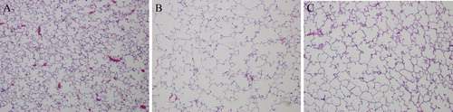
Table 2. Quantitative analyses of lung histomorphology in rats from each group.
SPA and SPC expression in the lung
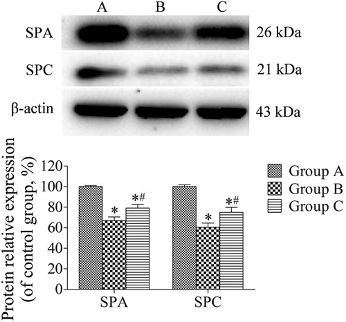
We performed quantitative real-time PCR and western blotting to detect mRNA and protein expression levels of SPA and SPC. The levels of SPA and SPC in lung samples from group B were significantly decreased compared to those from group A (both p < 0.05). By contrast, after the administration of SRT2104 to rats from group C, the expression levels of SPA and SPC were prominently higher than those in group B (both p < 0.05; and ).
Figure 2. SPA and SPC mRNA expression in lung tissues. All analyses were performed in triplicate. The relative expression of SPA and SPC mRNA was calculated with the 2−ΔΔCt method. Values are presented as mean ± SD (n = 15). *p < 0.05 vs. group A; #p < 0.05 vs. group B.
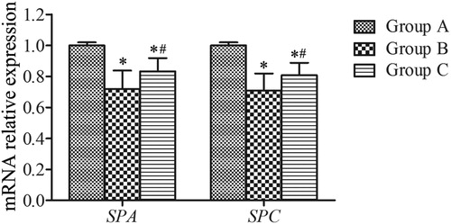
Figure 3. Western blotting analysis of the expression of SPA and SPC in lung tissues. β-actin was provided as the loading control. The expression of protein was analyzed by densitometry and normalized with β-actin. All analyses were performed in triplicate. Values are presented as mean ± SD (n = 15). *p < 0.05 vs. group A; #p < 0.05 vs. group B.
Immunohistochemical analysis of SPA and SPC
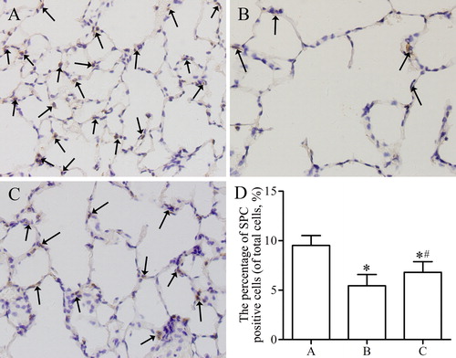
SPA- and SPC-positive cells presented yellowish-brown color in the cytoplasm, which was mainly located in the alveolar corner of AECII () and Citation5(A–C)). As shown in and , the percentages of SPA- and SPC-positive cells were prominently decreased in rats of group B compared with those of group A (both p < 0.05). However, after the administration of SRT2104, the percentages of SPA- and SPC-positive cells were significantly higher in group C than in group B (both p < 0.05).
Figure 4. Immunohistochemical analysis of the expression of SPA in lung parenchyma from control group (A), emphysema group (B), and emphysema + SRT2104 group (C). The arrows indicate the SPA positive cells. Magnification, ×400. (D) The percentage of SPA positive cells in each group. Values are presented as mean ± SD (n = 15). *p < 0.05 vs. group A; #p < 0.05 vs. group B.
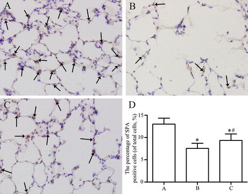
Figure 5. Immunohistochemical analysis of the expression of SPC in lung parenchyma from control group (A), emphysema group (B), and emphysema + SRT2104 group (C). The arrows indicate the SPC positive cells. Magnification, ×400. (D) The percentage of SPC positive cells in each group. Values are presented as mean ± SD (n = 15). *p < 0.05 vs. group A; #p < 0.05 vs. group B.
SA-β-gal activity
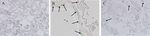
To detect cellular senescence, the SA-β-gal activity was assayed in murine lung tissues of each group. Blue-colored cells in lung samples were considered SA-β-gal-positive, indicating the presence of senescent cells. As shown in , the SA-β-gal activity was prominently increased in group B compared to group A (). Most SA-β-gal-positive cells were located at the corners of the alveoli. By contrast, the SA-β-gal activity was visibly decreased after the administration of SRT2104 to the rats of group C ().
Figure 6. SA-β-gal activity of lung tissues from control group (A), emphysema group (B), and emphysema + SRT2104 group (C). The cells in lung samples with blue color were considered SA-β-gal positive, which means senescent cells. The arrows indicate the SA-β-gal-positive cells. Magnification, ×200.
Quantification of SIRT1, FoxO3a, p53, and p21 expression
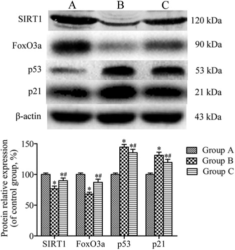
SIRT1, FoxO3a, p53, and p21 expression levels were quantified using western blotting. shows that the expression of SIRT1 and FoxO3a was conspicuously reduced in rats from group B compared to those from group A, while the expression levels of p53 and p21 were upregulated in group B in comparison to group A (all p < 0.05). However, SRT2104 treatment increased in the animals of group C the expression levels of SIRT1 and FoxO3a and downregulated p53 and p21 expression compared to group B (all p < 0.05).
Figure 7. Western blotting analysis of the expression of SIRT1, FoxO3a, p53, and p21 in lung tissues. β-actin was provided as the loading control. The expression of protein was analyzed by densitometry and normalized with β-actin. All analyses were performed in triplicate. Values are presented as mean ± SD (n = 15). *p < 0.05 vs. group A; #p < 0.05 vs. group B.
SIRT1 deacetylase activity
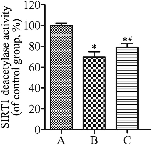
Accompanying the downregulation in SIRT1 protein expression, SIRT1 deacetylase activity was significantly decreased in the rats of group B compared to those of group A (p < 0.05). As shown in , SRT2104 treatment increased the SIRT1 deacetylase activity in group C compared to the untreated group B (p < 0.05).
Figure 8. SIRT1 deacetylase activity of lung tissues from control group (A), emphysema group (B), and emphysema + SRT2104 group (C). All analyses were performed in triplicate. Values are presented as mean ± SD (n = 15). *p < 0.05 vs. group A; #p < 0.05 vs. group B.
Discussion
Internationally, COPD prevalence continues to increase. Cigarette smoking is considered to be the central risk factor in the development of COPD [Citation13]. LPS plays vital roles in activating inflammatory cells to secrete factors that injure the epithelium, contributing to the pathogenesis and development of emphysema [Citation28]. Cellular senescence is considered an important mechanism promoting chronic lung diseases, such as COPD. Cellular senescence is caused by stress-related and replicative senescence involving activation of p53 and p16, respectively, leading to the activation of p21 and cell cycle arrest [Citation29]. COPD is regarded as a disease of accelerated lung aging and exhibits typical signs of aging, including cellular senescence, telomere shortening, and mitochondrial dysfunction [Citation30].
Previously, we confirmed that lncRNA-mediated SIRT1/p53 and FoxO3a signaling pathways may regulate AECII senescence in patients with COPD [Citation31]. In the present study, we generated a rat model of emphysema based on cigarette smoke exposure and intratracheal LPS instillation. We demonstrated that treatment with SRT2104 reduced the pathological characteristics of emphysema and improved lung function parameters, including PR, Cdyn, and PEF. Moreover, SRT2104 treatment upregulated the expression levels of SPA, SPC, SIRT1, and FoxO3a, decreased SA-β-gal activity, increased SIRT1 deacetylase activity, and downregulated the levels of p53 and p21 expression in the lung.
Previous studies in mice demonstrated that a SIRT1 activator protects against emphysema via a reduction in cellular senescence [Citation13,Citation23]. Sundar et al. revealed that cellular senescence in lung epithelial cells plays a vital role in the pathogenesis and development of COPD caused by cigarette smoking [Citation3]. In the current study, we confirmed that the pathological characteristics of emphysema were reduced and the lung function indexes, including PR, Cdyn, and PEF, were improved after SRT2104 treatment. However, the pathogenesis of COPD/emphysema is very complicated. AECII possess the unique ability to synthesize SPA and SPC, play important roles in maintaining alveolar homeostasis, and are involved in lung tissue repair [Citation4,Citation32]. As a SIRT1 activator SRT2104 exhibits therapeutic effects in several diseases such as cardiac fibrosis, osteoarthritis, nephropathy, and psoriasis [Citation14,Citation16,Citation17,Citation19]. It was also revealed that SIRT1 is an antiaging molecule involved in the response to chronic inflammation and oxidative stress [Citation33]. SIRT1 is recognized as a longevity gene with remarkable abilities to reverse aspects of aging [Citation34]. miR-570-3p inhibition reverses cellular senescence by restoring the levels of the antiaging molecule SIRT1 [Citation35]. In COPD, reduction in shelterin telomere protection protein 1 causes cellular senescence involving SIRT1 deacetylase [Citation36]. Cellular senescence mediated by SIRT1 participates in the development of COPD, but the specific effects of SRT2104 on AECII senescence in rats with emphysema remain elusive.
We performed quantitative real-time PCR, western blotting, and immunohistochemical analyses to detect the expression levels of SPA and SPC in lung samples. The mRNA and protein expression levels of SPA and SPC in lung samples from group B were substantially decreased compared to those from group A. This confirmed that AECII were damaged in lung samples derived from group B rats. To analyze whether cellular senescence was present in rats of each group, the SA-β-gal activity was assayed in lung tissues. The number of AECII is twice that of AECI, which are mainly located at the corners of the alveoli [Citation37]. As shown in , most SA-β-gal-positive cells in our experiments were also located at the corners of the alveoli. Furthermore, the SA-β-gal activity in group B was prominently increased relative to that in group A. Following SRT2104 administration, the SA-β-gal activity was substantially decreased in rats from group C. Based on these findings, we suggest that SRT2104 reduces AECII senescence.
FoxO3a is a non-histone substrate of SIRT1. Protein complexes with the combination of FoxO3a and SIRT1 may participate in cellular senescence [Citation38]. Upon stimulation by stress, p53 induces the expression of the cyclin-dependent kinase inhibitor p21 [Citation39]. In the current study, the effects of SRT2104 on AECII senescence in rats with emphysema may be associated with the activation of SIRT1/FoxO3a and SIRT1/p53 signaling pathways. Moreover, the reduction in AECII senescence may accelerate lung tissue repair and lung function improvement.
Conclusion
SRT2104 treatment reduced the pathological characteristics associated with emphysema and improved the lung function. Moreover, SRT2104 treatment increased the SIRT1 level and activity, which are associated with upregulated FoxO3a expression, downregulated levels of p53 and p21, and reduced AECII senescence. Therefore, SRT2104 might promote beneficial effect in emphysema by suppressing AECII senescence and provide a novel potential therapeutic strategy for COPD.
Declaration of interest
The authors declare that there is no conflict of interest. The authors are responsible for the content and writing of this paper.
Additional information
Funding
References
- Global Initiative for Chronic Obstructive Lung Disease. Global strategy for the diagnosis, management, and prevention of chronic obstructive pulmonary disease. Seattle: GOLD; 2020. Available from: http://www.goldcopd.com/
- Zheng H, Liu Y, Huang T, et al. Development and characterization of a rat model of chronic obstructive pulmonary disease (COPD) induced by sidestream cigarette smoke. Toxicol Lett. 2009;189(3):225–234. DOI:10.1016/j.toxlet.2009.06.850
- Sundar IK, Rashid K, Gerloff J, et al. Genetic ablation of p16INK4a does not protect against cellular senescence in mouse models of chronic obstructive pulmonary disease/emphysema. Am J Respir Cell Mol Biol. 2018;59(2):189–199. DOI:10.1165/rcmb.2017-0390OC
- Whitsett JA, Wert SE, Weaver TE. Alveolar surfactant homeostasis and the pathogenesis of pulmonary disease. Annu Rev Med. 2010;61:105–119. DOI:10.1146/annurev.med.60.041807.123500
- Hoffman AM, Ingenito EP. Alveolar epithelial stem and progenitor cells: emerging evidence for their role in lung regeneration. Curr Med Chem. 2012;19(35):6003–6008. DOI:10.2174/092986712804485872
- Bordo D. Structure and evolution of human sirtuins. Curr Drug Targets. 2013;14(6):662–665. DOI:10.2174/1389450111314060007
- Guarente L, Franklin H. Epstein lecture: Sirtuins, aging, and medicine. N Engl J Med. 2011;364(23):2235–2244. DOI:10.1056/NEJMra1100831
- Salminen A, Kaarniranta K, Kauppinen A. Crosstalk between oxidative stress and SIRT1: impact on the aging process. Int J Mol Sci. 2013;14(2):3834–3859. DOI:10.3390/ijms14023834
- Finkel T, Deng CX, Mostoslavsky R. Recent progress in the biology and physiology of sirtuins. Nature. 2009;460(7255):587–591. DOI:10.1038/nature08197
- Satoh A, Stein L, Imai S. The role of mammalian sirtuins in the regulation of metabolism, aging, and longevity. Handb Exp Pharmacol. 2011;206:125–162. DOI:10.1007/978-3-642-21631-2_7
- Schultz MB, Rinaldi C, Lu Y, et al. Molecular and cellular characterization of SIRT1 allosteric activators. Methods Mol Biol. 2019;1983:133–149. DOI:10.1007/978-1-4939-9434-2_8
- Guterres FA, Martinez GR, Rocha ME, et al. Simvastatin rises reactive oxygen species levels and induces senescence in human melanoma cells by activation of p53/p21 pathway. Exp Cell Res. 2013;319(19):2977–2988. DOI:10.1016/j.yexcr.2013.07.026
- Hamsanathan S, Alder JK, Sellares J, et al. Cellular senescence: the Trojan horse in chronic lung diseases. Am J Respir Cell Mol Biol. 2019;61(1):21–30. DOI:10.1165/rcmb.2018-0410TR
- Miyaji N, Nishida K, Tanaka T, et al. Inhibition of knee osteoarthritis progression in mice by administering SRT2014, an activator of silent information regulator 2 ortholog 1. Cartilage. 2020. DOI:10.1177/1947603519900795
- Duan CM, Zhang JR, Wan TF, et al. SRT2104 attenuates chronic unpredictable mild stress-induced depressive-like behaviors and imbalance between microglial M1 and M2 phenotypes in the mice. Behav Brain Res. 2020;378:112296. DOI:10.1016/j.bbr.2019.112296
- Zhang J, Bi R, Meng Q, et al. Catalpol alleviates adriamycin-induced nephropathy by activating the SIRT1 signalling pathway in vivo and in vitro. Br J Pharmacol. 2019;176(23):4558–4573. DOI:10.1111/bph.14822
- Liu ZH, Zhang Y, Wang X, et al. SIRT1 activation attenuates cardiac fibrosis by endothelial-to-mesenchymal transition. Biomed Pharmacother. 2019;118:109227. DOI:10.1016/j.biopha.2019.109227
- Wu H, Wu J, Zhou S, et al. SRT2104 attenuates diabetes-induced aortic endothelial dysfunction via inhibition of P53. J Endocrinol. 2018;237(1):1–14. DOI:10.1530/JOE-17-0672
- Krueger JG, Suárez-Fariñas M, Cueto I, et al. A randomized, placebo-controlled study of SRT2104, a SIRT1 activator, in patients with moderate to severe psoriasis. PLoS One. 2015;10(11):e0142081. DOI:10.1371/journal.pone.0142081
- van der Meer AJ, Scicluna BP, Moerland PD, et al. The selective Sirtuin 1 activator SRT2104 reduces endotoxin-induced cytokine release and coagulation activation in humans. Crit Care Med. 2015;43(6):e199–e202. DOI:10.1097/CCM.0000000000000949
- Rabelo MAE, Lucinda LMF, Reboredo MM, et al. Acute lung injury in response to intratracheal instillation of lipopolysaccharide in an animal model of emphysema induced by elastase. Inflammation. 2018;41(1):174–182. DOI:10.1007/s10753-017-0675-5
- Gu C, Li Y, Xu WL, et al. Sirtuin 1 activator SRT1720 protects against lung injury via reduction of type II alveolar epithelial cells apoptosis in emphysema. COPD. 2015;12(4):444–452. DOI:10.3109/15412555.2014.974740
- Yao H, Chung S, Hwang JW, et al. SIRT1 protects against emphysema via FOXO3-mediated reduction of premature senescence in nescence in mice. J Clin Invest. 2012;122(6):2032–2045. DOI:10.1172/JCI60132
- Agarwal AR, Yin F, Cadenas E. Short-term cigarette smoke exposure leads to metabolic alterations in lung alveolar cells. Am J Respir Cell Mol Biol. 2014;51(2):284–293. DOI:10.1165/rcmb.2013-0523OC
- Li Y, Xu W, Yan J, et al. Differentiation of human amniotic fluid-derived mesenchymal stem cells into type II alveolar epithelial cells in vitro. Int J Mol Med. 2014;33(6):1507–1513. DOI:10.3892/ijmm.2014.1705
- Livak KJ, Schmittgen TD. Analysis of relative gene expression data using real-time quantitative PCR and the 2(-delta delta C(T)) method. Methods. 2001;25(4):402–408. DOI:10.1006/meth.2001.1262
- Caito S, Rajendrasozhan S, Cook S, et al. SIRT1 is a redox-sensitive deacetylase that is post-translationally modified by oxidants and carbonyl stress. FASEB J. 2010;24(9):3145–3159. DOI:10.1096/fj.09-151308
- Martin TR. Recognition of bacterial endotoxin in the lungs. Am J Respir Cell Mol Biol. 2000;23(2):128–132. DOI:10.1165/ajrcmb.23.2.f189
- Barnes PJ, Baker J, Donnelly LE. Cellular senescence as a mechanism and target in chronic lung diseases. Am J Respir Crit Care Med. 2019;200(5):556–564. DOI:10.1164/rccm.201810-1975TR
- Barnes PJ. Pulmonary diseases and ageing. Subcell Biochem. 2019;91:45–74. DOI:10.1007/978-981-13-3681-2_3
- Gu C, Li Y, Liu J, et al. LncRNA-mediated SIRT1/FoxO3a and SIRT1/p53 signaling pathways regulate type II alveolar epithelial cell senescence in patients with chronic obstructive pulmonary disease. Mol Med Rep. 2017;15(5):3129–3134. DOI:10.3892/mmr.2017.6367
- Fujino N, Kubo H, Suzuki T, et al. Isolation of alveolar epithelial type II progenitor cells from adult human lungs. Lab Invest. 2011;91(3):363–378. DOI:10.1038/labinvest.2010.187
- Conti V, Corbi G, Manzo V, et al. Sirtuin 1 and aging theory for chronic obstructive pulmonary disease. Anal Cell Pathol (Amst). 2015;2015:897327. DOI:10.1155/2015/897327
- Bonkowski MS, Sinclair DA. Slowing ageing by design: the rise of NAD+ and sirtuin-activating compounds. Nat Rev Mol Cell Biol. 2016;17(11):679–690. DOI:10.1038/nrm.2016.93
- Baker JR, Vuppusetty C, Colley T, et al. MicroRNA-570 is a novel regulator of cellular senescence and inflammaging. FASEB J. 2019;33(2):1605–1616. DOI:10.1096/fj.201800965R
- Ahmad T, Sundar IK, Tormos AM, et al. Shelterin telomere protection protein 1 reduction causes telomere attrition and cellular senescence via Sirtuin 1 deacetylase in chronic obstructive pulmonary disease. Am J Respir Cell Mol Biol. 2017;56(1):38–49. DOI:10.1165/rcmb.2016-0198OC
- Guillot L, Nathan N, Tabary O, et al. Alveolar epithelial cells: master regulators of lung homeostasis. Int J Biochem Cell Biol. 2013;45(11):2568–2573. DOI:10.1016/j.biocel.2013.08.009
- Ganesan S, Unger BL, Comstock AT, et al. Aberrantly activated EGFR contributes to enhanced IL-8 expression in COPD airways epithelial cells via regulation of nuclear FoxO3A. Thorax. 2013;68(2):131–141. DOI:10.1136/thoraxjnl-2012-201719
- Bargonetti J, Manfredi JJ. Multiple roles of the tumor suppressor p53. Curr Opin Oncol. 2002;14(1):86–91. DOI:10.1097/00001622-200201000-00015