
Abstract
Chronic obstructive pulmonary disease (COPD) is a common chronic disease characterized by airflow obstruction, which seriously threatens people’s health. The COPD mouse model was established with cigarette smoke induction. Hematoxylin-eosin staining and Masson staining were carried out to observe the pathological changes of lung tissues in COPD mice. RTEL1 was silenced in COPD mice, and immunohistochemistry was used to detect RTEL1, ki67 and Caspase-3 expression. The role of RTEL1 in inflammation were evaluated by ELISA, and the impacts of RTEL1 on M1 and M2 macrophage markers (iNOS and CD206) were evaluated by qPCR and western blotting. In COPD model, there was an increase in the number of inflammatory cells, with slightly disorganized cell arrangement, unclear hierarchy, condensed and solidified nuclei, while knockdown of RTEL1 improved the inflammatory infiltration. Moreover, knockdown of RTEL1 reduced ki67-positive cells and increased Caspase-3 positive cells in COPD group. The increased inflammatory factors (IL-1β, MMP-9, TNF-α, IL-4, IL-6, and IL-23) in COPD were suppressed by knockdown of RTEL1, while iNOS was raised and CD206 was inhibited. In conclusion, knockdown of RTEL1 promoted M1 and inhibited M2 macrophage polarization and inflammation to alleviate COPD.
Introduction
Chronic obstructive pulmonary disease (COPD) is characterized by airflow obstruction, which is a common chronic disease and can further evolve into pulmonary heart disease and respiratory failure[Citation1, Citation2]. Smoking is one of the main risk factors for COPD, and the disease is also associated with air pollution, occupation, and infection [Citation3, Citation4]. COPD has developed into a rigorous global health problem and is one of the major risk factors for global population death. At present, the pathogenesis of COPD is constantly exploring, and it is very important to seek strategies that can treat and prevent the disease.
Airway inflammation is a characteristic of COPD, which is associated with the pathogenesis and progression of COPD [Citation5]. In recent years, evidence has shown that neutrophil inflammation disrupts elastic fiber assembly components during lung development and increases the susceptibility of adult mice to COPD [Citation6]. Therefore, reducing inflammation can effectively prevent and alleviate the deterioration of COPD [Citation7]. Ergosterol, a biologically active ingredient, inhibits inflammation and oxidative stress and apoptosis through the NF-κB/p65 pathway, and alleviates cigarette smoke-induced COPD [Citation8]. Hydrogen sulfide can prevent emphysema by inhibiting the PHD2/HIF-1α/MAPK axis, thereby curbing inflammation, epithelial cell injury and apoptosis [Citation9]. Melatonin alleviates the development of COPD, possibly by attenuating NLRP1 inflammasome and IL-1β to inhibit airway inflammation [Citation10].
Regulator of telomere elongation 1 (RTEL1) is an indispensable DNA helicase, which is very important for genome stability [Citation11], and its mutation can increase the susceptibility to diseases such as high-grade glioma and astrocytoma [Citation12]. COPD is considered to be a disease of accelerated lung aging, characterized by cellular senescence, mitochondrial dysfunction, shortened telomeres and epigenetic changes, etc [Citation13]. Therefore, RTEL1, as a telomere regulator, may play a role in COPD, but the action mechanism of RTEL1 in COPD is still unknown.
Macrophages belong to innate immune cells that perform phagocytosis and eliminate pathogens [Citation14]. In the lung microenvironment, plenty of macrophages, including alveolar macrophages and interstitial macrophages, are present [Citation15]. Macrophages have been reported to be of vital significance in a variety of respiratory diseases, such as allergic asthma, pulmonary fibrosis, pulmonary hypertension and so on [Citation16–18]. In our study, COPD mouse model was constructed and staining was carried out to observe the pathological changes of lung tissues in mice. In addition, the effects of RTEL1 on inflammation and macrophage markers in COPD was also explored, which provided a basis for the following research of RTEL1 on COPD and a new direction for the therapy of COPD.
Materials and methods
Establishment of COPD mouse model
BALB/c mice (6–8 weeks, 18–20 g) were obtained from SPF Biotechnology (Beijing, China) and grouped into Control and COPD model group (n = 6). On the first day of the experiment, mice in the model group were challenged with 2 μL of lipopolysaccharides (LPS, 5 μg/μL) by nasal drip, followed by cigarette smoke (CS) induction. Ten cigarettes were lit each day and the smoke was allowed to remain in the closed box environment, after which the mice were placed in the box for half an hour. The CS induction was done continuously for a month [Citation19]. On the last day of modeling, LPS was added nasally to increase infection efficiency.
Afterwards, another 18 BALB/c mice (SPF Biotechnology) were taken and randomly divided into Control, lv-sh-NC (injected with lentiviral particles encoding negative control shRNA) and lv-sh-RTEL1 group (injected with lentiviral particles encoding RTEL1 shRNA) (n = 6). The lentiviral particles (1.0 × 10⁹ TU, 50 μL) were subcutaneously injected into the shaved dorsal skin of the mice to establish the RTEL1 knockdown model, and then mice of the lv-sh-NC and lv-sh-RTEL1 group were received CS and LPS treatment. All mice were anesthetized by isoflurane inhalation and sacrificed by cervical dislocation. Lung tissues and serum of mice were taken for following experiments.
All animal experiments complied with the Guide for the Care and Use of Laboratory Animals and were approved by Hainan General Hospital Medical Ethics Committee (Approval No. Med-Eth-Re [2023] 347).
Hematoxylin-eosin (HE) staining
Left lung tissues were obtained and fixed in paraformaldehyde (4%), and then dehydrated with ethanol gradient. The tissues were embedded in paraffin and cut into 4 μm thick slices. Paraffin slices were routinely dewaxed with xylene and ethanol (Sigma, USA). Then slices were stained with hematoxylin and eosin in turn. Eventually, the slices were dehydrated and sealed with neutral gum. The staining results were observed under an optical microscope.
Masson staining
Paraffin sections were dewaxed, washed, and dyed with Regaud’s hematoxylin (Sigma) for 5–10 min and Masson Ponceau S acid fuchsin stain (Sigma) for 5–10 min. Subsequently, sections were washed in 2% aqueous glacial acetic acid and then differentiated in 1% aqueous phosphomolybdic acid for 3–5 min. Afterwards, sections were stained with aniline blue for 5 min and washed. The slices were dehydrated and finally sealed with neutral gum.
Immunohistochemistry (IHC)
The paraffin-embedded lung tissues were dewaxed and rehydrated, then incubated with anti-RTEL1 (ab85557, Abcam, Shanghai, China), anti-ki67 antibody (ab15580, 1:1,000, Abcam) and anti-Caspase-3 (ab184787, 1: 1,000, Abcam) overnight at 4 °C. Afterwards, slices were incubated with horseradish peroxidase (HRP)-labeled secondary antibody at 37 °C for 1 h. Then slices were stained with DAB and hematoxylin, and finally photographed under a microscope. ImageJ and IHC Profiler were used for calculate IHC score as previous reported [Citation20]. IHC score = (Number of pixels in zone) × (Score of the zone)/Total number of pixels in the image. The nuclear-positive staining cells was scored as follows: high proportion, 4, moderate proportion, 3, low proportion, 2, and negative staining, 1.
Real-time fluorescent quantitative PCR (RT-qPCR)
TRIzol reagent (Invitrogen, USA) was applied to extract total RNA from lung tissues. The concentration of RNA was determined by UV spectrophotometer, and the ratio of OD260/OD280 between 1.9 and 2.0 indicated a high purity. The cDNA template was synthesized by reverse transcription in a PCR amplifier, and RT-qPCR was run on an ABI7500 quantitative PCR instrument (Applied Biosystems, USA) under the following conditions: pre-denaturation at 95 °C for 30 s, denaturation at 95 °C for 10 s, and annealing at 60 °C for 30 s for 40 cycles. Glyceraldehyde-3-phosphate dehydrogenase (GAPDH) was used as an internal reference. The Ct values obtained were analyzed by the 2−ΔΔCt method. The experiment was repeated three times, and the primer sequences were shown in .
Table 1. Primer sequences for RT-qPCR.
Western blotting (WB)
Tissues were treated with RIPA lysis buffer to separate total protein samples. The Pierce BCA Protein Assay Kit (Thermo Fisher Scientific, Waltham, MA, USA) was applied to evaluate protein concentration. Proteins were separated using a 10% sodium dodecyl sulfonate-polyacrylamide gel electrophoresis (SDS-PAGE) and then transferred to a polyvinylidene fluoride (PVDF) membrane. After blocking the membrane in 5% skim milk, it was incubated with primary antibody at 4 °C overnight, including anti-RTEL1 (PA5-72995, 1:1,000, Thermo Fisher Scientific), anti-iNOS (ab178945, 1:1,000, Abcam), anti-CD206 (ab64693,1:1,000, Abcam), and anti-GAPDH (ab9485, 1:1,000, Abcam). The membrane was then incubated with appropriate goat anti-rabbit secondary antibodies at room temperature for 1 h. On the incubation plate of Tanon 5200 chemiluminescence imaging system, the enhanced chemiluminescence (ECL) liquid was used for developing, and the images were collected by exposure.
Detection of enzyme linked immunosorbent assay (ELISA)
According to the manufacturer’s instructions, the levels of TNF-α, IL-1β, MMP-9, IL-4, IL-6, and IL-33 in COPD mice were measured using the corresponding ELISA kits (Esebio, Shanghai, China). Total 50 μL of diluted samples and standards were added to the wells of the microtiter plate. Afterwards, 100 μL of HRP-labeled antibody was added to each well, and the plates were incubated for 60 min at 37 °C. The OD value of each well was measured at 450 nm within 15 min. The concentration of the samples was calculated by standard curve.
Statistical analysis
Statistical analysis was performed using GraphPad Prism 8.0. The data were expressed as mean ± standard deviation. t-test was used to analyze the quantitative data for comparison between the two groups. One-way ANOVA and Tukey’s test was used for comparison between multiple groups. p < .05 was regarded as statistical significance.
Results
Successful construction of COPD mouse model
The lung tissues of mice in both groups were obtained and subjected to HE staining and Masson staining. As we can see, there was a clear alveolar tissue structure and cellular layers in Control group. Moreover, the cells were arranged in a neat and orderly manner, and no inflammatory cell infiltration was observed. Compared with the Control group, the COPD group showed a significant elevation in the number of inflammatory cells, slightly disorganized cell arrangement, unclear hierarchy, and condensed and solidified nuclei ().
Figure 1. Successful construction of COPD mouse model. Hematoxylin-eosin staining and Masson staining of lung tissues in Control and COPD. (A) Hematoxylin-eosin staining of lung tissues in each group (200×, 100 μm). (B) Masson staining of lung tissues in each group (200×, 100 μm). (C–D) Detection of ki67 and Caspase-3 by immunohistochemistry (200×, 100 μm).
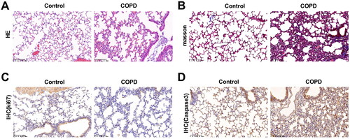
IHC was executed to detect the levels of ki67 and Caspase-3 in lung tissues, demonstrating that compared with Control group, ki67-positive cells were significantly reduced in COPD group, as well as an increased Caspase-3 positive cells ().
The expressions of RTEL1, inflammatory factors and macrophage markers in COPD mouse
The expression levels of inflammatory factors in both groups were detected by ELISA. In comparison with the Control group, the levels of MMP9, IL-1β, and TNF-α were significantly elevated in the COPD group of mice (p < .01, ).
Figure 2. Detection of inflammatory factors in serum and RTEL1 expression in lung tissues of COPD mice. (A) ELISA was applied to detect TNF-α, IL-1β and MMP-9 in mice. (B) RT-qPCR was used to detect the mRNA level of RTEL1. (C) Western blotting was performed to detect the protein expression of RTEL1. **p < .01 vs. control.
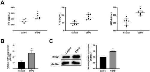
The expression levels of RTEL1 in the lung tissues of mice in two groups were detected by qPCR and WB. In comparison with the Control group, the expression level of RTEL1 was significantly higher in the COPD group ().
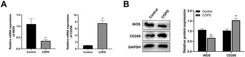
RT-qPCR and WB were performed to detect the expression levels of M1 and M2 macrophage markers iNOS and CD206 in the lung tissues of mice in both groups, demonstrating that compared with the Control group, there was a decreased iNOS expression and an increased CD206 expression in the COPD group (p < .01, ).
Figure 3. Detection of the expression levels of M1 and M2 macrophage markers, iNOS and CD206 in Control and COPD group by RT-qPCR (A) and western blotting (B). **p < .01 vs. control.
Knockdown of RTEL1 alleviates the development of COPD
The knockdown efficiency of RTEL1 in mice was verified by RT-qPCR, showing that compared with the lv-sh-NC group, the expression level of RTEL1 in the lv-sh-RTEL1 group was significantly decreased ().
Figure 4. Down-regulation of RTEL1 can improve the development of COPD. (A) RT-qPCR was used to detect the knockdown efficiency of RTEL1. (B) Hematoxylin-eosin staining of lung tissues in each group (200×, 100 μm). (C) Masson staining of lung tissues in each group (200×, 100 μm). (D–F) Detection of RTEL1, ki67 and Caspase-3 by immunohistochemistry (200×, 100 μm) and IHC scores. F. ELISA detection of inflammatory factors (TNF-α, IL-1β, MMP9, IL-4, IL-6, and IL-33) in mice. **p < .01 vs. control ##p < .01 vs. lv-sh-NC.
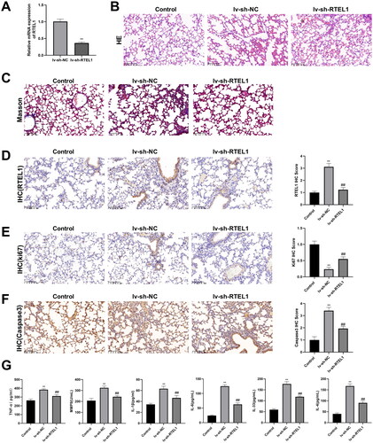
The lung tissues of the three groups were obtained for HE and Masson staining. In Control group, the alveolar tissue structure and cell level were clear, the cells were arranged neatly and orderly, and no inflammatory cell infiltration was observed. Compared with the Control group, the number of inflammatory cells in the alveolar tissue of the lv-sh-NC group was significantly increased, the cell arrangement was slightly disordered, the level was not obvious, and the nucleus was concentrated and solidified, while RTEL1 silencing alleviated it (). The phenomenon indicated that knockdown of RTEL1 significantly improved inflammatory infiltration and cell arrangement in COPD.
IHC was carried out to measure the expression of RTEL1, showing that compared with Control group, CS induction increased RTEL1 positive cells in the lv-sh-NC group, and after knocking down RTEL1, RTEL1 positive cells were reduced (). Furthermore, compared with the Control group, there were less ki67 positive cells and more Caspase3 positive cells in the lv-sh-NC group (). Compared with the lv-sh-NC group, the ki67 positive cells in the lv-sh-RTEL1 group were significantly increased, and the Caspase3 positive cells were significantly decreased.
In addition, knockdown of RTEL1 dramatically reduced the increased inflammatory factors caused by CS + LPS. Compared with the Control group, the concentrations of inflammatory factors (TNF-α, IL-1β and MMP-9, IL-4, IL-6, and IL-33) in the lv-sh-NC group were significantly increased, while knockdown of RTEL1 inhibited the levels of inflammatory factors ().
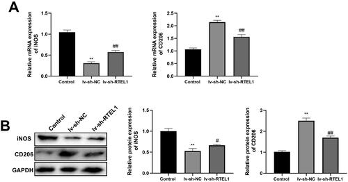
Knockdown of RTEL1 regulates M1 and M2 macrophage polarization in COPD
Compared with the Control group, there was a lower iNOS and higher CD206 expression in the lv-sh-NC group (p < .01, ). After knockdown of RTEL1, the expression of iNOS in the lv-sh-RTEL1 group was significantly increased, while CD206 was significantly decreased (p < .01, ).
Figure 5. Down-regulation of RTEL1 can promote M1 and inhibit M2 macrophage polarization in COPD mice. (A) RT-qPCR was used to detect the expression levels of macrophage markers iNOS and CD206 in mice. (B) Western blotting was performed to detect the protein expression of macrophage markers iNOS and CD206 in mice. **p < .01 vs. control #p < .05 vs. lv-sh-NC ##p < .01 vs. lv-sh-NC.
Discussion
As a chronic inflammatory disease, COPD causes a large number of deaths worldwide every year [Citation21, Citation22]. At present, progress has been made in improving severe cases, however, due to the complex pathogenesis of COPD, it is still necessary to find reliable targets for the treatment of COPD. Herein, COPD mouse model was constructed to observe the pathological features of lung tissues and detect the expression of inflammatory factors. Meanwhile, the roles of RTEL1 in inflammation and macrophage-related markers were inspected.
In previous studies, many therapeutic targets have been shown to dramatically attenuate the progression of COPD. For example, inhibition of circXPO1 alleviates inflammation caused by CS in COPD [Citation23]. Calpeptin suppresses calpain/IκBα signaling to improve inflammation in CS-induced mice and BEAS-2B cells [Citation24]. Receptor-interacting protein 2 silencing may disrupt NF-κB activity to improve acute lung injury in COPD mice [Citation25]. RTEL1 is a kind of DNA helicase, which is substantial for telomeres and genome integrity [Citation26]. It has been confirmed that the mutation of RTEL1 is related to heterogeneous lung and extrapulmonary phenotype [Citation27]. Moreover, RTEL1 mutation may be associated with the pathogenesis of pulmonary fibrosis[Citation28]. Peljto et al. also prove that rare variants in telomerase reverse transcriptase (TERT) and RTEL1 are significantly associated with idiopathic pulmonary fibrosis and have a greater impact on the disease [Citation29]. In our study, we found that RTEL1 was overexpressed in COPD mice, and after knockdown of RTEL1, the inflammation was significantly improved in COPD mice, indicating that RTEL1 can be used as a potential therapeutic target for COPD.
Inflammation is also closely associated with the occurrence and progression of COPD. Chronic inflammation of the lung parenchyma and peripheral airways can largely cause irreversible airflow limitation and an increase in inflammatory cells [Citation30]. TNF-α and IL-1β are potential biomarkers for assessing lung function and inflammation in patients with COPD [Citation31]. In this study, inflammatory factors TNF-α, IL-1β, MMP-9, IL-4, IL-6, and IL-23 were significantly increased in COPD mice, indicating that inflammation played a crucial role in COPD. It is found that IL-1β induces pro-inflammation to alter the fibroblast microenvironment and impair the function of lung progenitor cells, leading to chronic inflammatory diseases such as COPD [Citation32]. Fu et al. find that IL-1β and systemic inflammation in the airways are associated with exacerbations of COPD, which may lead to a vicious circle of the disease, and the scheme of inhibiting inflammation to alleviate COPD needs to be further explored [Citation33]. MMP-9 is also of vital importance in the development of COPD. It has been reported that significant activation of MMP-9 is associated with acute exacerbation of COPD [Citation34]. Macrophage-specific NOX2 is involved in emphysema progression by regulating the SIRT1/MMP-9 pathway [Citation35]. Herein, RTEL1 knockdown significantly curbed the expression of inflammatory factors and inflammatory infiltration, which further verified the targeted therapeutic effect of RTEL1 on COPD.
Alveolar macrophages in asthma mice exhibit significant M2 polarization, and targeting M2 macrophages can reduce airway inflammation and remodeling through the miR-378a-3p/GRB2 pathway [Citation36]. Mycobacterium tuberculosis infection promotes the polarization of CS-exposed macrophages to M1 and M2 phenotypes, while promoting the expression of MMP9 and MMP12 [Citation37]. Naringenin significantly inhibits the expression of iNOS in BEAS-2B-derived macrophages induced by cigarette smoke extract, thereby inhibiting M1 macrophage polarization [Citation38]. Airway epithelial cell-derived exosomes contribute to M1 macrophage polarization to exacerbate the progression of COPD through the upregulation of triggering receptor expressed on myeloid cells-1 (TREM-1) expression [Citation39]. In our study, COPD mice exhibited an increased CD206 and a decreased iNOS, while knockdown of RTEL1 reversed the phenomenon, indicating that knockdown of RTEL1 may suppress the occurrence and development of COPD by promoting M1 and inhibiting M2 macrophage polarization.
In summary, through the in vivo experiment, we found that RTEL1 was up-regulated in COPD mouse model, and knockdown of RTEL1 significantly inhibited inflammation and M2 macrophage polarization in COPD, while promoted M1 macrophage polarization, resulting in improving inflammation infiltration to alleviate COPD development, which provided a new orientation for the prevention and therapy for the disease.
Ethics approval
All animal experiments were in accordance with ARRIVE Guidelines and were approved by Hainan General Hospital Medical Ethics Committee (Approval No. Med-Eth-Re [2023] 347).
Author contributions
Conceptualization, HPX and HN; Methodology, HW; Investigation, JL and HW; Formal Analysis, HPX and HN; Writing - Original Draft, HPX and HN; Writing - Review & Editing, HW, JL and JJY. All authors had full access to the data in the study and take responsibility for the integrity of the data and the accuracy of the data analysis.
Disclosure statement
No potential conflict of interest was reported by the author(s).
Data availability statement
The datasets used and/or analyzed during the current study are available from the corresponding author on reasonable request.
Additional information
Funding
References
- Miravitlles M, Ribera A. Understanding the impact of symptoms on the burden of COPD. Respir Res. 2017;18(1):1. doi: 10.1186/s12931-017-0548-3.
- Baraldo S, Turato G, Saetta M. Pathophysiology of the small airways in chronic obstructive pulmonary disease. Respiration. 2012;84(2):89–8. doi: 10.1159/000341382.
- Raherison C, Girodet PO. Epidemiology of COPD. Eur Respir Rev. 2009;18(114):213–221. doi: 10.1183/09059180.00003609.
- Labaki WW, Rosenberg SR. Chronic obstructive pulmonary disease. Ann Intern Med. 2020;173(3):Itc17–itc32. doi: 10.7326/AITC202008040.
- Cavaillès A, Brinchault-Rabin G, Dixmier A, et al. Comorbidities of COPD. Eur Respir Rev. 2013;22(130):454–475. doi: 10.1183/09059180.00008612.
- Benjamin JT, Plosa EJ, Sucre JM, et al. Neutrophilic inflammation during lung development disrupts elastin assembly and predisposes adult mice to COPD. J Clin Invest. 2021;131(1):e139481. doi: 10.1172/JCI139481.
- Tsoumakidou M, Siafakas NM. Novel insights into the aetiology and pathophysiology of increased airway inflammation during COPD exacerbations. Respir Res. 2006;7(1):80. doi: 10.1186/1465-9921-7-80.
- Sun X, Feng X, Zheng D, et al. Ergosterol attenuates cigarette smoke extract-induced COPD by modulating inflammation, oxidative stress and apoptosis in vitro and in vivo. Clin Sci. 2019;133(13):1523–1536. doi: 10.1042/CS20190331.
- Guan R, Wang J, Li D, et al. Hydrogen sulfide inhibits cigarette smoke-induced inflammation and injury in alveolar epithelial cells by suppressing PHD2/HIF-1α/MAPK signaling pathway. Int Immunopharmacol. 2020;81:105979. doi: 10.1016/j.intimp.2019.105979.
- Peng Z, Zhang W, Qiao J, et al. Melatonin attenuates airway inflammation via SIRT1 dependent inhibition of NLRP3 inflammasome and IL-1β in rats with COPD. Int Immunopharmacol. 2018;62:23–28. doi: 10.1016/j.intimp.2018.06.033.
- Olivier M, Hesketh A, Pouch-Pélissier MN, et al. RTEL1 is required for silencing and epigenome stability. Nucleic Acids Res. 2023;51(16):8463–8479. doi: 10.1093/nar/gkad610.
- Vannier JB, Sarek G, Boulton SJ. RTEL1: functions of a disease-associated helicase. Trends Cell Biol. 2014;24(7):416–425. doi: 10.1016/j.tcb.2014.01.004.
- Barnes PJ. Senescence in COPD and its comorbidities. Annu Rev Physiol. 2017;79(1):517–539. doi: 10.1146/annurev-physiol-022516-034314.
- Zhang L, Wang CC. Inflammatory response of macrophages in infection. Hepatobiliary Pancreat Dis Int. 2014;13(2):138–152. doi: 10.1016/s1499-3872(14)60024-2.
- Bain CC, MacDonald AS. The impact of the lung environment on macrophage development, activation and function: diversity in the face of adversity. Mucosal Immunol. 2022;15(2):223–234. doi: 10.1038/s41385-021-00480-w.
- Saradna A, Do DC, Kumar S, et al. Macrophage polarization and allergic asthma. Transl Res. 2018;191:1–14. doi: 10.1016/j.trsl.2017.09.002.
- Kishore A, Petrek M. Roles of macrophage polarization and macrophage-derived miRNAs in pulmonary fibrosis. Front Immunol. 2021;12:678457. doi: 10.3389/fimmu.2021.678457.
- Li M, Riddle S, Kumar S, et al. Microenvironmental regulation of macrophage transcriptomic and metabolomic profiles in pulmonary hypertension. Front Immunol. 2021;12:640718. doi: 10.3389/fimmu.2021.640718.
- Cai Q, Chen S, Zhu Y, et al. Knockdown of GNL3L alleviates the progression of COPD through inhibiting the ATM/p53 pathway. Int J Chron Obstruct Pulmon Dis. 2023;18:2645–2659. doi: 10.2147/COPD.S424431.
- Huang X, Guan W, Xiang B, et al. MUC5B regulates goblet cell differentiation and reduces inflammation in a murine COPD model. Respir Res. 2022;23(1):11. doi: 10.1186/s12931-021-01920-8.
- Rabe KF, Watz H. Chronic obstructive pulmonary disease. Lancet. 2017;389(10082):1931–1940. doi: 10.1016/S0140-6736(17)31222-9.
- Duffy SP, Criner GJ. Chronic obstructive pulmonary disease: evaluation and management. Med Clin North Am. 2019;103(3):453–461. doi: 10.1016/j.mcna.2018.12.005.
- Du Y, Ding Y, Shi T, et al. Suppression of circXPO1 attenuates cigarette smoke-induced inflammation and cellular senescence of alveolar epithelial cells in chronic obstructive pulmonary disease. Int Immunopharmacol. 2022;111:109086. doi: 10.1016/j.intimp.2022.109086.
- Zuo J, Hu Z, Liu T, et al. Calpeptin attenuates cigarette smoke-induced pulmonary inflammation via suppressing calpain/IκBα signaling in mice and BEAS-2B cells. Pathol Res Pract. 2018;214(8):1199–1209. doi: 10.1016/j.prp.2018.06.019.
- Dong J, Liao W, Tan LH, et al. Gene silencing of receptor-interacting protein 2 protects against cigarette smoke-induced acute lung injury. Pharmacol Res. 2019;139:560–568. doi: 10.1016/j.phrs.2018.10.016.
- Hourvitz N, Awad A, Tzfati Y. The many faces of the helicase RTEL1 at telomeres and beyond. Trends Cell Biol. 2023;34(2):109–121. doi: 10.1016/j.tcb.2023.07.002.
- Borie R, Bouvry D, Cottin V, et al. Regulator of telomere length 1 (RTEL1) mutations are associated with heterogeneous pulmonary and extra-pulmonary phenotypes. Eur Respir J. 2019;53(2):1800508. doi: 10.1183/13993003.00508-2018.
- Kropski JA, Loyd JE. Telomeres revisited: RTEL1 variants in pulmonary fibrosis. Eur Respir J. 2015;46(2):312–314. doi: 10.1183/13993003.00710-2015.
- Peljto AL, Blumhagen RZ, Walts AD, et al. Idiopathic pulmonary fibrosis is associated with common genetic variants and limited rare variants. Am J Respir Crit Care Med. 2023;207(9):1194–1202. doi: 10.1164/rccm.202207-1331OC.
- Barnes PJ. Inflammatory mechanisms in patients with chronic obstructive pulmonary disease. J Allergy Clin Immunol. 2016;138(1):16–27. doi: 10.1016/j.jaci.2016.05.011.
- Lin X, Fan Y, Wang X, et al. Correlation between tumor necrosis factor-α and interleukin-1β in exhaled breath condensate and pulmonary function. Am J Med Sci. 2017;354(4):388–394. doi: 10.1016/j.amjms.2017.06.004.
- Ciminieri C, Woest ME, Reynaert NL, et al. IL-1β induces a proinflammatory fibroblast microenvironment that impairs lung progenitors’ function. Am J Respir Cell Mol Biol. 2023;68(4):444–455. doi: 10.1165/rcmb.2022-0209OC.
- Fu JJ, McDonald VM, Baines KJ, et al. Airway IL-1β and systemic inflammation as predictors of future exacerbation risk in asthma and COPD. Chest. 2015;148(3):618–629. doi: 10.1378/chest.14-2337.
- Papakonstantinou E, Karakiulakis G, Batzios S, et al. Acute exacerbations of COPD are associated with significant activation of matrix metalloproteinase 9 irrespectively of airway obstruction, emphysema and infection. Respir Res. 2015;16(1):78. doi: 10.1186/s12931-015-0240-4.
- Trocme C, Deffert C, Cachat J, et al. Macrophage-specific NOX2 contributes to the development of lung emphysema through modulation of SIRT1/MMP-9 pathways. J Pathol. 2015;235(1):65–78. doi: 10.1002/path.4423.
- Wang Q, Hong L, Chen M, et al. Targeting M2 macrophages alleviates airway inflammation and remodeling in asthmatic mice via miR-378a-3p/GRB2 pathway. Front Mol Biosci. 2021;8:717969. doi: 10.3389/fmolb.2021.717969.
- Le Y, Cao W, Zhou L, et al. Infection of Mycobacterium tuberculosis promotes both M1/M2 polarization and MMP production in cigarette smoke-Exposed macrophages. Front Immunol. 2020;11:1902. doi: 10.3389/fimmu.2020.01902.
- Chen Z, Wu H, Fan W, et al. Naringenin suppresses BEAS-2B-derived extracellular vesicular cargoes disorder caused by cigarette smoke extract thereby inhibiting M1 macrophage polarization. Front Immunol. 2022;13:930476. doi: 10.3389/fimmu.2022.930476.
- Wang L, Chen Q, Yu Q, et al. Cigarette smoke extract-treated airway epithelial cells-derived exosomes promote M1 macrophage polarization in chronic obstructive pulmonary disease. Int Immunopharmacol. 2021;96:107700. doi: 10.1016/j.intimp.2021.107700.