Abstract
Pannexins have been proposed to play a role in gap junctional intercellular communication and as single-membrane channels, although many of their molecular characteristics differ from connexins. Localization of untagged Panx1 and Panx3 exogenously expressed in five cultured cell lines revealed a cell surface distribution profile with limited evidence of cell surface clustering and variable levels of intracellular pools. However, N-glycosylation–defective mutants of pannexins exhibited a more prominent intracellular distribution with decreased cell surface labeling, suggesting an important role for pannexin glycosylation in trafficking. Similar to wild-type pannexins, the glycosylation-defective mutants failed to noticeably transfer microinjected fluorescent dyes to neighboring cells, suggesting that few, or no functional intercellular channels were formed. Finally, varied distribution patterns of endogenous Panx1 and Panx3 were observed in cells of osteoblast origin and Madin-Darby canine kidney cells. Collectively, diverse expression and distribution profiles of Panx1 and Panx3 suggest that they may have multiple cellular functions.
INTRODUCTION
Upon their original discovery, the three-member pannexin (Panx) family was proposed to have a role in gap junctional intercellular communication (GJIC) due to their sequence homology to invertebrate gap junction proteins (innexins) (Panchin et al. Citation2000; Phelan Citation2005). Although they have no sequence homology to connexin family members, their topology is predicted to be similar to connexins, with four transmembrane domains, two cysteine-containing extracellular loops and both the amino and the carboxyl termini exposed to the cytoplasm. As recently reported, Panx1 (Boassa et al. Citation2007; Penuela et al. Citation2007) and Panx3 (Penuela et al. Citation2007) are glycoproteins, a characteristic not shared by any connexin family member. Moreover, their ectopic expression profile in normal rat kidney (NRK) and Madin-Darby canine kidney (MDCK) cells showed limited evidence of clustering, and ultrastructural analysis of Panx1 expressing cells revealed no evidence for gap junction–like structures (Boassa et al. Citation2007). However, Panx1 was found to also exist in a hexameric arrangement like connexins, supporting its role in forming single-membrane channels, which are also referred to in the literature as hemichannels or pannexons (Boassa et al. Citation2007). Panx1 was first reported to form single-membrane channels in unpaired Xenopus oocytes (Bruzzone et al. Citation2005) and this finding has now been reported in erythrocytes that are devoid of connexins and gap junctions (Locovei et al. Citation2006). Cell surface pannexin single-membrane channels have been shown to be permeable to tracer molecules like carboxyfluorescein and act as mechanosensitive ATP-permeable channels (Bao et al. Citation2004; Locovei et al. Citation2006). Moreover, Panx1 single-membrane channels have been linked to the release of interleukin 1β (Pelegrin and Surprenant Citation2006) and as part of a complex with P2X7 to mediate apoptotic cell death (Locovei et al. Citation2007). Recent evidence suggests that Panx1 channels might be involved in ATP release from taste cells in a Ca2 +-independent manner (Romanov et al. Citation2007), and their channel opening during stroke increases neuronal cell death (Thompson et al. Citation2006). Far less is known about the functional status of Panx3, although we have recently shown that it forms cell surface channels permeable to sulforhodamine B dye under conditions of physiological Ca2 +(Penuela et al. Citation2007).
The role of pannexins in GJIC has been the subject of considerable debate, with evidence being presented for both their capacity and incapacity to form intercellular channels. Bruzzone et al. (Citation2003) reported that in paired Xenopus oocytes, rat Panx1 alone or in combination with Panx2 induced the formation of intercellular channels under conditions of overexpression and after 24 to 48 h of oocyte pairing. However, Boassa et al. (Citation2007) found that rat and mouse Panx1 only minimally established junctional conductances in paired oocytes even after 24 h. In the same study, MDCK cells expressing Panx1 failed to transfer Lucifer yellow. Conversely, Lai et al. (Citation2007) reported limited sulforhodamine dye transfer in GJIC-deficient C6 glioma cells expressing Panx1-GFP (green fluorescent protein), whereas Vanden Abeele et al. (Citation2006) implicated functional Panx1 intercellular channels in the transfer of Ca2 + to adjacent cells. Dual–whole-cell patch clamp electrophysiological recordings from paired untagged and GFP-tagged Panx1 and Panx3 expressing mouse neuroblastoma (N2A) cells revealed no significant increase in junctional conductances above background (Huang et al. Citation2007a; Penuela et al. Citation2007). Thus, although the role of pannexins in forming single-membrane channels is uncontested, additional studies are required to fully elucidate their role in GJIC.
In the present study, we focus on examining the distribution and cellular organization of both ectopically expressed Panx1 and Panx3 and their glycosylation-defective mutants as well as the localization of endogenously expressed Panx1 and Panx3. We hypothesize that profiling the distribution of Panx1 and Panx3 in cells obtained from multiple tissue sources will provide insights and leads into their putative functions.
MATERIALS AND METHODS
Mouse Panx1 and Panx3 Constructs, Mutants, and Pannexin-Specific Antibodies
Constructs encoding full-length untagged Panx1 and Panx3 were described previously (Penuela et al. Citation2007). Site-directed mutagenesis was used to generate the Panx1N254Q and Panx3N71Q mutants as described (Penuela et al. Citation2007). Pannexin-specific antibodies were reported previously (Penuela et al. Citation2007).
Cell Lines and Culture Conditions
Normal rat kidney (NRK), human embryonic kidney (293T), human HeLa, Madin-Darby canine kidney (MDCK) and mouse osteoblastic (MC3T3-E1) cells were all obtained from ATCC (Manassas, VA) and grown in Dulbecco's modified Eagle's medium (DMEM) supplemented with 10% fetal bovine serum (FBS), 100 units/ml penicillin, 100 μ g/ml streptomycin, and 2 mM glutamine or Alpha-MEM (minimum essential medium) (for MC3T3-E1). Rat epidermal keratinocytes (REKs) were originally obtained from Dr. Vincent Hascall. Mouse neuroblastoma (N2A) cells, also from ATCC, were grown in Eagle's minimum medium, supplemented with 10% heat inactivated FBS, 100 units/ml penicillin, and 100 μ g/ml of streptomycin. All media and reagents were obtained from Invitrogen (Burlington, ON), Sigma (St. Louis, MO), and BD Biosciences (Mississauga, ON). Mouse osteoblasts from neonatal calvaria were cultured as described previously (McLachlan et al. Citation2005).
Transfections and Immunocytochemistry
For transient transfections, mammalian cells grown to 50% to 75% confluency in 35-or 60-mm culture dishes were transfected in Opti-MEM1 medium (Invitrogen) containing Lipofectamine 2000 and 1 μ g of plasmid DNA, and evaluated after 48 h as described previously (Penuela et al. Citation2007). For functional assays of untagged pannexins, each pannexin encoding construct was cotransfected in a 5:1 ratio with a vector encoding red or green fluorescent protein (pDsRed/pEGFP, Clontech) for selection of live cells projected to also coexpress untagged Panx1 or Panx3. Functional assays were performed 48 h post transfection.
Cells grown on coverslips were immunolabeled as previously described (Penuela et al. Citation2007). Briefly, coverslips were fixed with cold 80% methanol and 20% acetone at 4°C for 20 min. Affinity purified anti-Panx1 or anti-Panx3 antibodies were used at 1 μ g/ml. For peptide preadsorption assays, the diluted antibody was preincubated with a 10:1 molar excess of the purified cognate peptide for 30 min prior to labeling the cells. Primary antibodies were incubated with the cells for 1 h at room temperature prior to washing and reincubation with a donkey anti-rabbit antibody conjugated to Texas Red (Jackson ImmunoResearch, West Grove, PA). Coverslips were rinsed in phosphate-buffered saline (PBS) and water, counterstained with Hoechst 33342 to denote the nuclei, and mounted. Analysis was performed on a Zeiss (Thornwood, NY) LSM 510 inverted confocal microscope.
Fluorescent Dye Microinjection Studies
GJIC-deficient HeLa cells were cotransfected with cDNAs encoding Cx32, Panx1, Panx3, Panx1N254Q, or Panx3N71Q, together with an expression vector encoding free GFP or DsRed to denote the Panx-expressing cells. For microinjection of 1% Lucifer yellow dye (suspended in 0.15 M LiCl) or 2 mg/ml sulforhodamine B, clusters of two or more contacting pannexin expressing cells (as denoted by the red or green fluorescence) were selected as sites of injection. Dye injection was performed until the impinged cell was brightly fluorescent, using a Leica microsystems fluorescent microscope equipped with an Eppendorf microinjection system (McHenry, IL). After 2 min, the incidence of transfer was photographed and recorded. As negative controls, wild-type HeLa cells were microinjected in parallel experiments.
RESULTS
Ectopic Distribution Profiles of Panx1 and Panx3
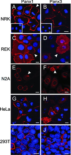
In order to assess if Panx1 and Panx3 exhibited distinct distribution profiles when expressed in different cells, we expressed untagged Panx1 and Panx3 in five cell lines of distinct origin. As previously shown (Penuela et al. Citation2007), both Panx1 and Panx3 exhibited a prevalent cell surface distribution with limited clustering when expressed in NRK cells (, ). It was notable that the distribution profile did not change substantially whether the contacting cell expressed pannexins or not. Doubling of the signal intensity was observed in areas of cell-to-cell contact, but no plaques or aggregates indicative of gap junctions were clearly observed (, ). Importantly, the preincubation with the cognate peptides used to generate these antibodies eliminated the immunostaining, providing good evidence that these antibodies have a high affinity and avidity for their respective pannexin family member (, , inserts). Interestingly, when Panx1 and Panx3 were expressed in REKs, both pannexins were localized to the cell surface with a marked increase in fluorescent intensity due to signals coming from two opposing cell membranes (, ). In addition, these keratinocytes retained considerably more Panx1 and Panx3 within intracellular compartments. Panx1 was localized to the cell surface and a perinuclear compartment in mouse N2A cells (, arrowhead), whereas Panx3 exhibited a more pleomorphic profile when expressed in the same cell type (, ). Typically, N2A cells present mixed populations representing varying degrees of differentiation, and Panx3 appeared more intracellular in spread N2A cells. Ectopic expression of Panx1 and Panx3 in GJIC-deficient HeLa cells resulted in both pannexins being localized to the cell surface and what appears to be an array of intracellular compartments (, ). Finally, 293T cells, originally isolated for their ability to amplify the expression vectors, localized Panx1 and Panx3 to the cell surface with possibly a population of pannexins being retained in the compressed areas representing the cytoplasm (, ). The specificity of all of the observed subcellular distribution profiles were confirmed by peptide preadsorption assays (data not shown).
Figure 1 Immunocytochemistry of cultured cell lines engineered to ectopically express untagged Panx1 or Panx3. The localization of Panx1 (A, C, E, G, and I; red) and Panx3 (B, D, F, H and J; red) were revealed by immunolabeling with purified anti-Panx1 and anti-Panx3 antibodies, respectively. Inserts in A and B depict peptide pre-adsorption assays for each pannexin antibody. Arrowheads denote different pannexin cell surface distribution profiles in NRK (A) and REKs (C, D); Panx1 localization in a perinuclear compartment (E) and Panx3 at the cell surface (F). Nuclei are depicted in blue with Hoechst 33342 (A–D; G–J). Bars = 10 μ m
Trafficking of Glycosylation-Defective Panx1 and Panx3 Mutants
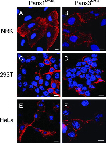
To examine the role of glycosylation in the subcellular localization of pannexins, we previously generated Panx1N254Q and Panx3N71Q mutants that were devoid of predicted glycosylation sites (Penuela et al. Citation2007). When expressed in NRK cells, as previously shown (Penuela et al. Citation2007), these mutants were not as frequently found at the cell surface as their wild type counterparts, resulting in a substantial increase in intracellular pannexins (, ). In the case of 293T cells, cells expressing high levels of the mutants revealed further evidence of intracellular distribution of the glycosylation-defective pannexins (, ). Similar to NRK cells, HeLa cells expressing the pannexin mutants displayed a mixed profile, with a subpopulation of the proteins localized to the cell surface as well as intracellular compartments (, ).
Figure 2 Subcellular expression profiles of N-glycosylation mutants Panx1N254Q and Panx3N71Q. Mutant pannexins were expressed in NRK (A, B), 293T (C, D), and HeLa (E, F) cells prior to immunolabeling for Panx1 (A, C, E; red) or Panx3 (B, D, F; red). Note the presence of both mutants at the cell surface as well as within intracellular compartments. Nuclei were stained with Hoechst 33342 (blue). Bars = 10 μ m.
Evaluation of Intercellular Channel Function of Pannexins and Mutants
The functional status of pannexins in relationship to their putative role in forming intercellular channels is an issue of current debate. To test the possibility that carbohydrate additions to Panx1 and Panx3 may inhibit their ability to form intercellular channels, Panx1N254Q and Panx3N71Q were expressed in GJIC-deficient HeLa cells in parallel with their wild-type counterparts and Cx32, as positive control. Microinjection of paired cells expressing either free GFP or DsRed as indicators of successful transfection of pannexins, mutants, or Cx32 were performed in three independent transfections with two different dyes. Sulforhodamine B transfer was observed in 100% of Cx32-expressing cells injected, but was not observed for any cells expressing the pannexins or the glycosylation-defective mutants (). A few instances of low levels of Lucifer yellow transfer was detected between Panx1 and Panx3 expressing cells, but a similar low level of dye transfer was observed in wild-type HeLa cells. Unlike Cx32-expressing cells, which were extensively coupled, cells expressing either pannexin mutants exhibited no evidence of Lucifer yellow transfer ().
TABLE 1 Incidences of dye transfer in pannexin and mutant expressing HeLa cells
Distribution of Endogenous Panx1 and Panx3 in Cultured Cell Lines
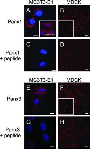
Our previous study revealed that both Panx1 and Panx3 exhibited diverse localization patterns in situ, raising the possibility that endogenous pannexins may also have diverse localization profiles in cultured cells. In the current study, two cell lines, namely MC3T3-E1 (a mouse osteoblastic cell line) and MDCK (Madin-Darby canine kidney) cells, together with primary cultures of mouse osteoblasts expressed detectable levels of endogenous Panx1 and/or Panx3 in immunocytochemistry assays. MC3T3-E1 cells typically revealed an intracellular distribution profile for both Panx1 and Panx3 (, ), whereas Panx1 was also detected in primary osteoblasts (, insert). All antibody binding was effectively competed with cognate peptides ensuring the validity of the signal (, ). Panx1 and Panx3 were both found in MDCK cells at the cell surface with limited evidence for clustering (, ). Intriguingly, MDCK cells varied in their Panx1 and Panx3 expression levels, which may reflect clonal variations or differences in growth densities, and in some instances presented a clear stitching pattern at the cell surface (, inserts). For both pannexins, the antibody labeling was virtually eliminated by competing with the cognate peptide, with the exception of some non-specific nuclear staining (, ).
Figure 3 Expression of endogenous Panx1 and Panx3 in cultured cell lines and primary osteoblasts. Immunolabeling of the mouse osteoblast cell line MC3T3-E1 (A, C, E, G; red) for Panx1 or Panx3, or primary osteoblasts for Panx1 (A, insert) revealed that these pannexins were localized to intracellular compartments. Immunolabeling of MDCK cells (B, D, F, H) for Panx1 or Panx3 revealed that these pannexins were evenly distributed at the cell surface in most cells while some cells exhibited a cell surface stitching pattern (B, F, inserts). When the antibodies were premixed with the cognate peptides used to generate the anti-pannexin antibodies, the vast majority of labeling was eliminated (C, D, G, H). Nuclei were stained with Hoechst 33342 (A, C, E, G). Bars = 10 μ m.
DISCUSSION
Up until the beginning of the new century, only connexins were thought to be capable of forming gap junctions and mediating GJIC. The identification of the pannexin family of proteins resulted in this position being revisited, as their sequence homology to the invertebrate innexins was compelling. In recent years, rat and mouse pannexins have been cloned and expressed in a number of cellular systems. summarizes many of the major findings for two of the three pannexin family members (Panx1 and Panx3) and highlights features that are shared with connexin family members and many others that are unique to pannexins. It is notable that these two pannexins are glycoproteins and are robust in forming functional single-membrane channels, whereas their diverse cellular and subcellular distribution profiles and inconsistencies in forming intercellular channels remain as areas of considerable interest. In the present study, we further address the distribution of ectopic and endogenously expressed Panx1 and Panx3 and evaluate the possibility that glycosylation-defective mutants may form robust GJIC.
TABLE 2 Summary of pannexin 1 and pannexin 3 characteristics
Consistent with the in situ distribution of Panx1 and Panx3 (Penuela et al. Citation2007), diverse subcellular distribution profiles were observed for both wild-type Panx1 and Panx3 when expressed in reference cell lines of different tissue origin. Although in most instances a clear cell surface distribution was observed, the subpopulation of pannexins that remained within intracellular compartments was quite varied. This diversity of subcellular localization was also recapitulated for endogenous pannexins, as MDCK cells clearly localized Panx1 and Panx3 to the cell surface, whereas cells of osteoblastic origin localized Panx1 and Panx3 to intracellular compartments. Previous reports of endogenous Panx1 in astrocytes (Locovei et al. Citation2007), hippocampal neurons (Zoidl et al. Citation2007), oligodendrocytes, epithelial cells, spinal motoneurons, CA1 pyramidal cells in the hippocampus, among others (Zappala et al. Citation2006; Huang et al. Citation2007a), point towards a predominantly intracellular distribution of Panx1 in these primary cell cultures. This distribution profile may reflect a role for pannexins as single-membrane channels, as suggested by the reported evidence for Panx1 forming Ca2 +-permeable channels in the endoplasmic reticulum that contribute to Ca2 + leak and participate in calcium signaling and homeostasis (Vanden Abeele et al. Citation2006). Distinct pannexin localization patterns have also been demonstrated in situ. For instance, keratinocytes within the human epidermis express both Panx1 and Panx3 with the distribution of Panx1 being more punctate at what appears to be cell surfaces, whereas Panx3 was more intracellular (Penuela et al. Citation2007). Here we observe in cultured REKs that both Panx1 and Panx3 have similar distributions when ectopically expressed, with an enrichment of fluorescence at sites of cell-to-cell contact that far exceeds the intensity seen at locations where the contacting cells do not overexpress pannexin. Moreover, both Panx1 and Panx3 exhibit a diffuse background in the cytoplasm of the cell. This reference cell model provides support for the possibility that pannexins in adjacent cells possibly interact to stabilize their localization. Thus, this raises the further likelihood that pannexins are regulated differentially depending on the cellular origin and environment.
HeLa cells have been reported to express Panx1 endogenously (Dvoriantchikova et al. Citation2006b) and colocalized with Golgi-specific markers. However, in our studies, immunolabeling of wild-type HeLa cells failed to detect endogenous Panx1 or Panx3, suggesting that there is variability amongst HeLa cells clones. Upon ectopic expression of Panx1 or Panx3, HeLa cells revealed a distribution pattern similar to NRK, N2A, and 293T cells. However, Panx3 was found to exhibit a cytoplasmic distribution more frequently than Panx1, suggesting that some trafficking differences may exist between these two pannexins. Previous reports of ectopic expression of rat Panx1-GFP in human bone marrow endothelial cells resulted in its localization to the endoplasmic reticulum and Golgi apparatus (Dvoriantchikova et al. Citation2006a). Alternatively, mouse Panx1-GFP was observed to traffic efficiently to the cell surface of NRK cells, whereas only Panx3-GFP was retained in the endoplasmic reticulum (Penuela et al. Citation2007). In C6 glioma cells, Panx1-GFP was found both at the cell surface and intracellular compartments (Lai et al. Citation2007) and was found to have a delayed delivery to the cell surface in human prostate cells (LNCaP) cells (Vanden Abeele et al. Citation2006). Thus, caution needs to be exercised in the use of GFP-tagged pannexins, because it is not yet clear that the GFP tag is completely benign as it may inhibit or delay the maturation of Panx1 or Panx3 as it traffics to the cell surface.
Recently, the glycosylation-defective mutants of Panx1 and Panx3 have been shown to significantly increase the intracellular population of these pannexins, possibly reflecting a decrease in mutant transport to the cell surface (Penuela et al. Citation2007). This observation is consistent with that reported for 293T cells by Boassa et al. (Citation2007) where the Panx1N254Q-myc mutant was not found to localize to the plasma membrane. However, expression of the Panx1N254Q and Panx3N71Q mutants in NRK and HeLa cells consistently revealed a notable cell surface population, with a significant intracellular retention of the mutants, again highlighting some differences in reference cell models. These differences could be linked to the presence of low levels of endogenous pannexins in some reference cells lines and not others, or possible differences in pannexin binding partners that have yet to be determined. Interesting, endogenous Panx1 exhibits polymorphic Western blot banding profiles indicative of diverse levels of N-glycosylation in different tissues (Penuela et al. Citation2007), suggesting that there is considerable heterogeneity in the glycosylation status of Panx1. These observations lead to the possibility that different glycosylation levels could control the distribution profiles and ultimately the function of pannexins in vivo. Other glycoproteins like the Fc receptor have been shown to exhibit trafficking defects when N-glycosylation is inhibited (Gut et al. Citation1998). N-glycan motifs have also been documented as apical sorting determinants for several glycoproteins expressed in epithelial cells (Potter et al., Citation2006). It remains to be determined if the carbohydrate motifs of pannexins direct their trafficking to specific cell surface domains in polarized cells. It also requires further experimentation to determine whether unglycosylated pannexin mutants are more rapidly internalized and degraded than their wild-type counterparts, resulting in an increased intracellular steady-state population.
In the current study, we tested the hypothesis that the carbohydrate moieties of Panx1 and Panx3 might act as negative determinants to intercellular channel formation. This hypothesis is based on the fact that connexins are not glycosylated, and that perhaps the sugars present at the cell surface would act as an impediment to proper docking of the pannexin channels, especially if pannexins are glycosylated to include negatively charged sugars (e.g., sialic acid). However, our use of a coexpression strategy where fluorescent proteins were used as reporter proteins for pannexin and mutant expression found no meaningful increases in GJIC as assessed by dye transfer. This finding is consistent with the lack of junctional conductance observed for the Panx1 glycosylation-defective mutant in paired oocytes (Boassa et al. Citation2007). Thus, the presence or absence of the carbohydrate moieties of pannexins alone does not appear to be sufficient to explain why GJIC was observed in some, but not all experimental settings. Panx1 homomeric channels have been reported to form large pores with single channel conductances of 550 pS (Bao et al. Citation2004) and thus are predicted to be large enough to allow the passage of molecules up to 1000 Da (Bruzzone et al. Citation2005; Locovei et al. Citation2006). Therefore, solely based on size, functional Panx1 intercellular channels would be expected to allow the passage of Lucifer yellow (MW 457) and sulforhodamine B (MW 558). However, three independent groups found no significant evidence for dye or electrical coupling in vitro under similar culture conditions (Boassa et al. Citation2007; Penuela et al. Citation2007; Huang et al Citation2007a). It is still possible that different cell backgrounds, tissues, or conditions may allow for the formation of functional intercellular channels of homo-or heteromeric pannexins.
In summary, our findings support the hypothesis that Panx1 and Panx3 have multiple subcellular distribution profiles when endogenously or ectopically expressed. Thus, the functional status of pannexins in forming single-membrane channels, or possibly intercellular channels, is not necessarily expected to be the same in all cell types. Future research will need to continue to focus on the regulatory role that glycosylation plays, the conditions by which pannexin family members intermix, and the role putative pannexin binding proteins may play in determining their ultimate fate and function.
This work was supported by the Canadian Institutes of Health Research to D.W.L.
REFERENCES
- Bao L, Locovei S, Dahl G. Pannexin membrane channels are mechanosensitive conduits for ATP. FEBS Lett 2004; 572: 65–68
- Baranova A, Ivanov D, Petrash N, Pestova A, Skoblov M, Kelmanson I, Shagin D, Nazarenko S, Geraymovych E, Litvin O, Tiunova A, Born T L, Usman N, Staroverov D, Lukyanov S, Panchin Y. The mammalian pannexin family is homologous to the invertebrate innexin gap junction proteins. Genomics 2004; 83: 706–716
- Boassa D, Ambrosi C, Qiu F, Dahl G, Gaietta G, Sosinsky G. Pannexin1 channels contain a glycosylation site that targets the hexamer to the plasma membrane. J Biol Chem 2007; 282: 31733–31743
- Bruzzone R, Barbe M T, Jakob N J, Monyer H. Pharmacological properties of homomeric and heteromeric pannexin hemichannels expressed in Xenopus oocytes. J Neurochem 2005; 92: 1033–1043
- Bruzzone R, Hormuzdi S G, Barbe M T, Herb A, Monyer H. Pannexins, a family of gap junction proteins expressed in brain. Proc Natl Acad Sci U S A 2003; 100: 13644–13649
- Dvoriantchikova G, Ivanov D, Panchin Y, Shestopalov V I. Expression of pannexin family of proteins in the retina. FEBS Lett 2006a; 580: 2178–2182
- Dvoriantchikova G, Ivanov D, Pestova A, Shestopalov V. Molecular characterization of pannexins in the lens. Mol Vis 2006b; 12: 1417–1426
- Gut A, Kappeler F, Hyka N, Balda M S, Hauri H P, Matter K. Carbohydrate-mediated Golgi to cell surface transport and apical targeting of membrane proteins. EMBO J 1998; 17: 1919–1929
- Huang Y, Grinspan J B, Abrams C K, Scherer S S. Pannexin1 is expressed by neurons and glia but does not form functional gap junctions. Glia 2007a; 55: 46–56
- Huang Y-J, Maruyama Y, Dvoryanchikov G, Pereira E, Chaudhari N, Roper S D. The role of pannexin 1 hemichannels in ATP release and cell-cell communication in mouse taste buds. Proc Natl Acad Sci U S A 2007b; 104: 6436–6441
- Lai C PK, Bechberger J F, Thompson R J, MacVicar B A, Bruzzone R, Naus C C. Tumor-suppressive effects of pannexin 1 in C6 glioma cells. Cancer Res 2007; 67: 1545–1554
- Locovei S, Bao L, Dahl G. Pannexin 1 in erythrocytes: Function without a gap. Proc Natl Acad Sci USA 2006; 103: 7655–7659
- Locovei S, Scemes E, Qiu F, Spray D C, Dahl G. Pannexin1 is part of the pore forming unit of the P2X7 receptor death complex. FEBS Lett 2007; 58: 483–488
- McLachlan E, Manias J, Gong X, Lounsbury C, Shao Q, Bernier S, Bai D, Laird D. Functional characterization of oculodentodigital dysplasia-associated Cx43 mutants. Cell Commun Adhes. 2005; 12: 279–292
- Panchin Y, Kelmanson I, Matz M, Lukyanov K, Usman N, Lukyanov S. A ubiquitous family of putative gap junction molecules. Curr Biol 2000; 10: R473–R474
- Pelegrin P, Surprenant A. Pannexin-1 mediates large pore formation, interleukin-1beta release by the ATP-gated P2X7 receptor. EMBO J 2006; 25: 5071–5082
- Penuela S, Bhalla R, Gong Q-X, Cowan K N, Celetti S J, Cowan B J, Bai D, Shao Q, Laird D W. Pannexin 1 and pannexin 3 are glycoproteins that exhibit many distinct characteristics from the connexin family of gap junction proteins. J Cell Sci 2007; 120: 3772–3783
- Phelan P. Innexins: members of an evolutionarily conserved family of gap-junction proteins. Biochim Biophys Acta 2005; 1711: 225–245
- Potter B A, Hughey R P, Weisz O A. Role of N-and O-glycans in polarized biosynthetic sorting. Am J Physiol Cell Physiol 2006; 290: C1–C10
- Romanov R A, Rogachevskaja O A, Bystrova M F, Jiang P, Margolskee R F, Kolesnikov S S. Afferent neurotransmission mediated by hemichannels in mammalian taste cells. EMBO J 2007; 26: 657–667
- Thompson R J, Zhou N, MacVicar B A. Ischemia opens neuronal gap junction hemichannels. Science 2006; 312: 924–927
- Vanden Abeele F, Bidaux G, Gordienko D, Beck B, Panchin Y V, Baranova A V, Ivanov D V, Skryma R, Prevarskaya N. Functional implications of calcium permeability of the channel formed by pannexin 1. J Cell Biol 2006; 174: 535–546
- Zappala A, Cicero D, Serapide M F, Paz C, Catania M V, Falchi M, Parenti R, Panto M R, La Delia F, Cicirata F. Expression of pannexin1 in the CNS of adult mouse: Cellular localization and effect of 4-aminopyridine-induced seizures. Neuroscience 2006; 141: 167–178
- Zoidl G, Petrasch-Parwez E, Ray A, Meier C, Bunse S, Habbes H-W, Dahl G, Dermietzel R. Localization of the pannexin1 protein at postsynaptic sites in the cerebral cortex and hippocampus. Neuroscience 2007; 146: 9–16