
Abstract
To assess the effects of prolonged exposure to arsenic (As, as arsenate) on host immune competence overall and resistance to Newcastle disease (ND) viral infection in particular, a study was carried out in broiler chicks. At 7 days of age, chicks were assigned to groups that would undergo varying vaccination, challenge, and/or As exposures; Group 1 was a control; Group 2 was to receive Newcastle disease virus (NDV) only; two groups (Groups 3, 4) were to be given As daily (50 mg/kg, by gavage) from Days 7–35 of the experiment. All groups underwent normal vaccination on Days 5, 23, and 32 against live NDV (B1 type, LaSota strain); two groups (Groups 2, 4) were challenged with field-isolated NDV at Day 24. At Days 14, 21, 28, and 35 of age, subsets of chicks in each group were evaluated. The results showed feed intake and weight gain were lower in As-treated and NDV-challenged chicks. In As-treated chicks, absolute and relative spleen weights were significantly greater, whereas those of the thymus significantly lower, over the entire 35-day period. Effects on bursa weights (absolute, relative) were only significantly reduced through Day 21. Antibody titers against ND were significantly reduced (vs. control) over the whole 35 days in birds that received As alone, but only significantly depressed through the first 21 days in birds that received As + NDV; thereafter, titers were significantly greater (in parallel with effects in birds that received NDV alone). In contrast, antibody responses to T-dependent antigen (Sheep red blood cells [SRBC]) were significantly lower in As only- and As + NDV-treated chicks throughout the study period. Among birds exposed to As (alone or with NDV), in situ phagocytic activity was elevated and cutaneous sensitivity responses decreased during the period from Day 28 to Day 35. NDV alone had spurious effects on phagocytic activity but did cause significant reductions in cutaneous sensitivity responses. It was concluded that arsenic decreased immunity in broiler chicks, thereby making them prone to ND.
Introduction
Heavy metal toxicity is a major abiotic stress leading to hazardous outcomes in exposed organisms (Hossain et al. Citation2012; Yang et al. Citation2014; Kousar & Javed Citation2015); in many cases, because metals can bioaccumulate in water and food (Palaniappan & Vijayasundaram Citation2009; Kousar & Javed Citation2014; Javed Citation2015), the risk of greater levels of exposures can increase over time. Arsenic – the twelfth most common element present in nature – is a semi-metallic element found in soil, groundwater, surface water, air, and in various foods. Arsenic occurs naturally in the Earth’s crust, with higher concentrations found in specific geographic areas, rocks, and minerals (Duker et al. Citation2005; Ravenscroft et al. Citation2009; Zhang et al. Citation2014). Although arsenic is found in inorganic and organic forms in the environment, it is the inorganic forms that present the more serious health concern (Ventura-Lima et al. Citation2010; Kousar & Javed Citation2015). Of the many potential effects in situ, arsenic inhibits DNA repair, induces chromosomal aberrations, and disrupts gene expression (Tong et al. Citation2015).
Arsenic compounds such as roxarsone (trivalent derivative of phenylarsonic acid: C6H5As(O)(OH)2) have been extensively used as a growth promoter in the poultry industry (Jones Citation2007). Under such circumstances, the concentrations of arsenic in biowaste is increased, and this, in turn, leads to contamination of soils and increased levels of the metal in nearby plants and vegetables (Andra et al. Citation2010; Khan et al. Citation2013). The clinical signs of arsenic toxicity can include decreased weight gain/feed intake, generalized dullness, as well as increases in skin lesions and salivation (Wu et al. Citation2008; Sharaf et al. Citation2013). Arsenic, both in vivo and in vitro, also acts as an immunosuppressive to impair the function of several key immune cells (Martin-Chouly et al. Citation2011). As a result, general decreases in cellular and humoral immune response have been documented; in one specific case, the number of splenic antibody-forming cells generated against a T-dependent antigen (like sheep erythrocytes) was decreased (Nain & Smits Citation2012).
Newcastle disease (ND), a devastating viral disease among poultry, causes a fatal enteric, respiratory and neurological pathology (Gowthaman et al. Citation2011; Munir et al. Citation2015), often leading to high morbidity and mortality (Miller et al. Citation2010; Ge et al. Citation2015). Heavy metal exposure of birds decreased antibody titers against Newcastle disease virus (NDV) – even in vaccinated flocks – and significantly increased the mortality percentages in the exposed Japanese quail (Nain & Smits Citation2011). In Pakistan, particularly in southern and central areas, groundwater levels of arsenic are high (up to 100 μg/L) as compared to permissible levels recorded by the World Health Organization (10 μg/L; WHO Citation2001). In Sindh and Punjab, ≈ 20–36% of the population is routinely exposed to arsenic (Islam et al. Citation2009) through consumption of contaminated poultry, animal products, drinking water, and other dietary sources with high concentrations of the metal (i.e. mushrooms and rice) (Datta et al. Citation2012).
Although there is information in the literature concerning effects of arsenic toxicity and host responses to the virus (Dangleben et al. Citation2013), there is no information regarding the situation in Pakistan – in particular, immunotoxicological findings in and arsenic-exposed broiler chicks concurrently infected with the NDV. Therefore, the present study describes the effect of arsenic on the immune system of broiler chicks exposed to ND vaccine and field virus.
Material and methods
Chemicals and other reagents
Arsenic (as disodium hydrogen arsenate, Na2HAsO4 7H2O) was purchased from Merck (Darmstadt, Germany). Black India ink was obtained from Pelikan (Hanover, Germany). Avian tuberculin was bought from the Veterinary Research Institute (Lahore, Pakistan). Sheep red blood cells (SRBC) were prepared from blood obtained from a local abattoir. After centrifuging (150 × g, 15 min, room temperature), the SRBC were suspended in normal saline to yield a 3% suspension. For the hemagglutination inhibition (HI) assay, NDV antigen was purchased from M/S Intervet (Kempton Park, South Africa).
Newcastle disease virus isolation and determination of50% embryo lethal dose (ELD50)
Previously-isolated NDV – following the method described by Pansota et al. (Citation2013) – was used to challenge some of the broiler chicks. In brief, organs (lungs, trachea, spleen) were collected from chicks in areas of suspected field outbreaks of ND. Tissues (≈1 g) were homogenized in normal saline (10 mL) and gentamycin was added (1 mg/mL) (Alexander Citation1989). The homogenate was centrifuged (1000 × g, 20 min, 4 °C) and the supernatant was stored in aliquots at –20 °C. After thawing, the supernatant was inoculated into 11-day-old embryonated eggs via the chorioallantoic route (Hitchner Citation1980). Allantoic fluid of dead embryos was then harvested and tested for viral presence using a spot agglutination test/hemagglutination test (Allan Citation1978) and confirmed by a HI test (MAAF 1984). Confirmed isolates were further processed for pathogenicity in assays based on Mean Death Time; isolates were characterized as Lentogenic, Mesogenic, or Velogenic strains (Alexander Citation1989). The 50% embryo lethal dose (ELD50) of Velogenic strains of NDV was determined by the method of Villegas and Purchase (Citation1989). Fertilized eggs were incubated at 37 °C for 9 days before being inoculated with 10 μL mL of 10-fold serial dilutions (10−3, 10−4, 10−5, and 10−6; 5 eggs/dilution) of allantoic fluid of the virus. The eggs were then incubated at 37 °C to permit determination of the ELD50. Embryo mortality was evaluated for 7-day post-inoculation. The ELD50 was found to be 10−8 and so was selected as the dose used in the current experiment.
Experimental design
A total of 96 1-day-old broiler chicks were procured from a hatchery and kept in metal wire cages in a sterile facility maintained at ambient temperature (26 ± 2 °C) with a 75% relative humidity. The chicks were provided ad libitum access to a corn soy basal feed containing 21% total protein (Abu-Akkada & Awad Citation2015) and filtered water throughout the experiment. All chicks were vaccinated against hydropericardium syndrome (Day 17), infectious bronchitis (Day 5), and infectious bursal disease (Days 10 and 20) over the course of the experiment (see below).
After 6 days of acclimatization, the chicks were divided randomly into four equal groups (n = 24/group; ). All the groups were vaccinated according to standard approved vaccination schedule. Chicks in Groups 3 and 4 received arsenate (50 mg/kg, by crop tubing of stock solution that was freshly prepared in distilled water) daily from Day 7- to Day 35 of age. All chicks in Groups 2 and 4 were challenged by field-isolated NDV on Day 24 of age. The dose of arsenate used here was based on already established findings in broiler chicks by Mashkoor et al. (Citation2013).
Table 1. Groupings and treatments of broiler chicks.
This experiment was planned in accordance with all national legislation concerning the protection of animal welfare and followed guidelines set by the University Graduate Studies and Research Board (GSRB); the GSRB approved all experimental protocols.
Feed intake, body weight, absolute and relative organ weights
Because several birds within each treatment group were to receive exogenous agents, e.g. SRBC, tuberculin antigen, India ink, at various timepoints over the course of the study, a dedicated stand-alone subset of birds in each group were used solely for monitoring of feed intake (calculated as g/bird/day), body weight (BW) assessments (on weekly basis), for antibody titer levels vs. ND vaccine (see below), and ultimately, for assessment of various immune system organ relative weights. At Days 14, 21, 28, and 35 of the experiment, immediately after being bled from the wing vein, random chicks within each experimental group subset were euthanized by cervical dislocation. After obtaining their final BW, specific organs (i.e. spleen, bursa of Fabricius, thymus) were removed and absolute weights obtained. Relative weights (i.e. as percentage of BW) of each organ were subsequently calculated.
Hemagglutination inhibition for Newcastle diseasevaccine virus
The blood collected above was used to assess the extent of host humoral responses against the ND vaccine virus. Subsequently isolated serum was used in a HI assay (Chukwudi et al. Citation2012) wherein normal saline (50 μL) was added to each well of a round-bottom microtiter plate after which test serum (50 mL) was added to the first well of a dedicated row and serial 1:1 dilutions made until a 1:1024 dilution was attained. Diluted (4HA) NDV antigen (50 μL) was then added to each well and the plate incubated for 30 min at room temperature. Washed naïve chicken RBC suspension (1%, 50 μL) was then added to each well and the plate was incubated at 37 °C for 30 min. Antibody titers were then interpreted based on the standard protocol (Buxton & Fraser Citation1977) of identifying the final well in a row that showed deposition of agglutinated materials.
Antibody titer against sheep red blood cells
At Day 7 of age, 1 mL of a 3% SRBC suspension was injected intravenously into a separate subset of three chicks in each group (i.e. primary dosing). A booster dose was then injected into these chicks 14 days later (i.e. at Day 21 of age). Wing vein blood samples were collected into tubes without anti-coagulant from these chicks at 7, 14, 21, and 28 days after the primary dose. Each time, serum was isolated using standard protocols and stored at −20 °C until analysis of the antibody titers. At that time, each serum sample was inactivated at 56 °C for 30 min and then analyzed for total anti-SRBC antibodies using the method of Delhanty and Solomon (Citation1966). In brief, each inactivated serum sample was titrated for total and mercaptoethanol (ME)-resistant (IgG) anti-SRBC antibody titers. ME-sensitive (IgM) antibody titers were obtained by subtracting the level (titer) of ME-resistant antibodies from that of the total antibodies measured. All titer data were expressed in terms of log2.
Cutaneous sensitivity response to avian tuberculin
At Day 32 of age, a different subset of three chicks/group received 0.2 mL avian tuberculin (0.5 mg/mL saline) by injection into the intra-digital space between the 3rd and 4th digit of their right foot. Normal saline (0.2 mL) was injected into the same space in the left foot as the “self”-control. In all cases, skin thickness between the digits (of both right and left foot) was measured via a micrometer screw gauge (to assess host cell-mediated immune response) 24, 48, and 72-h post-injection. Thickness was also measured immediately prior to either injection to provide a baseline value (Zahid et al. Citation2015). The cutaneous hypersensitivity response at each timepoint was calculated as [toe web thickness after injection – before injection] OR [avian tuberculin response right foot – saline response left foot] (Akhtar et al. Citation2008).
Carbon clearance assay
A supernatant fraction of Black India ink was obtained by centrifugation (3000 × g) for 30 min. At Day 14 of age, another different subset of three chicks/group received ink (1 mL/kg) via injection in the right wing vein. An identical treatment was carried out on another subset set of three chicks/group at Day 28 of age. On each of those days, blood samples were collected from the left wing vein prior to (0 min) and 3- and 15-min post-ink injection. Each blood sample was immediately transferred into 4 mL of 1% sodium citrate and centrifuged (50 × g, 25 °C) for 4 min. The relative amount of carbon particles remaining in the supernatant was then measured at a wavelength of 640 nm in a Spectro 20D Plus RS-232C spectrophotometer (Labmed Inc., Los Angeles, CA), using the 0 min sample of each bird as a zero value for its respective sample (Sarker et al. Citation2000).
Statistical analysis
All data are reported as means ± SD. Data were subjected to a one-way analysis of variance (ANOVA) and the means of different groups were compared by least significant difference using a Statistix statistical package (M/S Analytical Software, Tallahassee, FL). Statistical significance was accepted at a p values <0.05.
Results
General host parameters
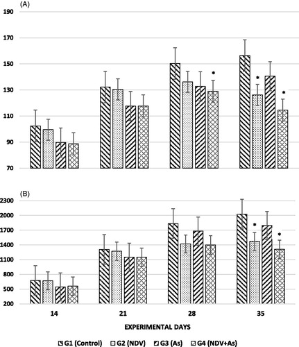
Feed intake at Days 14 and 21 was not significantly different among all experimental chicks when compared to that of the control chicks (Group 1). However, significantly lower feed intake at Day 28 among chicks in Group 4 was noted. Chicks in Groups 2 and 4 showed significantly lower feed intake at Day 35 (). At Days 7 (data not shown), 14, 21, and 28, a non-significant difference was seen in the BWs of all groups in comparison to those of the control chicks. Significantly (p < 0.05) lower BWs as compared with control chicks were noted in chicks in Groups 2 and 4 at Day 35 ().
Figure 1. Effects of exposures on feeding and body mass of broiler chicks. (A) Feed intake (in terms of g/day). (B) Body weight (g) of test birds. Group 1 (G1): Control. Group 2 (G2): NDV-vaccine alone. Group 3 (G3): Arsenic-treated and non-vaccinated. Group 4 (G4): Arsenic-treated and NDV-vaccinated. All chicks in Groups 2 and 4 were challenged by field-isolated ND virus when they reached Day 24 of age. Values shown are means (±SD) in grams; N = 3/group. *Value significantly different from control on specific day (p < 0.05).
The absolute and relative weights of the spleen on all experimental days (except in Group 4 at Day 14) were significantly (p < 0.05) higher in birds in Groups 3 and 4 as compared to values for the controls. The absolute and relative weights of the spleen in Group 2 hosts at Day 35 were also higher relative to that of the control ( and ). In comparison, absolute bursa weights were significantly lower (vs. control chick values) in chicks in Groups 3 and 4 on Days 14 and 21; on all other days, in all Groups the differences in weights from control bird values were not significant (). A similar trend was observed in relative weights of the bursa () except that on Day 21, the significance of differences were no longer apparent – likely due in part to what appeared to be a certain (albeit insignificant) loss in BW in the Group 3 and 4 birds. Lastly, the absolute and relative weights of each thymus were significantly lower (vs. control chick values) in birds in Groups 3 and 4 throughout the experiment (). In addition, relative thymus weights were also significantly lower in Group 2 chicks as compared to control chicks at Days 28 and 35.
Figure 2. Absolute lymphoid organ weights (g). Values were obtained from organs harvested from birds treated as outlined in Figure 1 legend. (A) Spleen, (B) Bursa, and (C) Thymus. Values shown are means (±SD) of organ weights in grams; N = 3/group. *Value significantly different from control on specific day (p < 0.05).
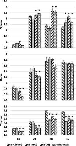
Figure 3. Relative lymphoid organ weights. Values were obtained from organs harvested from birds treated as outlined in Figure 1 legend. (A) Spleen, (B) Bursa, and (C) Thymus. Values shown are means (±SD) of organ weight:body weight ratios (unit-less); N = 3/group. *Value significantly different from control on specific day (p < 0.05).
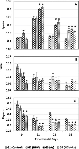
It is unsurprising that while all experimental birds had significantly lower (vs. control) absolute thymic weights at Days 28 and 35 – given the BWs among the chicks in Groups 2 and 4 only at Day 35 were significantly lower than the controls – the relative weights of the thymus in these hosts on that day were less dramatically lower than control relative values when compared to the differences among the groups at Day 28. Given this same “boostive” effect of a lower BW on Day 35, it is clearer why the relative weights of the spleen and bursa of the Group 2 and Group 4 birds were somewhat elevated relative to the trends noted among the absolute weights on that day (compare final columns, and ).
Immune responses
At Days 14 and 21 of the experiment, antibody titers against NDV were significantly lower in arsenic-treated vaccinated chicks (Groups 3 and 4) as compared to in the control vaccinated chicks. On Days 28 and 35, the titers in the challenged groups (Groups 2 and 4) were significantly higher compared to control levels, oddly, on these days, arsenic-treated chicks in Group 3 had significantly lower titers compared to control chicks (). Host antibody titers against SRBC on all experimental days were significantly lower in the arsenic-treated chicks (Groups 3 and 4) as compared to in the controls. However, antibody titers against SRBC on all days did not differ between chicks in Group 2 and the controls – except on Day 35, where the titers against SRBC were significantly lower ().
Figure 4. Antibody titers. (A) Antibody responses against Newcastle disease. (B) Antibody titers against sheep red blood cells. Birds were treated as outlined in Figure 1 legend and then bled on different experimental days. Separate sets of birds were inoculated with SRBC (see Methods) and then bled on indicated days of the experiment. Values shown are mean (±SD) inverse log2 titers. N = 3/group for values against NDV; N = 3/group for values against SRBC. *Value significantly different from control (in given ND or SRBC study) on specific day (p < 0.05).
Among chicks injected with avian tuberculin at Day 32 of age, in situ cutaneous sensitivity responses were significantly consistently decreased in challenged and arsenic-treated chicks at 24-, 48-, and 72-h post-antigen injection (Groups 2, 3, and 4) (). On Days 14 and 28, in situ phagocytic activity of mononuclear cells (via measures of clearance of injected carbon particles) after 3 and 15 min was significantly higher in arsenic-treated chicks in Groups 3 and 4 relative to that seen in control chicks (). Phagocytic activity at 15 min on Day 14 and at 3 min at Day 28 was significantly lower (vs. in control chicks) in ND-challenged chicks (Group 2), however, activity was significantly higher (vs. in control chicks) in these same challenged Group 2 chicks at 15 min on Day 28 ().
Figure 5. Cutaneous sensitivity responses. Responses on different experimental days in birds treated as outlined in Figure 1 legend. Values shown are mean changes (±SD) in mm; N = 3/group. *Value significantly different from control on specific day (p < 0.05).
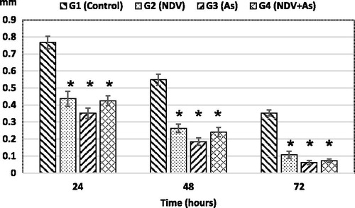
Figure 6. In vivo phagocytic potential. Phagocytic index (PI%) was evaluated in vivo on different experimental days in birds treated as outlined in Figure 1 legend. Bars shown are mean (±SD) PI% values; N = 3/group each at Days 14 and 28. *Value significantly different from control on specific day (p < 0.05).
Discussion
Arsenic, a heavy metal, causes tissue damage of various systems including the immune system (Khan et al. Citation2014). Arsenic may affect activities of immune cells, i.e. monocyte, lymphocyte, and macrophages) resulting in immunosuppression (Duker et al. Citation2005). Arsenic interferes with splenic macrophage ability to present antigen, thereby altering the capacity of antibody-forming cells to generate IgM and IgG against sheep erythrocytes and proliferative response of lymphocytes (Sikorski et al. Citation1991). Moreover, macrophage phagocytic activity was significantly decreased by arsenic exposure in European starlings (Fairbrother et al. Citation1994) and broiler chickens (Vodela et al. Citation1997). In the literature, it is noted that arsenic is an immunosuppressant (Chakraborty et al. Citation2013). With this background on the immunotoxic potential of arsenic, the present study was conducted to explore how arsenic could impact on ND-infected broiler chicks.
In the present study, clinical signs of arsenic toxicity were noted, e.g. decreased BW gain, reduced feed intake, emaciation, lacrimation, open-mouth breathing, pale comb, tremors, and convulsions. Decreased feed intake and weight gain have also been previously reported in other models, i.e. broilers, and fish (Vodela et al. Citation1997; Pedlar et al. Citation2002). In addition to changes in food intake/weight gain, emaciation and lacrimation in arsenic-exposed Mallard ducklings has been documented (Hoffman et al. Citation1992). Decreases in BW gain/feed intake could be due to gastrointestinal disturbances and renal toxicity from the arsenic (Mashkoor et al. Citation2013). In ducks, a decrease in weight, less feed intake, and increased salivation were reported as arsenic toxicities (Islam et al. Citation2009).
In the current study, relative weights of the spleen in arsenic-exposed chicks were significantly higher compared to that in control chicks, while relative weights of the bursa of Fabricius and thymus were lower in all treated groups compared to values in controls. Youssef et al. (Citation1996) also noted decreased bursa and thymus relative weights in ND-challenged arsenic-exposed chicks. In contrast, Aggarwal et al. (Citation2008) did not observe any effect on relative weights of the spleen, bursa, or thymus in arsenic-treated rats. In comparison, Jadhav et al. (Citation2007) and Ai-Zhi and Zhen-Yong (Citation2007) noted increased relative weights of the spleen and thymus in Wister rats and chickens exposed to arsenic via their drinking water. Oxidative stress generated by arsenic results in less feed intake, decreased metabolism, poor absorption of metabolites, and ultimately less BW and organ weights (Sharaf et al. Citation2013). Moreover, oxidative stress also causes cell death in arsenic intoxicated animals. Pyruvate dehydrogenase enzyme, used in the citric acid cycle, is inhibited by arsenic metabolites; as a result of disturbed cell energy production (Aposhian & Aposhian Citation2006), hosts would experience lack of weight gain.
In chicks administered arsenic and challenged with NDV here, there was a significantly lower antibody response against SRBC and ND vaccine virus. The lower antibody titers could be due to decreased overall immune functions in these chicks. In addition to general toxicity, arsenic is known to affect lymphocyte, monocyte, and macrophage activities in many mammals, resulting in immunosuppression (Lage et al. Citation2006; Sakurai et al. Citation2006). Likewise, phagocytic activity of macrophages and other immune responses have been reported to be significantly reduced in arsenic-treated broiler chickens (Vodela et al. Citation1997). Generally, arsenic can disrupt glucocorticoid regulation of immune function (Kaltreider et al. Citation2001) and arsenic-mediated apoptosis may lead to a reduced/diminished immune response in mice (Harrison & McCoy Citation2001), rats (Bustamante et al. Citation1997), and humans (Gonzales-Rangel et al. Citation2005). Li et al. (Citation2010) and Cho et al. (Citation2012) also noted a decrease in humoral immunity to SRBC (T-lymphocyte macrophage-dependent response) in rats exposed to arsenic. Arsenic trioxide treatment of mice caused suppressed primary humoral responses to SRBC (Burchiel et al. Citation2009). Lastly, intratracheal administration of gallium arsenide to mice led to decreased primary IgM antibody responses against SRBC (Sikorski et al. Citation1991). Thus, the results here with the arsenic-treated chicks are in keeping with outcomes already documented in mammals exposed to arsenicals.
The present study showed that the response to intradermal administration of avian tuberculin was significantly lower in birds treated with NDV and/or arsenic (alone or in combination) compared to that by the control birds. In normal reactions, T-cells (and their functional sub-populations), B-cells, and mononuclear phagocytes all are key to the tuberculin reactions. Notably, there are time-related shifts in T-cell subtypes during the course of this response (Konttinen et al. Citation1983; Morzadec et al. 2012), with variations in the local proportion/numbers of CD4+ and CD8+ cells playing an active role in the generation of any positive tuberculin reaction. Intra-dermal administration of avian tuberculin here would be expected to stimulate T-cells with minimal effects on B-cells and result in increased cytokine/chemokine release and recruitment of an array of immune cells (mostly macrophages, neutrophils, etc.) to the injection site, thereby increased skin thickness at the site of administration would occur.
Among the birds exposed to arsenic here, reduced cutaneous responses could be attributable in part to effects of arsenic that resulted in decreased T- and B-cell numbers and/or activity. Burchiel et al. (Citation2009) reported a decrease in T-cell population levels in arsenic-exposed mice and Claudie et al. (Citation2012) noted that humans exposed to arsenic yielded T-cells with demonstrably lower proliferative activities (ex vivo). One could surmise the effect seen here might also be attributed to changes in macrophages/granulocytes processing the tuberculin antigen or in the degree of cells migrating to the tissue. Of these two options, the former would seem unlikely. This is because the phagocytic activity data at Day 28 indicated the treated birds (NDV and/or arsenic, each alone or in combination) displayed increased functionality among their phagocytic cells and it seems unlikely within 4 days this effect would be lost (i.e. recall that tuberculin injection occurred at Day 32 of age). Still, the increases in phagocytic activity were somewhat unexpected in that other studies with various host species reported that phagocytic activity of isolated macrophages/cells in situ were significantly decreased by arsenic exposure (Vodela et al. Citation1997; Nayak et al. Citation2007; Aggarwal et al. Citation2008; Das et al. Citation2010).
Instead, it would seem the amounts of cells migrating to the tissue were more likely impacted by arsenic exposure. A loss of ability to recruit such cells to the injection site is highly plausible in that cytokine/chemokine production by the lymphocytes was likely impacted by the arsenic. Depressed production of various cytokines by immune cells (macrophages, neutrophils, lymphocytes) in broilers as a result of arsenic toxicity was previously reported by Khan et al. (Citation2014). This decrease in production of cytokines/chemokines (and other immune parameter changes) could be a response to an increased production of reactive oxygen and reactive nitrogen species (ROS and RNS) in response to the arsenic (Yang et al. Citation2013). Induction of oxidative stress has long been considered a mode of toxic action for arsenic. As a metal, arsenic can act as oxidant and so lead to a strong induction of ROS and RNS in cells and in bio-fluids (Lage et al. Citation2006). In addition, the lipophilic nature of arsenic makes membranes more prone to damage, for example lipid peroxidation, leading ultimately to cell death (Farombi et al. Citation2007). Elevated ROS levels in un-stimulated peripheral blood mononuclear cells (PBMC) after exposure to arsenic as well as release of the ROS by the PBMC could – apart from affecting these immune system cells – cause oxidative damage to tissues (Dangleben et al. Citation2013; Urbanska et al. Citation2014) and other immune cells (i.e. local macrophages/monocytes) that might reside in those tissues. Even in the absence of that particular toxic manifestation, it is also well established that arsenic directly impacts on the functions of lymphocytes, monocytes, macrophage, and other granulocytic cells in many mammals (and other models from non-mammalian species), resulting in immunosuppression (Gonsebatt et al. Citation1994; Lage et al. Citation2006; Sakurai et al. Citation2006). Thus, either directly (i.e. by affecting aspects of particular cell function) or indirectly (i.e. via ROS/RNS-induced effects on cell numbers), it is plausible that continued exposure of the chicks to arsenic yielded decreases in measurable production of cytokines/chemokines. Future studies with similarly treated birds will quantify many of the key cytokines related to the various responses measured in this study to validate these suspicions. While this could help explain the observed outcomes here among birds that received any exposures to arsenic, an explanation as to why the birds that received NDV alone had reduced cutaneous sensitivity responses remains uncertain.
Based on the results here, it was concluded by us that arsenic caused toxic effects in broiler chicks that manifest as decreased feed intake, BW, and changes in weights of selected organs (including those key to host immune function). Arsenic-treated broilers also had decreased antibody titers against ND and lowered responses to SRBC. Moreover, the phagocytic ability and lymphoproliferative response of isolated cells were decreased in the case of arsenic treatment and challenged groups. Hence, it was concluded that arsenic decreased immunity in broiler chicks, thereby making them prone to ND.
Acknowledgements
The authors wish to thank the University of Agriculture, Faisalabad, Pakistan is for providing financial assistance.
Disclosure statement
The authors declare no conflicts of interest. The authors alone are responsible for the content of this manuscript.
References
- Abu-Akkada S, Awad A. 2015. Protective effects of probiotics and prebiotics on Eimeria tenella-infected broiler chickens. Pak Vet J. 35:446–450.
- Aggarwal S, Naraharisetti M, Dandapat S, Degen G, Malik J. 2008. Concurrent subacute exposure to arsenic through drinking water and malathion via diet in male rats: Effects on hepatic drug-metabolizing enzymes. Arch Toxicol. 82:543–551.
- Ai-Zhi C, Zhen-Yong W. 2007. Effect of different supplemented arsenic preparation on growth of body weight and main immune organs in chickens. J Domest Animal Ecol. 2007:1.
- Akhtar M, Hafez M, Muhammad F, Haq A, Anwar I. 2008. Immunomodulatory and protective effects of sugar cane juice in chickens against Eimeria infection. Turk J Vet Anim Sci. 32:463–467.
- Alexander D, editor. 1989. A laboratory manual for isolation and identification of avian pathogens. 3rd ed. Philadelphia (PA): University of Pennsylvania.
- Allan W. 1978. Newcastle disease virus vaccines: Their production and uses. Rome: FAO (Food and Agriculture Organization of the United Nations).
- Andra S, Makris K, Quazi S, Sarkar D, Datta R, Bach S. 2010. Organo-copper complexes during roxarsone degradation in wastewater lagoons. Environ Sci Pollut Res Int. 17:1167–1173.
- Aposhian H, Aposhian M. 2006. Arsenic toxicology: Five questions. Chem Res Toxicol. 19:1–15.
- Burchiel S, Mitchell L, Lauer F, Sun X, McDonald J, Hudson L, Liu K. 2009. Immunotoxicity and biodistribution analysis of arsenic trioxide in C57Bl/6 mice following a 2-week inhalation exposure. Toxicol Appl Pharmacol. 241:253–259.
- Bustamante J, Dock L, Vahter M, Fowler B, Orrenius S. 1997. The semiconductor elements arsenic and indium induce apoptosis in rat thymocytes. Toxicology. 118:129–136.
- Buxton A, Fraser G, editors. 1977. Animal microbiology. 1st ed. Oxford: Blackwell Scientific Publications.
- Chakraborty S, Ray M, Ray S. 2013. Sajal cell to organ: Physiological, immunotoxic and oxidative stress responses of Lamellidens marginalis to inorganic arsenite. Ecotoxicol Environ Saf. 94:153–163.
- Cho Y, Ahn K, Back M, Choi J, Ji J, Won JH, Fu Z, Jang J, Kim D. 2012. Age-related effects of sodium arsenite on splenocyte proliferation and TH1/TH2 cytokine production. Arch Pharm Res. 35:375–382.
- Chukwudi O, Chukwuemeka E, Mary U. 2012. Newcastle disease virus shedding among healthy commercial chickens and its epidemiological importance. Pak Vet J. 32:354–356.
- Claudie M, Fidaa BEE, Mélinda M, Olivier F, Laurent V. 2012. Inorganic arsenic impairs proliferation and cytokine expression in human primary T lymphocytes. Toxicology. 300:46–56.
- Dangleben N, Skibola C, Smith M. 2013. Arsenic immunotoxicity: A review. Environ Health. 12:73.
- Das S, Pan D, Bera A, Rana T, Bhattacharya D, Bandyapadyay S, De S, Sreevatsava V, Bhattacharya S, Das S, et al. 2010. Sodium arsenite mediated immuno-disruption through alteration of transcription profile of cytokines in chicken splenocytes under in vitro system. Mol Biol Rep. 38:171–176.
- Datta B, Bhar M, Patra P, Majumdar D, Dey R, Sarkar S, Mandal T, Chakraborty A. 2012. Effect of environmental exposure of arsenic on cattle and poultry in Nadia district, West Bengal, India. Toxicol Int. 19:59–62.
- Delhanty J, Solomon J. 1966. The nature of antibodies to goat erythrocytes in the developing chicken. Immunology. 11:103–113.
- Duker A, Carranza E, Hale M. 2005. Arsenic geochemistry and health. Environ Int. 31:631–641.
- Fairbrother A, Fix M, O’Hara T, Ribic C. 1994. Impairment of growth and immune function of avocet chicks from sites with elevated selenium, arsenic, and boron. J Wildl Dis. 30:222–233.
- Farombi E, Adelowo O, Ajimoko Y. 2007. Biomarkers of oxidative stress and heavy metal levels as indicators of environmental pollution in African cat fish (Clarias gariepinus) from Nigeria Ogun River. Int J Environ Res Public Health. 4:158–165.
- Ge M, Zhang W, Shi G, Xiao C, Zhao X, Zhang R. 2015. Astragalus polysaccharide perseveres cytomembrane capacity against Newcastle disease virus infection. Pak Vet J. 35:382–384.
- Gonsebatt M, Vega L, Montero R, Garcia-Vargas G, DelRazo L, Albores A, Cebrian M, Ostrosky-Wegman P. 1994. Lymphocyte replicating ability in individuals exposed to arsenic via drinking water. Mutat Res. 313:293–299.
- Gonzales-Rangel Y, Portales-Perez D, Galicia-Cruz O, Escudero-Lourdes C. 2005. Chronic exposure to arsenic sensitizes CD3+ and CD56+ human cells to sodium arsenite-mediated apoptosis. Proc West Pharmacol Soc. 48:89–91.
- Gowthaman V, Singh S, Dhama K, Barathidasan R, Ramakrishnan M. 2011. Pathology and molecular diagnosis of Newcastle disease virus infection in broiler breeders. Indian J Vet Path. 35:168–170.
- Harrison M, McCoy K. 2001. Immunosuppression by arsenic: A comparison of cathepsin L inhibition and apoptosis. Int Immunopharmacol. 1:647–656.
- Hitchner SB. 1980. Virus propagation in embryonating eggs. In: Hitchner SB, Domermuth CH, Purchase HG, Williams JE, editors. Isolation and identification of avian pathogens. College Station, Texas, USA The American Association of Avian Pathologists, A&M University; p. 120–121.
- Hoffman D, Sanderson C, LeCaptain L, Cromartie E, Pendleton G. 1992. Interactive effects of arsenate, selenium, and dietary protein on survival, growth, and physiology in Mallard ducklings. Arch Environ Contam Toxicol. 22:55–62.
- Hossain M, Piyatida P, Teixeira da Silva J, Fujita M. 2012. Molecular mechanism of heavy metal toxicity and tolerance in plants: Central role of glutathione. J Botany. 2012:872875.
- Islam M, Awa M, Mostofa M, Begum F, Khair A, Myenuddin M. 2009. Effect of spirulina on toxic signs body weight and hematological parameters in arsenic-induced toxicities in ducks. Intl J Poult Sci. 8:75–79.
- Jadhav S, Sarkar S, Patil R, Tripathi H. 2007. Effects of subchronic exposure via drinking water contaminating metals: A biochemical and histopathological study in male rats. Arch Environ Contam Toxicol. 53:666–677.
- Javed M. 2015. Chronic dual exposure (waterborne + dietary) effects of cadmium, zinc and copper on growth and their bioaccumulation in Cirrhina mrigala. Pak Vet J. 35:143–146.
- Jones F. 2007. A broad view of arsenic. Poult Sci. 86:2–14.
- Kaltreider R, Davis A, Lariviere J, Hamilton J. 2001. Arsenic alters the function of the glucocorticoid receptor as a transcription factor. Environ Health Perspect. 109:245–251.
- Khan A, Hussain H, Sattar A, Khan M, Abbas R. 2014. Toxico-pathological aspects of arsenic in birds and mammals: A review. Intl J Agric Biol. 16:1213–1224.
- Khan A, Sharaf R, Khan M, Saleemi M, Mahmood F. 2013. Arsenic toxicity in broiler chicks and its alleviation with ascorbic acid: A toxico-patho-biochemical study. Intl J Agric Biol. 15:1105–1111.
- Konttinen Y, Bergroth V, Visa-Tolvanen K, Reitamo S, Förström L. 1983. Cellular infiltrate in situ and response kinetics of human intradermal and epicutaneous tuberculin reactions. Clin Immunol Immunopathol. 28:441–449.
- Kousar S, Javed M. 2014. Heavy metal toxicity and bioaccumulation patterns in body organs of four freshwater fish species. Pak Vet J. 34:161–164.
- Kousar S, Javed M. 2015. Diagnosis of metal-induced DNA damage in fish using Comet assay. Pak Vet J. 35:168–172.
- Lage C, Nayak A, Kim C. 2006. Arsenic ecotoxicology and innate immunity. Integr Comp Biol. 46:1040–1054.
- Li Q, Lauer F, Liu K, Hudson L, Burchiel S. 2010. Low-dose synergistic immunosuppression of T-dependent antibody responses by polycyclic aromatic hydrocarbons and arsenic in C57BL/6J murine spleen cells. Toxicol Appl Pharmacol. 245:344–351.
- Martin-Chouly C, Morzadec C, Bonvalet M, Galibert M, Fardel O, Vernhet L. 2011. Inorganic arsenic alters expression of immune and stress response genes in activated primary human T-lymphocytes. Mol Immunol. 48:956–965.
- Mashkoor J, Khan A, Khan M, Abbas R, Saleemi M, Mahmood F. 2013. Arsenic induced clinico-hemato-pathological alterations in broilers and its attenuation by Vitamin E and selenium. Pak J Agric Sci. 50:131–138.
- Miller P, Decanini E, Alfonso C. 2010. Newcastle disease: Evolution of genotypes and the related diagnostic challenges. Infect Genet Evol. 10:26–35.
- Morzadec C, Bouezzedine F, Macoch M, Fardel O, Vernhet L. 2012. Inorganic arsenic impairs proliferation and cytokine expression in human primary T-lymphocytes. Toxicology. 300:46–56.
- Munir T, Aslam A, Zahid B, Ahmed I, Imran M, Ijaz M. 2015. Potential of commonly resident wild birds towards Newcastle disease virus transmission. Pak Vet J. 35:106–107.
- Nain S, Smits J. 2011. Subchronic lead exposure, immunotoxicology, and increased disease resistance in Japanese quail (Corturnix coturnix japonica). Ecotoxicol Environ Saf. 74:787–792.
- Nain S, Smits J. 2012. Pathological, immunological and biochemical markers of subchronic arsenic toxicity in rats. Environ Toxicol. 27:244–254.
- Nayak A, Lage C, Kim C. 2007. Effects of low concentrations of arsenic on innate immune system of the zebrafish (Danio rerio). Toxicol Sci. 98:118–124.
- Palaniappan P, Vijayasundaram V. 2009. The effect of arsenic exposure and the efficacy of DMSA on the proteins and lipids of the gill tissues of Labeo rohita. Food Chem Toxicol. 47:1752–1759.
- Pansota F, Rizvi F, Javed M, Khan M, Khan A, Sharif A, Muhammad G. 2013. Use of hyper-immune serum for passive immunization of chicks experimentally infected with Newcastle disease virus. Pak J Agric Sci. 50:279–288.
- Pedlar R, Ptashynski M, Evans R, Klaverkamp J. 2002. Toxicological effects of dietary arsenic exposure in lake whitefish (Coregonus clupeaformis). Aquat Toxicol. 57:167–189.
- Ravenscroft P, Brammer H, Richards K, editors. 2009. Arsenic pollution: A global synthesis. London: Wiley-Blackwell.
- Sakurai T, Ohta T, Tomita N, Kojima C, Hariya Y, Mizukami A, Fujiwara K. 2006. Evaluation of immunotoxic and immunodisruptive effects of inorganic arsenite on human monocytes/macrophages. Intl Immunopharmacol. 6:304–315.
- Sarker N, Tsudzuki M, Nishibori M, Yasue H, Yamamoto Y. 2000. Cell-mediated and humoral immunity and phagocytic ability in chicken lines divergently selected for serum immunoglobulin M and G levels. J Poult Sci. 79:1705–1709.
- Sharaf R, Khan A, Khan M, Hussain I, Abbas R, Gul S, Mahmood F, Saleemi M. 2013. Arsenic induced toxicity in broiler chicks and its amelioration with ascorbic acid: Clinical, hematological and pathological study. Pak Vet J. 33:277–281.
- Sikorski E, Burns L, Stern M, Luster M, Munson A. 1991. Splenic cell targets in gallium arsenide-induced suppression of the primary antibody response. Toxicol Appl Pharmacol. 110:129–142.
- Tong D, Ortega J, Kim C, Huang J, Gu L, Li G. 2015. Arsenic Inhibits DNA mismatch repair by promoting EGFR expression and PCNA phosphorylation. J Biol Chem. 290:14536–14541.
- Urbanska A, Zolla V, Verzani P, Santambrogio L. 2014. Physiological and pathological role of reactive oxygen species in the immune cells. Chapter 23. In: Massoud A, Rezaei N, editors. Immunology of aging. Berlin: Springer-Verlag; p. 309–321.
- Ventura-Lima J, Bogo M, Monserrat J. 2010. Arsenic toxicity in mammals and aquatic animals: A comparative biochemical approach. Ecotoxicol Environ Saf. 74:211–218.
- Villegas P, Purchase H. 1989. Titration of biological suspensions. In: Alexander D, editor. A laboratory manual for isolation and identification of avian pathogens. 3rd ed. Philadelphia (PA): University of Pennsylvania.
- Vodela J, Renden J, Lenz S, McElhenney W, Kemppainen B. 1997. Drinking water contaminants (arsenic, cadmium, lead, benzene, and trichloroethylene). 1. Interaction of contaminants with nutritional status on general performance and immune function in broiler chickens. J Poult Sci. 76:1474–1492.
- WHO (World Health Organization). 2001. Arsenic and arsenic compounds. International Program on Chemical Safety. Geneva: World Health Organization.
- Wu J, Liu J, Waalkes M, Cheng M, Li L, Li C, Yang Q. 2008. High dietary fat exacerbates arsenic-induced liver fibrosis in mice. Exp Biol Med (Maywood). 233:377–384.
- Yang X, Zhang H, Fan G, Liu D, Ge Y, Jiang J, Wang Z. 2014. DNA damage of lung cells from immature cadmium-ingested mice. Pak Vet J. 34:73–77.
- Yang Y, Bazhin A, Werner J, Karakhanova S. 2013. Reactive oxygen species in the immune system. Int Rev Immunol. 32:249–270.
- Youssef S, El Sanousi A, Afifi N, El-Brawy A. 1996. Effect of subclinical lead toxicity on the immune response of chickens to Newcastle disease virus vaccine. Res Vet Sci. 60:13–16.
- Zahid B, Saleem G, Aslam A, Imran M, Younas M. 2015. Effect of immunostimulants on humoral response against infectious bursal disease in broilers. Pak Vet J. 35:227–230.
- Zhang W, Liu Y, Ge M, Yao C, Xue J, Zhang Z. 2014. Resveratrol reduces oxidative stress and improves arsenic efflux in rats exposed to arsenic trioxide. Pak Vet J. 34:251–253.