
Abstract
Introduction
Minimally invasive tissue sampling of the brain in newborns using the Bard Monopty needle helps to diagnose various neurological conditions by obtaining relevant brain cores. We designed a modified procedure to provide maximum diagnostic utility in brain tissue biopsies.
Method
Twenty newborns underwent postmortem minimally invasive tissue sampling of the brain through the anterior fontanelle and posterior approach, using the engraved lines on the needle labeled from mark 0 to 13. The cores were correlated with conventional autopsy findings.
Results
Meninges were best obtained at marks 0 and 1 from the anterior fontanelle and mark 1 from posterior fontenelle in 85% of cases. Periventricular brain parenchyma was best obtained from mark 3 and mark 1 from anterior and posterior fontanel, respectively in 90% cases. The sampling success in obtaining brain cores was 100%.
Discussion
This modified technique increases the yield of meninges and brain tissue in newborns and aids in diagnosis.
Introduction
Minimally invasive tissue sampling (MITS) is a feasible autopsy technique that is used to collect tissue cores from solid organs like the brain, lungs, and liver of all ages. The first historical reference to this technique was in an adult postmortem study by Dr. Kelly (1800s) in Maryland. Subsequently, in 1930, during the yellow fever outbreak, Parreiras and Genofre designed a needle to obtain tissue cores from target organs, which were used for histopathological and microbiological examination [Citation1]. In 2013, the Barcelona Institute for Global Health documented a similar accuracy as a complete diagnostic autopsy (CDA) with this technique in establishing the cause of death [Citation2]. The advantages of MITS are that it is feasible, less disfiguring, less time-consuming, and can be employed in resource-constrained settings by trained personnel. A report from the United Kingdom documented a consent frequency for autopsy from parents who have experienced stillbirths as less than 50% and 25% in neonatal deaths [Citation3]. Over the last decade, MITS has been considered a convenient replacement for CDA, as this technique is less disfiguring and has greater social acceptance [Citation4,Citation5]. The information gained through MITS can benefit the parents regarding diagnosis, risk of recurrence, and management of future pregnancies. This information benefits researchers, policymakers, and the community for audit surveys and planning health policies, including mortality surveillance [Citation6,Citation7].
Prematurity, low birth weight, neonatal infections, birth asphyxia, and birth trauma account for 78% of neonatal deaths in India [Citation8]. Neonatal hypoxic-ischemic encephalopathy [HIE] is the single most common cause of brain injury in newborns [Citation9]. Other common causes are birth asphyxia, neonatal seizures, intracranial hemorrhage, neural tube defects, hydrocephalus, and infections [Citation10,Citation11]. This study was undertaken with a specific aim to standardize the MITS technique of the brain using the Bard monopty needle to obtain relevant brain cores that would aid in a diagnosis, especially where parental consent cannot be obtained for CDA. The areas of interest are the meninges, periventricular region, and the white matter, especially in preterm babies, where maximum neurological injuries occur. This is a blind technique that has been standardized with reference to the depth of the needle, gestational age, and head circumference in neonates. This study was undertaken to refine this technique to obtain adequate, relevant brain cores, especially the meninges and periventricular parenchyma, which can contribute to the cause of death.
Materials and methods
Study setting and design
This was a one-year prospective observational study conducted in the Department of Pathology, Center for Fetal and Perinatal Pathology, KMC, Manipal. The study commenced after obtaining approval from the Institutional Ethics Committee (IEC 384/2020). Informed written consent was obtained from the guardian/parents.
Inclusion criteria
Twenty consecutive newborns with clinical features of neurological insults, respiratory distress, or asphyxia and sepsis in the neonatal intensive care unit (NICU) were included following their death.
Exclusion criteria
Macerated babies, fetuses, and preterm babies born before 22 weeks of gestational age (GA) were excluded from this study.
Method
The gestational ages of the twenty neonates ranged between 22 to 37 weeks. The neonates were grouped based on the GA and head circumference (HC). Group A included 7 preterm neonates with GA between 22 and 27 weeks and HC of 19–25 cm, and Group B had 13 neonates with GA between 28 and 37 weeks and HC of 23–33 cm. The clinical history of all cases was noted from the case records. After obtaining consent from the parents/guardians, all twenty newborns were subjected to MITS of the brain followed by CDA. All the MITS cores and autopsy materials were reviewed by two pathologists and a Ph.D scholar. The MITS core biopsies were reviewed and reported prior to the conventional autopsy biopsies, and the results were compared.
Bard Monopty needle for brain cores
The Bard Monopty needle is a 16 G, 160 mm needle specifically designed for collecting the brain cores (). The needle is lightweight with sharp tips and beveled trocar to collect samples with minimal crush artifacts and an angled deep sample notch for a high-quality core collection. The needle has a total of 13 markings. To standardize the technique, we designated the tip as a mark 0, followed by the next broad marking as mark 1, ending with mark 13. The double engraved lines after mark 9 were considered together as mark 10. The length of the sample notch where the tissue core is obtained is 15 mm with a penetration depth of 22 mm. The needle was charged prior to the procedure by rotating the handle. Two clockwise half-turns are required to lock the style into place. A white arrow appears in the ready window when the needle is loaded and ready to use.
Figure 1. Brain Bard Monopty needle with brain core.

Figure 2. Procedure for collection of brain cores from the anterior and posterior fontanel.
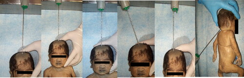
MITS Procedure ()
The deceased’s body was cleaned using 70% alcohol, and the area in the region of the anterior fontanel (AF) and the posterior fontanel (PF) (occipital protuberance) were shaved. The HC and GA of the neonate were noted. The area for puncture was marked at the center of the AF, and the charged needle was inserted on the skin surface at mark 0, and the cores were obtained. The needle was rotated a half-turn, and the cores were removed from the needle. The brain core tissue was observed for consistency, hemorrhage, and yellow discoloration. The sample was collected in a sterile container in 10% neutral buffered formalin. The needle was charged each time before collecting the next core, and the procedure was repeated for marks 1 and 2 at the same site. At mark 3, 0.5 cm right of the center of the AF, the needle was inserted at a 30° inclination toward the left side of the brain. At mark 4, the needle was re-inserted in the center and advanced parallel to the nose. At the PF, 2 cm below the occipital protuberance, three cores were obtained from marks 1, 2, and 3 with the direction of the needle pointing toward the orbit. A total of 8 brain cores were obtained. All cores were collected in separate containers, labeled according to the different marks, fixed in 10% neutral buffered formalin for four hours, and processed for histopathological examination. The cores were sent for histopathological examination.
CDA procedure
The brain was removed by giving an incision along the cranium from the mastoid to the other mastoid by flapping the scalp. The brain was fixed in 10% formalin, and one section each was taken from the meninges, right, and left periventricular area, including the cerebral cortex, cerebellum, and medulla.
Statistical analysis
The statistical analysis was done in Jamovi version 2.3.24 [Citation12,Citation13], and the results were presented as descriptive statistics. We evaluated the correlation between MITS and CDA. Descriptive statistics were performed on categorical variables focused on frequencies and proportions. The sensitivity, specificity, and 95% confidence interval were calculated.
Results
The brain cores were obtained from 20 newborns with a GA of 28.9 ± 4.11 weeks and HC of 26.1 ± 3.91 cm, using the modified MITS technique.
Descriptive analysis between HC, GA, and depth of the needle ()
Group A (GA – 22–27 weeks, HC 19–25 cm)
Anterior fontanel brain cores
The brain tissue obtained at the AF from mark 0 to 4 included the meninges, periventricular area with germinal matrix, cortical brain parenchyma, and white matter. Meninges were obtained from 3 out of 7 cases (42.8%) from marks 0, 1, 3 and 4. Periventricular parenchyma was obtained in all cases (100%), with a maximum yield at marks 2 and 3 (57%). Cortical parenchyma was obtained in 4 out of 7 cases (57%). At mark 4, further penetration was not possible due to the small size of the brain and contact with the base of the skull. Bone was obtained in 4 out of 5 cases (71%).
Table 1. Gestational age and head circumference of newborns.
Table 2. Histopathological findings of MITS from anterior and posterior fontanel.
Posterior fontanel brain cores
Brain tissues obtained from the PF from marks 1 to 3 were meninges, periventricular area with germinal matrix, choroid plexus, cortical parenchyma, occipital cortex, and white matter. Meninges were obtained in 5 out of 7 cases (71%) with maximum yield at mark 1 (60%). Periventricular parenchyma was obtained in 4 out of 7 cases (57%) and the maximum yield was at mark 1 and 3 (75%). Choroid plexus was obtained in 2 cases at mark 1.
Group B (GA – 28–37 weeks, HC 23–33 cm)
Anterior fontanel brain cores
Brain cores obtained from the AF were the meninges, periventricular area with germinal matrix, cortical parenchyma, white matter, and choroid plexus. Meninges were obtained in 11 cases out of 13 (84.6%), and the maximum yield was at mark 0 (72%) and mark 1 in 5 cases (45%). Periventricular parenchyma was obtained in 9 cases (69%), and the highest yield was obtained at mark 3 (77.7%). Cortical parenchyma was obtained in 10 cases (76.9%), and the maximum yield was at mark 4 (70%). Choroid plexus was noted in a single case at mark 3.
Posterior fontanel brain cores
Meninges, periventricular area with germinal matrix, cerebellum, cortical brain parenchyma, white matter, and choroid plexus were obtained. Meninges was obtained in 5 out of 13 cases (38%), with maximum yield at mark 1 and 2 (80%). Periventricular parenchyma was obtained in 5 cases (38%), with a high yield at mark 1 (60%). Cerebellar parenchyma was obtained in 5 cases (38%) and maximum yield at mark 1 (60%). Choroid plexus was obtained in 3 cases (23%), mark 1 showing the maximum yield (66.6%). Occipital cortex was obtained in 5 cases (38%) from marks 1, 2, and 3. Cortical tissue was obtained in 4 cases (30.7%), and maximum at marks 2 and 3.
Histopathological findings of MITS on brain cores
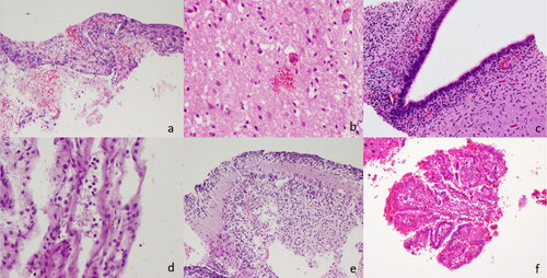
The various lesions identified included subarachnoid hemorrhage (SAH), white matter microhemorrhages, periventricular hemorrhage, intraventricular hemorrhage, germinal matrix hemorrhage, reactive astrocytes, anoxic neurons, and reactive glial cells (). Choroid plexus hemorrhage was seen in 3 cases, along with other grades of hemorrhage. SAH was seen in 9 cases, and subdural hemorrhage in 2 cases. Case 12 showed features of meningitis. Case 18 showed fungal hyphae in the parenchyma. Out of 20, 17 cases (85%) showed features of hypoxic-ischemic changes.
Figure 3. Histopathological findings (a) Subarachnoid hemorrhage, (b) Parenchymal microhemorrhage with anoxic neurons, (c) Periventricular area with germinal matrix with congestion, (d) Meningitis, (e) Cerebellum and (f) Choroid plexus hemorrhage.
In summary, meninges were obtained in 64% of cases at mark 0 anteriorly and in 70% of cases at mark 1 posteriorly. In 68.8% of cases, the periventricular area was obtained at mark 3 from the anterior fontenelle and marks 1 and 3 from the posterior approach in 55.6% and 33.3% of cases. The germinal matrix was obtained at mark 3 for 56.3% and in 50% of cases through the AF and PF approaches, respectively.
Histopathological findings of CDA
Meningeal hemorrhage, periventricular and intraventricular hemorrhage, cerebellar hemorrhage, and hypoxic changes were seen in CDA. Out of 20 cases, hypoxic changes were diagnosed in 4 cases (20%), meningeal hemorrhage in 10 cases (50%), periventricular hemorrhage in 4 cases (20%), intraventricular hemorrhage in 10 cases (50%), and cerebellar hemorrhage in 3 cases (15%). Features of meningitis was observed in one case, and intravascular fungal hyphae and spores were seen in a preterm baby (Case 18).
Comparison between CDA and MITS in the brain
The sensitivity, specificity, positive predictive value (PPV), negative predictive value (NPV), and its associated 95% confidence interval for MITS, when compared with CDA (gold standard) across meningeal hemorrhage, intraventricular hemorrhage, periventricular hemorrhage, and parenchymal hemorrhage were evaluated. We explored the association between the MITS and CDA findings based on correlation analysis. There was statistically significant moderate positive relationship between the findings from MITS and CDA (rs = 0.596, p = 0.006), which is statistically significant.
There was full concordance in diagnosis in 50% of the cases and partial concordance in the remaining 50% of the cases (). The modified MITS technique showed 62.5% sensitivity, 75% specificity, 90% PPV, and 33.3% NPV for diagnosing meningeal hemorrhage. For periventricular hemorrhage, there was 92.3% sensitivity, 85.7% specificity, 92.3% PPV, and 85.7% NPV; for intraventricular hemorrhage (including germinal matrix hemorrhage),100% sensitivity, 91.7% specificity, 88.9% PPV, and 100% NPV; and for brain parenchymal hemorrhage, 83.3% sensitivity, 100% specificity, 100% PPV, and 93.3% NPV ().
Table 3. Findings of MITS and CDA.
Table 4. Sensitivity, specificity, PPV, NPV, and associated 95% confidence interval of MITS when compared to CDA.
Discussion
MITS of the brain is a feasible technique in neonates as the fontanel remains open at birth, and hence, obtaining brain cores is relatively easy. In low-resource settings, parental consent for CDA is difficult due to religious and social factors. This technique can be a substitute to establish the type of neurological injuries. In a previous study, we documented the difficulty of obtaining meninges and the periventricular region [Citation14]. Similar difficulties were documented by Chawana et al. [Citation15] and Sebire [Citation16]. We attempted to standardize this technique for maximum diagnostic utility.
The depth of the Bard Monopty needle was used to target the meninges, periventricular area, intraventricular regions, and brain parenchyma, which were correlated with the GA and head circumference.
At GA 22–26 weeks and HC 19–25 cm, meninges were best obtained from marks 0, 1, 3, and 4 from the AF and mark 1 from the PF in the majority of the cases. In the case of massive parenchymal hemorrhage (Case 2), obtaining the meninges was difficult. The periventricular area was obtained at marks 2 and 3 from the AF and marks 1 and 3 from the PF, and the intraventricular area was obtained at marks 2 and 3 from the AF and at mark 3 from the PF. The brain parenchyma was best obtained at marks 2 and 3 from AF and PF in the majority of the cases.
At GA 28–37 weeks and HC 23–33 cm, meninges were best obtained at mark 0 from the AF and mark 1 and 2 from the PF. The periventricular parenchyma was obtained at mark 3 from the AF and at mark 1 from the posterior fontanel. The intraventricular area was obtained at mark 3 from AF and PF. Brain parenchyma was best obtained at mark 4 from the AF and marks 2 and 3 from the posterior fontanel.
A low sampling success in obtaining MITS brain cores in neonates has been documented in the literature. Celiloglu OS et al. (Turkey) recorded a 32% failure in extracting brain cores from the AF using postmortem needle biopsy [Citation17]. Nigam et al. (India) had a diagnostic yield of 42.8% in brain cores [Citation18]. A study by Garg et al. comparing CDA and MITS showed a yield of 68% of adequate brain cores [Citation19]. In our study, the meningeal layer was obtained in 85% of cases, the periventricular area in 90%, the intraventricular area in 90%, and brain parenchyma in 100% of cases. Hailu et al. (Ethiopia) discussed the difficulties in diagnosing IVH without the aid of imaging techniques and had reservations regarding diagnosing SAH and kernicterus on MITS [Citation4]. In our study, the major diagnoses, especially SAH and IVH, were identified in most cases. The MITS technique identified meningeal hemorrhage, including subarachnoid hemorrhage, in 90% of cases. 70% of IVH diagnosed on MITS were confirmed by CDA. A single case of bacterial meningitis and one fungal infection of the brain parenchyma was seen in both CDA and MITS. Ghanchi et.al documented one fungal and 52 bacterial infections in 346 MITS brain biopsies in preterm neonates [Citation20].
We observed that HIE changes more common in MITS compared to CDA. The possible explanation could be that multiple areas were sampled with the MITS technique. As only one representative area was sampled from the cerebral parenchyma and periventricular area, multiple sections from the brain could have increased the yield for hypoxic changes.
The major neurological lesions in neonates documented are IVH, hypoxic insults, meningitis, meningeal hemorrhage, germinal matrix hemorrhage (GMH), and white matter injury [Citation21–25]. Our study showed a sampling success of 100%, and we documented all the important neurological lesions that contributed to the cause of death by correlating with conventional diagnostic autopsy.
Recommendations
When using the Bard Monopty needle to obtain brain cores in neonates, meninges is best obtained at marks 0 and 1 from the AF in all neonates and mark 1 from the PF. Periventricular area, intraventricular region, and brain parenchyma are best obtained from mark 2, mark 3, and mark 4, respectively, at the AF and PF. In HC above 30 cm, additional cores for the periventricular area can be obtained from 2 and 3. Mark 4 from the AF is not recommended for HC below 23 cm as this results in penetration of the base of the skull bone. To increase the sensitivity and specificity, multiple brain cores from both sides of the brain in conjunction with cranial ultrasound (CUS) can be employed. This technique would be useful for obtaining brain tissue for bacterial and viral cultures.
Strengths of the study
To the best of our knowledge, this is the first study to standardize and refine the MITS technique using the Bard Monopty needle in neonates using the depth of the needle, gestational age, and head circumference. This technique can detect significant neurological findings in neonates that contribute to the cause of death. Lesions such as meningitis, meningeal hemorrhage, intraparenchymal brain lesions, loss of neurons, reactive glial cells, and brain hemorrhage can be demonstrated. The results show a good concordance with conventional diagnostic autopsy, which is the gold standard technique. This modified technique is ideal for low-resource settings where parental consent for a full autopsy is denied and imaging techniques are not available.
Challenges and limitations
As this is a blind technique, focal lesions and detection of congenital anomalies of the brain will be missed, and it is not a replacement for conventional diagnostic autopsy but serves as a substitute where consent for standard autopsy is denied. MITS can be used as an alternative to CDA, and in a low-resource setting where an autopsy cannot be performed. Parents may choose MITS over CDA as it is less disfiguring and less time-consuming.
Our study highlights the use of the modified MITS technique of the brain in neonates to target specific areas of interest in the brain for maximum diagnostic utility. This technique, when used in conjunction with CUS, aids in diagnosing various neurological insults. Studies with a larger cohort of neonates are required to validate this refined technique. We recommend that this technique can be used to determine the cause of death due to neurological injury in pediatric patients where the etiology of brain disease is not known.
Informed consent
Parents signed an informed consent regarding publishing their data and photographs.
Acknowledgments
The authors would like to thank all the families who participated in the study and Manipal Academy of Higher Education.
Disclosure statement
The authors have no financial or proprietary interests in any material discussed in this article. The authors report there are no competing interests to declare.
Data availability statement
The datasets used and analyzed during the current study are attached in the documents. Furthermore, information will be available from the corresponding author upon reasonable request.
Additional information
Funding
References
- Paganelli CR, Goco NJ, McClure EM, Banke KK, Blau DM, Breiman RF, Menéndez C, Rakislova N, Bassat Q. The evolution of minimally invasive tissue sampling in postmortem examination: a narrative review. Glob Health Action. 2020;13(1):1792682. doi:10.1080/16549716.2020.1792682.
- Health Europa. Minimally invasive tissue sampling part of new surveillance alliance. [accessed 2023 Jan 19]. Available from: https://www.healtheuropa.eu/minimally-invasive-tissue-sampling-new-global-surveillance-alliance/86402/.
- Manktelow BN, Smith LK, Evans TA, Hyman-Taylor P, Kurinczuk J, Field D, Smith PW, Mielewczyk F, Draper ES. MBRRACE-UK perinatal mortality surveillance report: UK perinatal death for births from January to December 2013-supplementary report: UK Trusts and Health Boards. Leicester: Leicester The Infant Mortality and Morbidity Studies, Department of Health Sciences, University of Leicester; 2015.
- Hailu R, Desta T, Bekuretsion Y, Bezabih M, Alemu A, Bekele T, Abebe B, Asefa M, Tigabu Z, Girma Y, et al. Minimally invasive tissue sampling in preterm deaths: a validation study. Glob Pediatr Health. 2020;7:2333794X20953263. doi:10.1177/2333794X20953263.
- Bassat Q, Castillo P, Martínez MJ, Jordao D, Lovane L, Hurtado JC, Nhampossa T, Santos Ritchie P, Bandeira S, Sambo C, et al. Validity of a minimally invasive autopsy tool for cause of death determination in pediatric deaths in Mozambique: an observational study. PLoS Med. 2017;14(6):e1002317. doi:10.1371/journal.pmed.1002317.
- Lewis C, Hutchinson JC, Riddington M, Hill M, Arthurs OJ, Fisher J, Wade A, Doré CJ, Chitty LS, Sebire NJ. Minimally invasive autopsy for fetuses and children based on a combination of post-mortem MRI and endoscopic examination: a feasibility study. Health Technol Assess. 2019; 23(46):1–104. doi:10.3310/hta23460.
- Morrison LT, Brown EG, Paganelli CR, Bhattarai S, Hailu R, Ntakirutimana G, Mbarushimana D, Subedi N, Goco N. Cost evaluation of minimally invasive tissue sampling (MITS) implementation in low-and middle-income countries. Clin Infect Dis. 2021;73(Suppl_5):S401–S407. doi:10.1093/cid/ciab828.
- Million Death Study Collaborators. Causes of neonatal and child mortality in India: a nationally representative mortality survey. The Lancet. 2010;376(9755):1853–60.
- Du Plessis AJ, Volpe JJ. Perinatal brain injury in the preterm and term newborn. Curr Opin Neurol. 2002;15(2):151–7. doi:10.1097/00019052-200204000-00005.
- Ferriero DM. The vulnerable newborn brain: imaging patterns of acquired perinatal injury. Neonatology. 2016; 109(4):345–51. doi:10.1159/000444896.
- Argyropoulou MI. Brain lesions in preterm infants: initial diagnosis and follow-up. Pediatr Radiol. 2010;40(6):811–8. doi:10.1007/s00247-010-1585-y.
- The jamovi project. jamovi. (Version 2.4) [Computer Software]. 2023. Available from: https://www.jamovi.org.
- R Core Team. R: a Language and environment for statistical computing. (Version 4.1) [Computer software]. (R packages retrieved from MRAN snapshot 2023-04-07). 2022. Available from: https://cran.r-project.org.
- Mathew M, Lewis L, Sreenivas A, Purkayastha J. Cause of death in neonates with neurological insults in the neonatal intensive care unit: insights from A MITS pilot study. Clin Infect Dis. 2021;73(Suppl_5):S408–S414. doi:10.1093/cid/ciab857.
- Chawana R, Baillie V, Izu A, Solomon F, Bassat Q, Blau DM, Breiman RF, Hale M, Houpt ER, Lala SG, et al. Potential of minimally invasive tissue sampling for attributing specific causes of childhood deaths in South Africa: a pilot, epidemiological study. Clin Infect Dis. 2019;69(Suppl 4):S361–S373. doi:10.1093/cid/ciz550.
- Sebire NJ. Towards the minimally invasive autopsy? Ultrasound Obstet Gynecol. 2006;28(7):865–7. doi:10.1002/uog.3869.
- Celıloğlu ÖS, Celıloğlu C, Kurnaz E, Özdemır R, Karadağ A. Diagnostic contribution of postmortem needle biopsies in neonates. Turk Patoloji Derg. 2013; 29(2):122–6. doi:10.5146/tjpath.2013.01162.
- Nigam N, Kumari N, Krishnani N, Ranade R. Diagnostic yield of post-mortem needle biopsies and their spectrum: experience from a tertiary care hospital. JCDR. 2019;13(7):13EC01–EC04. doi:10.7860/JCDR/2019/37907/13005.
- Garg S, Punia RP, Basu S, Mohan H, Bal A. Comparision of needle autopsy with conventional autopsy in neonates. Fetal Pediatr Pathol. 2009;28(3):139–50. doi:10.1080/15513810902772482.
- Ghanchi NK, Ahmed I, Kim J, Harakuni S, Somannavar MS, Zafar A, Tikmani SS, Saleem S, Goudar SS, Dhaded SM, et al. Pathogens identified by minimally invasive tissue sampling in India and Pakistan from preterm neonatal deaths: the PURPOSe study. Clin Infect Dis. 2023;76(3):e1004-11–e1011. doi:10.1093/cid/ciac747.
- Khwaja O, Volpe JJ. Pathogenesis of cerebral white matter injury of prematurity. Arch Dis Child Fetal Neonatal Ed. 2008;93(2):F153–61. doi:10.1136/adc.2006.108837.
- Starr R, De Jesus O, Shah SD. Periventricular and Intraventricular Hemorrhage. In: statPearls. Treasure Island (FL): StatPearls Publishing; 2023. [updated 2023 Mar 11]. Available from: https://www.ncbi.nlm.nih.gov/books/NBK538310/
- Cain DW, Dingman AL, Armstrong J, Stence NV, Jensen AM, Mirsky DM. Subpial hemorrhage of the neonate. Stroke. 2020; Jan; 51(1):315–8. doi:10.1161/STROKEAHA.119.025987.
- Tu YF, Chen CY, Lin YJ, Chang YC, Huang CC. Neonatal neurological disorders involving the brainstem: neurosonographic approaches through the squamous suture and the foramen magnum. Eur Radiol. 2005;15(9):1927–33. doi:10.1007/s00330-005-2737-6.
- Khalessi N, Afsharkhas L. Neonatal meningitis: risk factors, causes, and neurologic complications. Iran J Child Neurol. 2014;8(4):46–50.