
Abstract
Introduction
Methanol poisoning is a life-threatening emergency, in which the presence of a lentiform fork sign on magnetic resonance imaging, although not exclusive to methanol poisoning, can be of diagnostic assistance. This report highlights the additional value of diffusion-weighted imaging in methanol poisoning.
Case summary
A 38-year-old man was admitted to the hospital with severe metabolic acidosis, coma and visual disturbances due to methanol poisoning.
Images
Initial computed tomography of the brain was unremarkable. However, magnetic resonance imaging revealed the lentiform fork sign, with diffusion-weighted imaging changes in the lentiform nuclei.
Conclusion
Diffusion-weighted imaging sequences provide additional information compared to traditional magnetic resonance imaging in assessing methanol poisoning. Systematic evaluation is needed to fully understand and utilize the potential predictive value of diffusion-weighted imaging in this context.
Introduction
Methanol poisoning is a medical emergency that can lead to irreversible neurological complications and even death if not promptly managed [Citation1]. This report highlights the importance of the lentiform fork sign observed on magnetic resonance imaging (MRI) and explores the additional value of diffusion-weighted imaging in a patient with methanol poisoning.
Case summary
A 38-year-old man ingested approximately 500 mL of "spirits" and presented to hospital with gastrointestinal and visual symptoms, including the perception of rainbow-like halos, and coma. He underwent endotracheal intubation and was subsequently admitted to the intensive care unit (ICU). On admission to the ICU, the patient was hypothermic (34.3 °C), bradypneic (12 breaths/min), hypertensive (206/119 mmHg), and bradycardic (pulse rate 57 beats/min). Approximately 3 hours after admission, investigation revealed severe metabolic acidosis, with a pH of 6.72 and a lactate concentration of 12.3 mmol/L. The methanol concentration was 63 mmol/L (2,019 mg/L). The patient was mechanically ventilated, the metabolic acidosis was corrected, fluid replenishment was given, and ethanol was administered intravenously. On discharge, the patient exhibited a bilateral reduction in visual acuity and abnormalities in his visual fields, along with dystonia and attention deficits.
Images
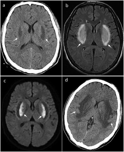
The first computed tomography (CT) scan of the brain, performed 37 min after the patient’s arrival at the emergency department, showed no significant abnormalities. Twenty hours post-admission, a repeat CT scan of the brain showed bilateral symmetrical hypodensities in the basal ganglia (). An MRI of the brain obtained at 80 hours after admission displayed bilateral symmetrical hyperintensities within the basal ganglia on T2 fluid attenuated inversion recovery (FLAIR) sequences, consistent with the lentiform fork sign. Diffusion-weighted imaging revealed high signal intensity within the lentiform nuclei, suggestive of cytotoxic edema; the calculated volume was approximately 18 cm³ (). On day 19, a follow-up CT of the brain showed progressive edematous changes and signs that were indicative of hemorrhagic conversion in the basal ganglia, suggesting an exacerbation of the initial brain injury ().
Figure 1. (a) A computed tomography scan taken 20 hours post-admission reveals symmetrical hypodensities in the bilateral basal ganglia (arrows). (b) A magnetic resonance image at 80 hours post-admission, T2-weighted-fluid-attenuated inversion recovery (T2-FLAIR) displays bilateral hyperintensities in the basal ganglia, indicative of the lentiform fork sign (arrows). (c) Diffusion-weighted image (DWI) shows diffusion restriction in the lentiform nuclei (arrows). (d) A follow-up computed tmography scan on day 19 post-admission demonstrates an expanded area of edema with evidence of hemorrhagic conversion (arrows).
These neuroimaging outcomes are consistent with previous reports in the literature that identify bilateral putaminal hemorrhagic necrosis as a consequence of methanol poisoning [Citation2].
The lentiform fork sign, characterized on MRI as bilateral symmetrical hyperintensities in the basal ganglia encircled by a hyperintense rim around the lentiform nucleus, is a neuroimaging feature first described by Kumar and Goyal in 2010 [Citation3]. This sign has since been recognized as an indicator of severe metabolic acidosis [Citation3] and has been observed in patients with uremic encephalopathy, metformin-induced encephalopathy, dialysis disequilibrium syndrome, and ethylene glycol and methanol poisoning [Citation3, Citation4].
The pathogenesis of the lentiform fork sign is thought to be closely tied to metabolic acidemia and its ability to disrupt the blood-brain barrier. This disruption may lead to vasogenic edema and subsequently to cytotoxic edema, the severity of which depends on the extent of acidemia.
Diffusion-weighted imaging is particularly valuable for distinguishing between cytotoxic and vasogenic edema, providing prognostic insights not readily apparent with other imaging techniques. The lentiform fork sign, suggestive of vasogenic edema, typically resolves on repeat imaging within 3–6 months [Citation3, Citation5]. However, high signal intensity on diffusion-weighted imaging has been linked with mortality in several instances [Citation6, Citation7], highlighting the critical nature of these findings. Moreover, there are limited studies incorporating diffusion-weighted imaging in the context of methanol poisoning [Citation7]. In methanol poisoning, restricted diffusion observed on diffusion-weighted MRI imaging is indicative of nonviable tissue, suggesting a decreased probability of functional recovery. The progression of cytotoxic edema in the basal ganglia to cystic degeneration can be monitored on subsequent MRI scans, offering valuable information for the assessment of long-term outcomes [Citation3].
Conclusion
Diffusion-weighted imaging sequences provide additional information compared to traditional MRI in assessing methanol poisoning. Systematic evaluation is needed to fully understand and utilize the potential predictive value of diffusion-weighted imaging in this context.
Disclosure statement
The authors report no conflict of interest.
Additional information
Funding
References
- Jangjou A, Moqadas M, Mohsenian L, et al. Awareness raising and dealing with methanol poisoning based on effective strategies. Environ Res. 2023;228:115886. doi: 10.1016/j.envres.2023.115886.
- Grasso D, Borreggine C, Perfetto F, et al. Lentiform fork sign: a magnetic resonance finding in a case of acute metabolic acidosis. Neuroradiol J. 2014;27(3):288–292. doi: 10.15274/NRJ-2014-10041.
- Kumar G, Goyal MK. Lentiform fork sign: a unique MRI picture. Is metabolic acidosis responsible? Clin Neurol Neurosurg. 2010;112(9):805–812. doi: 10.1016/j.clineuro.2010.06.006.
- Chang YA, Tarng DC, Yang CY. Lentiform fork sign in a uremic patient after inadvertent exposure to metformin. Clin Toxicol (Phila). 2022;60(3):406–407. doi: 10.1080/15563650.2021.1953519.
- Laespada-García MI, Azcárate-Díaz FJ, Méndez-Guerrero A. Lentiform fork sign’ as a radiological feature of posterior reversible encephalopathy syndrome. Acta Neurol Belg. 2021;121(3):757–759. doi: 10.1007/s13760-021-01643-z.
- Geboers K, Vanden Bossche S, Dekeyzer S. Lentiform fork sign in a girl with uremic encephalopathy. Acta Neurol Belg. 2022;122(2):535–536. doi: 10.1007/s13760-021-01764-5.
- Vara-Castrodeza A, Peréz-Castrillón JL, Dueñas-Laita A. Magnetic resonance imaging in methanol poisoning. Clin Toxicol (Phila). 2007;45(4):429–430. doi: 10.1080/15563650701285313.