Abstract
Introduction. Few cases of mercury sequestration in the appendix appear in the literature. Based on these, both prophylactic appendectomy and non-surgical management have been recommended. We report a case in which a patient with mercury retained in the appendix was managed conservatively without developing mercurialism or appendicitis. Case Report. A 43-year-old man ingested approximately one tablespoon of elemental mercury after an argument with his wife. An initial abdominal radiograph showed mercury in the pylorus of the stomach and a follow-up x-ray at 72 hours showed mercury localized to the appendix. The patient was treated as an outpatient and examined several times over a 37-day period. He never developed signs of appendicitis. On a follow-up examination 7 months after the ingestion, he was radiographically free of mercury. Periodically throughout his clinical course, blood mercury levels were obtained. Only one, 6 days after ingestion, showed an elevated mercury level of 68 mcg/L (reference range <10 mcg/L). Despite this, the patient never developed signs or symptoms of mercury poisoning. Conclusion. Patients in whom elemental mercury is retained in the appendix, who are without symptoms and have normal gastric mucosa, may be conservatively managed without surgery.
Keywords:
Introduction
Prior to the middle of the 19th century, physicians often orally administered elemental mercury as a treatment for acute bowel obstructions. It was thought, by virtue of its weight, to force open obstructions and to be non-toxic (Citation1,Citation2). The notion that ingestion of elemental mercury is non-toxic, however, has been challenged by case reports in which the rupture of balloons containing elemental mercury caused intestinal complications (Citation3–9). In several of these case reports, patients demonstrated elevated blood and urine mercury levels, and in two of them systemic intoxication was reported (Citation3,Citation4). A key feature in all of these cases is that mercury either spilled in the peritoneum during surgery or came in contact with exposed gastrointestinal tissues (Citation3–9). In patients with intact gastrointestinal (GI) mucosa, elemental mercury is typically excreted in the feces within a few days without resulting in local or systemic toxicity (Citation10,Citation11). In patients in whom mercury is not rapidly eliminated, but rather is retained within the appendix, there are conflicting reports as to development of mercury-related-complications and to the role of prophylactic appendectomy (Citation11–16). We report a case of elemental mercury retained in the appendix in which conservative management alone resulted in its elimination without the development of appendicitis or clinical signs of mercury poisoning.
Case report
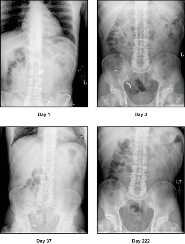
A 43-year-old man became depressed after arguing with his wife and, in a suicide gesture, drank an estimated tablespoon of elemental mercury. Within two hours of the ingestion, he was taken to a local Emergency Department. He denied any other ingestion but admitted to drinking alcohol and smoking two marijuana joints prior to the mercury ingestion. He reported no prior medical problems and admitted to drinking about a 6-pack of beer a day. His initial vital signs were unremarkable except hypertension (154/104 mm Hg) which resolved without treatment within a few hours of his arrival. His complete blood count, blood chemistries, and liver function tests were within the laboratory's normal values. His blood alcohol level was 188 mg/dL and neither acetaminophen nor salicylate was detected. A chest x-ray was normal but a plain film of the abdomen revealed an opacity in the region of the pylorus (, Day 1). The patient was given one dose of a cathartic (17 grams of polyethylene glycol in 8 ounces of water) and admitted overnight for observation and to facilitate psychiatric assessment in the morning. Within a few hours of his arrival, he was noted to have mild end expiratory wheezing. This was treated with an albuterol inhaler and attributed to his smoking 1½ packs of cigarettes daily. A repeat abdominal film in the morning showed radiopaque densities scattered throughout the small and large bowel. After consulting with the Psychiatry and Medical Toxicology services, the patient left the hospital agreeing to continue the cathartic as an outpatient and to return for a follow-up x-ray. Two days later (72 hours from ingestion), a repeat abdominal x-ray showed mercury scattered throughout his colon with a large amount retained within the appendix (, Day 3). A 24-hour urine collection, begun during his overnight hospitalization, came back at this time and revealed a normal mercury level of 15 mcg/L (laboratory reference < 20 mcg/L). The patient did not fill the prescription for the prescribed cathartic due to its cost. Therefore, he was recommended to take over-the-counter Metamucil and to sleep with blocks under the foot of his bed and on his left side. On his next follow-up, 6 days after the ingestion, a repeat x-ray showed continued retention of mercury within the appendix and a blood mercury level was elevated (68 mcg/L). Despite this, the patient had no subjective complaints and had none of the following findings of mercury intoxication: altered mental status, sweating, rash, ataxia, incoordination, tremor, diaphoresis, weakness, or by report personality changes. Neither chelation nor appendectomy was recommended; rather, the patient would watch for symptoms and would return for a follow-up in several weeks. On a repeat visit, 37 days from the ingestion, an abdominal x-ray showed continued retention of mercury in the appendix (, Day 37) and a blood mercury level of 8 mcg/L. Recommendations for Metamucil and Trendelenburg position at night were discontinued (although it is unclear if they were ever done by the patient) and follow-up would occur intermittently by phone. A follow-up radiograph, 7 months after the ingestion (, Day 222), showed no evidence of mercury within the appendix and a blood mercury level at this time was 3 mcg/L. At no point in the 7 months we followed the patient did he manifest or complain of symptoms suggestive of either mercury poisoning or appendicitis.
Fig. 1. Abdominal radiographs show the progression of mercury from the stomach pylorus (Day 1) to the appendix (Days 3 and 37) to its eventual evacuation (Day 222).
Discussion
Although often assumed to be benign, elemental mercury can cause intestinal complications – most commonly granulomas and abscesses (Citation4–9). A prerequisite to mercury causing complications is its spillage into the peritoneum during surgery or its contact with disrupted gastric mucosa (Citation4–9). In most cases in which elemental mercury was ingested by, or spilled in, patients with intact gastric mucosa, the mercury was safely evacuated without complication (Citation10,Citation11,Citation15–17). There are two cases in the literature in which mercury was reported to cause gastrointestinal problems in a patient with normal gastric mucosa (Citation12,Citation13). In both cases, mercury was retained within the appendix and led to appendicitis (Citation12,Citation13). The contribution of mercury to the appendicitis in these cases is unclear. In one, a Miller-Abbott tube placed in a 28-year-old man for an intestinal obstruction from a rectosigmoid carcinoma, ruptured filling the appendix with mercury. The appendix was subsequently removed and the pathology report listed the diagnosis as acute appendicitis secondary to elemental mercury (Citation13). In this case, no symptoms of appendicitis were reported and, although rare, left-sided colonic neoplasia can cause acute appendicitis (Citation18–21). In the other case, a 4-month-old, treated at home with elemental mercury for abdominal pain, had abdominal x-rays showing mercury within the appendix. Due to his symptoms, an appendectomy was performed. Although not inconsistent with appendicitis, the clinical course suggested that the symptoms preceded the mercury ingestion and a pathologic report confirming appendicitis was not given (Citation12).
In 5 other cases in which mercury was retained in the appendix, and in our own, no symptoms of appendicitis were reported (Citation11,Citation14–17). In one case, a prophylactic appendectomy was performed even though no symptoms of appendicitis developed, and the appendix was normal on pathologic report (Citation14). Although some authors recommend prophylactic appendectomy as a potential treatment, (Citation12,Citation14,Citation22) our case supports conservative management as a reasonable option in patients without symptoms.
Another concern for elemental mercury retained in the GI tract is its possible absorption and systemic toxicity. There have been no reports of persons with intact gastrointestinal mucosa developing systemic toxicity following ingestion of elemental mercury. In the cases in which elemental mercury was reported to have caused systemic toxicity, mercury either was spilled in the peritoneum or came in contact with exposed gastric mucosa (Citation3,Citation4). Despite an oral bioavailabity for elemental mercury of 0.04% (Citation23), our patient had an elevated blood mercury level of 68 mcg/L six days after his ingestion. While this suggests some initial absorption, further absorption did not exceed excretion because the blood mercury level was 8 mcg/L on day 37. In addition, no signs or symptoms of mercury intoxication developed. This is consistent with other patients having mildly elevated urine or blood mercury levels but no systemic symptoms (Citation6,Citation15).
Although we suggested laxatives and positional changes in our patient – based on similar strategies in other reports – the mercury appeared to have been evacuated at a time when neither measure was in use (Citation15,Citation16).
Conclusion
Ingested elemental mercury can be retained in the appendix. If the gastrointestinal mucosa is normal and the patient has no symptoms or signs of mercury toxicity or appendicitis, mercury retained in the appendix can be managed conservatively.
References
- MO Cantor. Mercury, its role in intestinal decompression tubes. Am J Surg 1947; 73:690–691.
- MO Cantor. Mercury lost in the gastrointestinal tract; report of an unusual case. JAMA 1951; 146:560–561.
- NS Haas, R Shih, and M Gochfeld. A patient with postoperative mercury contamination of the peritoneum. J Toxicol Clin Toxicol 2003; 41:175–180.
- JE Bredfeldt, and DD Moeller. Systemic mercury intoxication following rupture of a Miller-Abbott tube. Am J Gastroenterol 1978; 69:478–480.
- GF Crikelair, and T Hiratzka. Intraperitoneal mercury granuloma. Ann Surg 1953; 137:272–275.
- ME Efrusy, and WO Dobbins3rd. Persistent enterocutaneous fistula associated with intraperitoneal metallic mercury. Am J Dig Dis 1974; 19:373–377.
- MW Greenberg. Mercury-weighted nasogastric tube: its danger in intestinal surgery. South Med J 1972; 65:1154–1155.
- RR Larsen, KC Sawyer, RB Sawyer, and NS Saliba. Mercury Granuloma Complicating Gastrointestinal Tube Decompression. Surgery 1963; 54:442–447.
- WW Lindenmuth. Fecal fistula due to metallic mercury from a Miller-Abbott tube. JAMA 1949; 141:986–987.
- FI Harris. Intestinal intubation in bowel obstruction. Surg Gynecol Obstet 1945; 81:671–678.
- TB Hunter, GT Fon, and ME Silverstein. Complications of intestinal tubes. Am J Gastroenterol 1981; 76:256–261.
- MA Miller, TP Coon, J Greethong, and P Levy. Medicinal mercury presents as appendicitis. J Emerg Med 2005; 28:217.
- W Birnbaum. Inflammation of the vermiform appendix by metallic mercury. Am J Surg 1947; 74:494–496.
- E Ernst. Metallic mercury in the gastrointestinal tract. A case of ingested thermometer mercury. Acta Chir Scand 1985; 151:651–652.
- PE McKinney. Elemental mercury in the appendix: an unusual complication of a Mexican-American folk remedy. J Toxicol Clin Toxicol 1999; 37:103–107.
- N Wright, WB Yeoman, and GF Carter. Massive oral ingestion of elemental mercury without poisoning. Lancet 1980; 1:206.
- IL Hoffman. Spontaneous evacuation of metallic mercury from the vermiform appendix. Bull US Army Med Dept 1947; 8:802–803.
- E Arnbjornsson. Acute appendicitis as a sign of a colorectal carcinoma. J Surg Oncol 1982; 20:17–20.
- DC Collins. Left-sided colonic lesions masquerading as acute appendicitis. Am J Gastroenterol 1961; 36:521–524.
- DC Collins. Left-sided colonic diseases–masquerading as acute, fulminating appendicitis. Am J Proctol 1961; 12:304–308.
- HW Lai, CC Loong, LC Tai, CW Wu, and WY Lui. Incidence and odds ratio of appendicitis as first manifestation of colon cancer: a retrospective analysis of 1873 patients. J Gastroenterol Hepatol 2006; 21:1693–1696.
- L Yip, RC Dart, and JB Sullivan. Mercury. In: JB Sullivan, and GR Krieger. Clinical Environmental Health and Toxic Exposures. Philadelphia: Lippincott Williams and Wilkins, 2001; 867–879.
- E af Geijersstam, G Sandborgh-Englund, F Jonsson, and J Ekstrand. Mercury uptake and kinetics after ingestion of dental amalgam. J Dent Res 2001; 80:1793–1796.