
Abstract
Aim of the study
Following percutaneous exposure to the nerve agent VX, the remaining intact agent within the skin after decontamination is of great concern. Consequently, this leads to prolonged agent release to the blood circulation resulting in sustained intoxication, which may complicate the medical management. The decontamination procedure used should therefore possess the ability for agent removal both on and within the skin. The efficacy of three decontamination procedures was evaluated by measuring VX and the primary degradation product ethyl methyl phosphonic acid (EMPA) penetrated through human skin and the amount remaining within the skin.
Materials and methods
Decontamination was initiated 5 min post-exposure to VX on human dermatomed skin. Experiments were conducted using an in vitro skin penetration model and the amount remaining within the skin was determined by combining the tape-stripping technique and acetylcholinesterase activity measurements.
Results
In control experiments without decontamination, higher amounts of VX were recovered in the deeper layers of skin compared to EMPA, which was primarily located in the stratum corneum. Both Reactive Skin Decontamination Lotion (RSDL) and the RSDL training kit (TRSDL) significantly reduced the amount of VX within the skin and decreased the penetration through the skin. However, the degradation ability of RSDL was demonstrated to be beneficial by the reduction of intact agents remaining in the skin compared to TRSDL without agent degradation capability. Soapy water decontamination caused a “wash-in” effect of VX with decreased agent amounts within stratum corneum but increased the amount VX penetrated through the skin.
Conclusion
Efficient skin decontamination of VX requires skin decontaminants reaching deeper layers of the skin, and that both absorption and degradation properties are important. In addition, the “wash-in” effect by using soapy water may enhance VX release to the blood circulation.
1. Introduction
In medical management guidelines for victims percutaneously exposed to nerve agents, rapid skin decontamination is critical to prevent absorption and uptake in the blood circulation, as well as to avoid secondary contamination to prehospital responders and hospital personnelCitation1. In acute exposures, the skin contact risk is particularly high for low volatile nerve agents such as VX, as in the case of the alleged assassination of Kim Jong-Nam in 2017Citation2,Citation3. Studies of the skin penetration ability of VX in animal exposure models have shown that the maximum blood level of the agent is reached several hours after percutaneous exposure and is followed by a slow elimination due to the agent’s high persistencyCitation4–7. In addition, following percutaneous exposure to VX, a significant proportion of the applied dose is reported to remain in the deeper layers of the skinCitation8,Citation9. This depot effect may cause prolonged agent release and delayed symptoms as demonstrated by analysis of toxicokinetics and cholinesterase inhibition in guinea pigs, even when decontamination was performedCitation10. Consequently, extended preparedness for intensive medical care may be necessary post-exposure to the nerve agentCitation10–12.
Reactive Skin Decontamination Lotion (RSDL) has been shown to be the most efficient skin decontaminant of VX in vitro and in vivoCitation10,Citation13–17. To avoid prolonged release of VX, a repeated RSDL-decontamination procedure has been demonstrated to be efficientCitation17,Citation18. The product is currently fielded in many countries around the world, primarily in the armed forces. RSDL possesses both solubilizing and degrading properties and has low water content. Previous studies of VX degradation by RSDL have shown that the strong nucleophilic hydroxide ions formed at a high pH and that to a lesser extent the oximes, contribute to the fast degradation of the nerve agentCitation16,Citation19. The low water content prevents agent dilution and potential “wash-in,” a penetration-enhancing effect, previously observed for decontamination procedures using soapy waterCitation17,Citation20. The RSDL-kit includes two mechanisms for the absorption of agents from the skin; the plastic foam for physical removal and polyethylene glycol monomethyl ether (MPEG) which contributes to active agent absorptionCitation16. The decontamination kit can be obtained in a variant aimed for training (TRSDL) which only includes MPEG impregnated in the sponge without any active components to degrade nerve agents.
Tape-stripping is an efficient and well-established method to evaluate the kinetics and penetration depth of multiple agents, for example, pharmaceuticals, both in vivo and in vitroCitation21–24. The method has also been utilized to investigate the removal of stimulants to toxic agents by various decontamination protocols in vitroCitation25,Citation26. By repeatedly applying adhesive tape on the same skin area, certain amounts of stratum corneum (the outermost layer of epidermis) can be removed which enables the determination of agent distribution in tissueCitation27. This technique was proven to be highly valuable as a complement to in vitro penetration dataCitation28–30. However, tape-stripping has rarely been utilized in decontamination studies of live nerve agentsCitation31,Citation32.
This study aimed to evaluate the decontamination efficacy of RSDL, TRSDL, and soapy water following skin exposure to the nerve agent VX. VX and the main degradation product of VX, EMPA, were analyzed within the skin and in the receptor fluid following skin penetration. By studying the effect of the TRSDL in comparison to RSDL, the impact of agent degradation was evaluated. Experiments were performed using dermatomed human skin mounted in an in vitro diffusion cell followed by tape-stripping of the skin discs and subsequently acetylcholinesterase (AChE)-activity measurements to detect intact agent on the remaining skin surface.
2. Materials and methods
2.1. Materials
O-ethyl S-[2-(diisopropylamino)ethyl] methylphosphonothioate (VX; 95% pure measured by NMR; CAS No. 50782–69-9) was synthesized in-house. Reactive Skin Decontamination Lotion kits (RSDL) and the training kits (TRSDL) were obtained from Emergent BioSolutions (Gaithersburg, USA). Wet wipes were purchased from Coop (Solna, Sweden). DAX soap was acquired from CCS Healthcare AB (Borlänge, Sweden).
2.2. Human skin preparation
Full-thickness human Caucasian skin of abdominal origin was obtained from plastic surgery after informed consent according to the Declaration of HelsinkiCitation33. The study was approved by the Research Ethics Committee, Faculty of Medicine and Odontology, Umeå University, Umeå, Sweden (No. 03–161). The full-thickness skin was separated from subcutaneous fat and connective tissue and stored at −80 °C. Following acclimatization at −20 °C for 3–4 days, the skin was thawed at room temperature and dermatomed to a nominal thickness of 500 µm using the Humeca D80 dermatome (Humeca BV, Borne, The Netherlands). Small discs (1.9 cm2) were punched out and the discs were hydrated for approximately 24 h at +4 °C before use. The physical condition of skin samples was assessed visually to exclude any surface damage. All experiments were performed with skin from the same donor patient.
2.3. In vitro skin penetration experiments
The in vitro skin penetration experiments were performed utilizing a specifically designed diffusion cell containing a flow-through receptor solution compartmentCitation17. The diffusion chamber was maintained at 32 °C throughout the experiments (AccuBlock Digital Dry Bath; Labnet Int. Inc, Edison, USA) and the receptor solution was continuously pumped at a fixed flow rate of 20 μl min−1 using a syringe pump (CMA 400; CMA Microdialysis AB, Kista, Sweden). The receptor solution contained a mixture of ethanol and deionized water (1:3). Prior to the experiment, the complete system was allowed to reach an equilibrium of temperature and water balance for 30 min. The dermatomed disc was mounted in the diffusion cell and a Teflon seal was positioned upon to achieve a closed system. The nominal skin diffusion area was 1.13 cm2. Prior to adding donor solution, a zero sample of receptor solution was collected for 8 min (160 µl). In control experiments without decontamination, an infinite dose of neat VX (1 µl) was exposed to skin throughout the experimental time (60 min). Following exposure to a neat agent (1 µl) for 5 min, decontamination was performed according to the separate protocols described below (Section 2.4). The choice of exposure dose was based on the toxicity of the chemicalCitation11. The experimental time was 1 h and samples (200 µl) were collected every 10 min and kept at 10 °C in a fraction collector (CMA 470 Refrigerated Fraction Collector; CMA Microdialysis AB, Kista, Sweden). All vials were sealed immediately after the experiment was completed and stored at −20 °C until analysis. Each experiment was evaluated on four independent replicates (n = 4).
2.4. Decontamination protocols
2.4.1. RSDL and TRSDL decontamination
In experiments using the RSDL- or TRSDL-kit, the sponge was punched out to a size fitting the diffusion cell immediately prior to use. The swabbing was performed using a blunt tweezer. The protocol included swabbing with the RSDL- or TRSDL-sponge for 2 min and the remaining lotion was allowed to act on skin for 10 min prior to a second swabbing with a new RSDL- or TRSDL-sponge for 2 min. Wet wipe removal of RSDL- or TRSDL-lotion from the skin was conducted by using wipes cut to a size fitting the diffusion cell and exchanged twice to ensure thorough removal of the decontamination lotion.
2.4.2. Soapy water
The soapy water was an aqueous solution containing 2% soap in water, prepared immediately prior to decontamination. Decontamination was performed by washing the agent off repeatedly 10 times by adding 50 µl soapy water on the skin and instantly removed by careful vacuum suctioning. In total, the procedure lasted approximately 2 min.
2.5. Skin retention experiments (tape-stripping)
Following in vitro skin penetration experiments during 1 h, the dermatomed skin disc was gently removed from the diffusion cell and needled onto a plastic board. Adhesive tape (Fixomull® tape; Beiersdorf AG, Germany), cut in a standardized size, was applied over the skin disc and five distinct motions over the tape using the backside of a Hamilton syringe piston were performed. Tapes were removed from the skin tissue using clean forceps and each tape was placed in an individual glass vial. The procedure was repeated 10 times on each skin disc. All vials were stored at −20 °C until preparation for analysis. Before analysis, all vials were allowed to reach room temperature and 1 ml solvent (methanol and dichloromethane; 1:1 v:v) was added to each vial to extract VX and EMPA from the tape. Aliquots of 100 µl from each extract were transferred to 96-well analysis plates, or alternatively, 10 µl aliquots were diluted with 990 µl methanol in 1.5 ml analysis glass vials for extracts where concentrations of substance were above 50 µg ml−1.
After a completed tape-stripping procedure, the residual skin piece was swabbed for 10 s using a cotton bud. To enable detection of any remaining agent on the skin, the ChE check mobile (Securetec Detektions-Systeme AG, Germany) was utilized which is based on photometric measurements of cholinesterase activityCitation34. Firstly, the background value of the solution in the prefilled cuvette was measured followed by adding 10 µl of blood sample (AChE Check control high; Securetec Detektions-Systeme AG, Germany) and the haemoglobin content was determined. The cotton bud was then placed in the cuvette and shaken for 10 s to release the agent into the solution. Subsequently, the white cap was exchanged for the red cap containing the AChE substrate which was dissolved in solution by shaking and the AChE-activity was then determined. The cotton bud remained in the cuvette throughout the measurement.
2.6. Sample analysis
Skin penetration and tape samples were analyzed using an Acquity UPLC (I class) system coupled to a Xevo TQ-XS, tandem quadrupole mass spectrometer (Waters; Milford, MA, USA). The MS was fitted with an electrospray ionization probe and operated in both positive and negative ion modes. The capillary voltage was set to 2.0 kV and −2.5 kV respectively and argon was used as the collision gas. VX and EMPA were analyzed in multiple reaction monitoring (MRM) mode. MS/MS transitions for VX was m/z 268.3 > 128.2 and 79.0 in positive ion mode and MS/MS transitions for EMPA was m/z 123 > 95 and 77 in negative ion mode. The LC used an Acquity UPLC BEH C18 column, (2.1 mm × 50 mm, 1.7 µm particle size) from Waters and 0.1% formic acid in acetonitrile (v/v) and 0.1% formic acid in water (v/v) as eluents. The flow rate was 0.5 ml min−1, and the column temperature was 60 °C. Chromatographic separation was achieved by a linear gradient from 5% to 95% acetonitrile and total analysis time per sample of 2 min. The injection volume was 0.5 µl. Each sample batch was quantified using an external calibration curve. The standards and blanks were prepared in the same receptor solution as the samples. Calculated limit of quantification (LoQ), with the instrumental settings described above, for VX was 1 ng ml−1 and LoQ for EMPA was 6 ng ml−1Citation35.
2.7. Data analysis
All results are presented as the mean ± the standard error of the mean (SEM). The graphs were prepared and statistical analyses were performed using the GraphPad Prism program (version 6.0 GraphPad Software Inc., San Diego, USA).
The cumulative skin penetration following the completed experiment, defined as the cumulative amount, was calculated as µg cm−2 and the penetration rate as µg cm−2 h−1 in all experiments. For comparison of decontamination efficacy between products, the decontamination factor (DF) was defined as the ratio of the total cumulative amount of the control penetration experiments (without decontamination) and the total cumulative amount of the penetration experiments including decontamination.
The amount of VX and EMPA eluted from each tape is presented in µg. The agent distribution on and within the skin together with amounts detected in receptor solution is calculated as a percent of the agent amount exposed on the skin for the control experiments without decontamination. The agent percentage in the remaining skin was calculated from the amounts detected on tapes and receptor solution and was based on full recovery of the applied agent amount. Results from the ChE check mobile measurements are defined as percent activity of high range blood control value.
Statistical comparisons of the agent amounts or AChE-activity to one control group were performed by using one-way analysis of variance (ANOVA) followed by Dunnett’s multiple comparison test. Statistical significance of multiple pairs of means was assessed by using two-way ANOVA followed by Bonferroni multiple comparison tests to determine differences in penetration rates and agent amounts in individual tape samples. The null hypothesis, defined as identical mean values of compared groups, was discarded if the probability of identical mean values were below 0.05 (*p < 0.05). Probabilities of 0.01–0.001 and below 0.001 were additionally indicated (** and ***, respectively).
3. Results
The efficacy of three different decontamination protocols was evaluated following 5 min of in vitro human skin exposure to neat VX. The protocols for RSDL and TRSDL decontamination were based on the previously optimized procedure for decontamination of VXCitation17. To address the potential wash-in effect, decontamination by repeated washing with soapy water was included. In the control, 1 µl of neat VX was exposed to skin and allowed to absorb for 1 h. Samples of receptor fluid were collected every 10 min for analysis of penetrated VX and the degradation product EMPA. Tape-stripping and subsequent swabbing of the remaining skin surface for AChE-activity measurements were performed after completion of the absorption. In all experiments, decontamination was initiated 5 min post-exposure to VX.
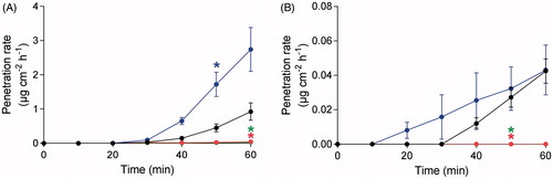
The penetration rates of VX and EMPA were determined for the three decontamination protocols and compared to the control without decontamination (). Decontamination using soapy water resulted in a significantly increased penetration rate of VX after 50 min to the end of the experiment compared to the control without decontamination, while both the RSDL- and TRSDL-protocols resulted in a significantly decreased penetration rate of VX at the end of the experiment. The lag-time of VX penetration was approximately 30 min in all experiments. The penetration rate of EMPA though the skin was significantly decreased following decontamination with RSDL and TRSDL at time point 50 min compared to the control without decontamination. No significant difference in the penetration rate of EMPA was observed for the soapy water procedure. The lag-time for EMPA was 10 min after soapy water decontamination compared to approximately 30 min for the other three experimental set-ups.
Figure 1. Penetration rate of (A) VX and (B) ethyl methylphosphonic acid (EMPA) through human dermatomed skin during 1 h experimental time. Decontamination was initiated 5 min post-exposure of VX. Decontamination with RSDL (green; bottom line), TRSDL (red; bottom line), soapy water (blue; top line) and control without decontamination (black; center line). Values are presented as mean ± SEM (n = 4). *p < 0.05 indicates significantly increased or decreased penetration rate from the indicated time-point until the end of the experiment compared to experiment without decontamination (two-way ANOVA).
The cumulative amount of VX in the receptor fluid was significantly decreased compared to the control following decontamination with RSDL and TRSDL (). The decontamination factor was 18 for RSDL and 13 for TRSDL, respectively. Soapy water decontamination resulted in a significantly increased cumulative amount of VX in the receptor fluid compared to the control.
Table 1. Cumulative amount of VX penetrated through human dermatomed skin with or without decontamination.
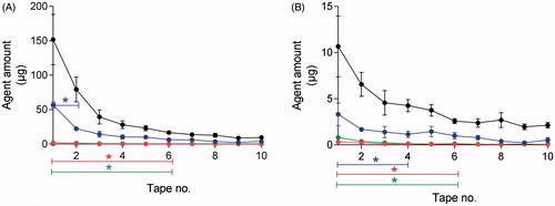
The amount of VX and EMPA on the individual tapes from each dermatomed skin disc was determined by agent extraction followed by LC-MS analysis (). The control experiments without decontamination resulted in the largest amounts of VX and EMPA on the first tapes of each set. The RSDL- and TRSDL-procedures demonstrated significantly decreased amounts of VX and EMPA on the 6 first tapes compared to the control. Decontamination with soapy water resulted in a significantly decreased amount of VX on tapes 1 and 2 compared to the control. Correspondingly, the amount of EMPA was significantly decreased on tape 1–4 after decontamination with soapy water.
Figure 2. Agent amount of (A) VX and (B) ethyl methylphosphonic acid (EMPA) on individual tapes. Decontamination with RSDL (green; bottom line), TRSDL (red; bottom line), soapy water (blue; center line) and control without decontamination (black; top line). Values are presented as mean ± SEM (n = 4). *p < 0.05 indicates significantly decreased amounts compared to experiments without decontamination (two-way ANOVA).
In control experiments without decontamination, the recovery of VX and EMPA was calculated for the entire skin tissue (). For both VX and EMPA, only a small proportion of the applied agent was recovered in the receptor solution 60 min post-exposure (0.03% for both VX and EMPA). The largest amount of VX remained in the deeper layers of skin (59.6%) and in the tape-stripped stratum corneum (40.3%). For EMPA, the largest amount was recovered in the tapes (83.9%) followed by the remaining skin (17%).
Figure 3. Recovery of skin tissue distribution of VX and ethyl methylphosphonic acid (EMPA) for control experiments without decontamination. The recovery is based on the initial concentration of 95% VX and 5% EMPA exposed on skin. Total amount in 10 tapes (black bar; left), remaining skin (green bar; middle) and penetrated skin (blue bar; right). The VX and EMPA amounts in the remaining skin was calculated by subtraction of the amounts detected in tapes and penetrated skin from the applied amount. Note the two segmented Y-axis. Values are presented as mean ± SEM (n = 4).
Following tape-stripping, the residual skin surface was swabbed with a cotton bud to detect the presence of intact agent as defined by inhibition of AChE using the Securetec ChE mobile (). Swabbed tissue from VX-exposed skin without decontamination and skin decontaminated with TRSDL or soapy water significantly inhibited AChE-activity compared to the blood control. Only RSDL showed a significant efficacy to remove the AChE inhibitor from the tissue (p < 0.01 compared to VX-exposed tissue without decontamination; data not shown).
Figure 4. Acetylcholinesterase (AChE) activity measurements by the ChE check mobile for skin surface samples after 10 sets of tape-stripping. Values are presented as mean ± SEM (n = 4). ***p < 0.001 indicates significantly decreased AChE-activity vs the blood control (one-way ANOVA).
4. Discussion
Medical management of victims percutaneously exposed to nerve agents might be greatly affected by the remaining intact agent within the skin after decontaminationCitation36,Citation37. Low volatile nerve agents, such as VX, are suggested to form depots in the deeper layers of the skin, which may cause continuous agent release and sustained symptomsCitation7–9. Therefore, efficient removal of nerve agents requires skin decontamination procedures that have the ability to reach agents within the skin. This study aimed to evaluate procedures using RSDL, TRSDL, and soapy water for their ability to remove VX from human skin and to reduce the agent amount penetrating through the skin.
Following 1 h of skin exposure to neat VX, the largest agent amounts were found in the deeper layers of the skin and only small amounts were recovered in the receptor solution. Similar data on tissue distribution has been reported with larger volumes of VX percutaneously exposed on pig skin for 24 hCitation29. For VX exposed to water dilution on the skin, the absorption rate through pig skin was found to be higher than for neat agent and consequently, the amount recovered in the skin periphery was lowerCitation28. The large proportion of VX remaining within the skin after 1 h, complies with the delayed symptom development following percutaneous exposure to VX demonstrated in vivo in humans and animalsCitation11,Citation38,Citation39.
Of the three decontamination procedures evaluated, RSDL resulted in the highest decontamination factor (DF = 18). The TRSDL-procedure also displayed significant decontamination efficacy compared to the control without decontamination (DF = 13). These data confirm the previously reported efficacy of RSDL to decontaminate skin contaminated with VX, especially the improvement achieved by the slightly extended procedure compared to the protocol recommended by the manufacturerCitation10,Citation13,Citation14,Citation17. The training kit, TRSDL, containing only MPEG impregnated in the sponge and lacking active degradation ingredients, has previously never been evaluated for the skin removal ability of live agents. Both RSDL and TRSDL reduced the VX-amounts in the top layers of stratum corneum and in the receptor fluid of the diffusion chamber, indicating that the solubilizing MPEG component of RSDL and TRSDL significantly contribute to the removal of VX from the skin. However, the agent degradation capacity of RSDL was demonstrated to be superior to reduce intact agents remaining in the skin compared to TRSDL as shown by AChE-activity measurements of skin swabs. This is consistent with the rapid nerve degradation by RSDL previously shown by NMR-spectroscopyCitation16,Citation19. To improve decontamination efficacy following skin exposure to VX and to potentially reaching the remaining agent in the deeper layers of skin, repeated decontamination has been shown to be advantageousCitation10,Citation17. It should, though, be considered that this may result in the instantaneous release of the remaining agent in the skin and risk for symptom aggravation caused by a transiently increased uptake in the blood circulation, but this method of decontamination would likely lead to lower nerve agent uptake over time and improved patient recoveryCitation17.
Washing the contaminated skin with soapy water decreased the VX amounts in the top layers of stratum corneum but increased the amount of agent penetrating skin compared to the control without decontamination. This finding strongly supports the suggested “wash-in” effect for VX following decontamination by washing with water or soapy waterCitation16,Citation17. In previous in vitro studies including various exposure chemicals, similar evidence for “wash-in” effects has also been reportedCitation20,Citation40,Citation41. The removal of decontaminants differed between the RSDL/TRSDL and soapy water protocols by the use of wet wipes for RSDL/TRSDL compared to vacuum suctioning for soapy water. The significant “wash-in” effect for soapy water could potentially be stronger following vacuum suctioning due to remaining moist on the skin. However, decontamination using wet wipes was previously shown to result in an enhanced penetration rate and amount of methyl salicylate through the skinCitation42. The presence of solvents and detergents in the wipes was considered to cause enhanced skin penetration.
The increased penetration rate of VX following soapy water decontamination could potentially result in a shorter time window for medical treatment following percutaneous exposure to VX compared to decontamination procedures with high efficacy to neutralize the agentCitation18. It is likely that the decrease of remaining agent amount within stratum corneum following soapy water decontamination is due to both the “wash-in” effect and the agent removal on top of skinCitation41. To enable an evaluation of the impact of decontamination procedures on extended agent release from the skin, prolonged experimental times would though be required.
EMPA is a hydrolysis product of VX, and is the main degradation product of VX in RSDLCitation16,Citation19. In the 1 µl sample exposed on skin, approximately 5% consisted of EMPA, which was also analyzed in individual tapes and in the receptor solution. Differently from VX, larger amounts of EMPA were recovered in the tapes after 1 h than in the remaining skin but similarly to VX, the amount in the receptor solution was very small also for EMPA. The recovery of EMPA primarily on the skin and in the stratum corneum likely reflects the higher hydrophilicity of EMPA compared to VX (logP −0.85 ± 0.62 and 2.05 ± 0.36 for EMPA and VX, respectively), resulting in a lower skin permeabilityCitation43,Citation44. All three decontamination protocols significantly decreased the EMPA amount in the tapes representing the main part of stratum corneum. RSDL and TRSDL also significantly reduced the agent amount in receptor solution samples, while the amounts following soapy water decontamination were unaffected compared to the control without decontamination. This indicates that the risk for a “wash-in” effect is lower for the hydrophilic EMPA than for the more lipophilic VX. Decontamination by RSDL could potentially result in increased amounts of EMPA due to degradation of VX, however, no such effect was observed during the relatively short 1 h experimental time. No information is available regarding the acute toxicity of EMPACitation45. However, it can be used to identify the exposure of VX in both serum and urine samples from victimsCitation46,Citation47.
The impact of in vitro results in an in vivo situation requires extrapolation of data which is challenging as physiological parameters cannot be addressed in vitroCitation48,Citation49. In in vivo-studies including guinea pigs or swine, RSDL has been shown to provide greater decontamination efficacy following VX exposure compared to several other decontamination procedures, for example, soapy water and Fuller’s EarthCitation14,Citation50. The higher efficacy of RSDL compared to soapy water decontamination is consistent with the present study. Soapy water decontamination has been demonstrated to be effective in vivo, also when decontamination was delayed to 1 h post-exposure to VXCitation14,Citation51. However, contradictory results have been demonstrated for soapy water decontamination delayed 45 min post-exposure to VX in a similar modelCitation13. Animals exposed to VX and decontaminated with soapy water have demonstrated limited or pronounced signs of nerve agent poisoningCitation13,Citation51. In comparison, animals decontaminated with RSDL only exhibited miosisCitation13. The “wash-in” effects for VX following soapy water decontamination displayed in vitro may therefore be associated with a higher probability for the onset of cholinergic signs rather than considering the decontamination as ineffective. Consequently, nerve agent-exposed patients decontaminated with soapy water may require medical management to a greater extent compared to, for example, RSDL-decontaminated patients.
In conclusion, the importance of skin decontamination procedures including both efficient absorption and degradation was demonstrated by combining data on skin retention and penetration of percutaneously exposed VX. In addition, the “wash-in” effect of soapy water decontamination was further elucidated, showing that despite a lowered amount of VX in stratum corneum this decontamination procedure may enhance VX release to the blood circulation. Decontamination procedures aiming at reaching deeper layers in the skin would be desirable following percutaneous exposure to highly toxic agents such as VX. Furthermore, the need for repeated decontamination was strengthened and should be further studied.
Acknowledgements
The authors would like to thank Specialistkliniken (Umeå, Sweden) for the human skin supply. This work was supported by the Swedish Ministry of Defence.
Disclosure statement
No potential conflict of interest was reported by the author(s).
Additional information
Funding
References
- Amend N, Niessen KV, Seeger T, et al. Diagnostics and treatment of nerve agent poisoning-current status and future developments. Ann NY Acad Sci. 2020;1479:13–28.
- Chai PR, Boyer EW, Al-Nahhas H, et al. Toxic chemical weapons of assassination and warfare: nerve agents VX and sarin. Toxicol Commun. 2017;1:21–23.
- Yusof AN. Statement by H.E. ambassador Ahmad Nazri Yusof permanent representative of Malaysia to the OPCW at the ninetieth session of the executive council. Organisation for the Prohibition of Chemical Weapons; 2019.
- Genovese RF, Benton BJ, Oubre JL, et al. Determination of threshold adverse effect doses of percutaneous VX exposure in African green monkeys. Toxicology. 2011;279:65–72.
- Mann TM, Price ME, Whitmore CL, et al. Bioscavenger is effective as a delayed therapeutic intervention following percutaneous VX poisoning in the guinea-pig. Toxicol Lett. 2018;293:198–206.
- Reiter G, Müller S, Hill I, et al. In vitro and in vivo toxicological studies of V nerve agents: molecular and stereoselective aspects. Toxicol Lett 2015;232:438–448.
- van der Schans MJ, Lander BJ, van der Wiel H, et al. Toxicokinetics of the nerve agent (+/−)-VX in anesthetized and atropinized hairless guinea pigs and marmosets after intravenous and percutaneous administration. Toxicol Appl Pharmacol 2003;191:48–62.
- Chilcott RP, Dalton CH, Hill I, et al. In vivo skin absorption and distribution of the nerve agent VX (O-ethyl-S-[2(diisopropylamino)ethyl] methylphosphonothioate) in the domestic white pig. Hum Exp Toxicol 2005;24:347–352.
- Wetherell JR, Armstrong SJ, Read RW, et al. VX penetration following percutaneous poisoning: a dermal microdialysis study in the guinea pig. Toxicol Mech Methods 2008;18:313–321.
- Joosen MJ, van den Berg RM, de Jong AL, et al. The impact of skin decontamination on the time window for effective treatment of percutaneous VX exposure. Chem Biol Interact 2017;267:48–56.
- Hamilton MG, Hill I, Conley J, et al. Clinical aspects of percutaneous poisoning by the chemical warfare agent VX: effects of application site and decontamination. Mil Med 2004;169:856–862.
- Thiermann H, Worek F, Kehe K. Limitations and challenges in treatment of acute chemical warfare agent poisoning. Chem Biol Interact 2013;206:435–443.
- Bjarnason S, Mikler J, Hill I, et al. Comparison of selected skin decontaminant products and regimens against VX in domestic swine. Hum Exp Toxicol 2008;27:253–261.
- Braue EH, Smith KH, Doxzon BF, et al. Efficacy studies of reactive skin decontamination lotion, M291 skin decontamination kit, 0.5% bleach, 1% soapy water, and skin exposure reduction paste against chemical warfare agents, part 1: guinea pigs challenged with VX. Cutaneous Ocular Toxicol 2011;30:15–28.
- Taysse L, Daulon S, Delamanche S, et al. Skin decontamination of mustards and organophosphates: comparative efficiency of RSDL and Fuller’s earth in domestic swine. Hum Exp Toxicol 2007;26:135–141.
- Thors L, Koch M, Wigenstam E, et al. Comparison of skin decontamination efficacy of commercial decontamination products following exposure to VX on human skin. Chem Biol Interact 2017;273:82–89.
- Thors L, Wigenstam E, Qvarnström J, et al. Improved skin decontamination efficacy for the nerve agent VX. Chem Biol Interact 2020;325:109135.
- Joosen MJ, van der Schans MJ, Kuijpers WC, et al. Timing of decontamination and treatment in case of percutaneous VX poisoning: a mini review. Chem Biol Interact 2013;203:149–153.
- Elsinghorst PW, Worek F, Koller M. Detoxification of organophosphorus pesticides and nerve agents through RSDL: efficacy evaluation by (31)P NMR spectroscopy. Toxicol Lett 2015;233:207–213.
- Moody RP, Maibach HI. Skin decontamination: importance of the wash-in effect. Food Chem Toxicol 2006;44:1783–1788.
- Cordery SF, Pensado A, Chiu WS, et al. Topical bioavailability of diclofenac from locally-acting, dermatological formulations. Int J Pharm 2017;529:55–64.
- Escobar-Chávez JJ, Merino-Sanjuán V, López-Cervantes M, et al. The tape-stripping technique as a method for drug quantification in skin. J Pharm Pharm Sci 2008;11:104–130.
- Fantini A, Demurtas A, Nicoli S, et al. In vitro skin retention of crisaborole after topical application. Pharmaceutics 2020;12:491.
- Paterson DA, Hallier J, Jenkins E, et al. Is the skin absorption of hydrocortisone modified by the variability in dosing topical products? Pharmaceutics 2018;10:9.
- Cao Y, Hui X, Elmahdy A, et al. In vitro human skin permeation and decontamination of diisopropyl methylphosphonate (DIMP) using dermal decontamination gel (DDGel) and reactive skin decontamination lotion (RSDL) at different timepoints. Toxicol Lett 2018;299:118–123.
- Cao Y, Hui X, Zhu H, et al. In vitro human skin permeation and decontamination of 2-chloroethyl ethyl sulfide (CEES) using dermal decontamination gel (DDGel) and reactive skin decontamination lotion (RSDL). Toxicol Lett 2018;291:86–91.
- Lademann J, Jacobi U, Surber C, et al. The tape stripping procedure-evaluation of some critical parameters. Eur J Pharm Biopharm 2009;72:317–323.
- Dalton C, Graham S, Jenner J. Effect of aqueous dilution on the absorption of the nerve agent VX through skin in vitro. Toxicol in Vitro 2018;53:121–125.
- Dalton C, Graham S, Jenner J. Effect of exposure area on nerve agent absorption through skin in vitro. Toxicol in Vitro 2015;30:454–461.
- Rolland P, Bolzinger MA, Cruz C, et al. Human scalp permeability to the chemical warfare agent VX. Toxicol in Vitro 2011;25:1974–1980.
- Rolland P, Bolzinger MA, Cruz C, et al. Hairy skin exposure to VX in vitro: effectiveness of delayed decontamination. Toxicol in Vitro 2013;27:358–366.
- Salerno A, Bolzinger MA, Rolland P, et al. Pickering emulsions for skin decontamination. Toxicol in Vitro 2016;34:45–54.
- WMA. World Medical Association Declaration of Helsinki: ethical principles for medical research involving human subjects. J Am Med Assoc 2013;310:2191–2194.
- John M, Ely EW, Halfkann D, et al. Acetylcholinesterase and butyrylcholinesterase in cardiosurgical patients with postoperative delirium. J Intensive Care 2017;5:29.
- Wenzl T, Haedrich J, Schaechtele A, et al. Guidance document on the estimation of LOD and LOQ for measurements in the field of contaminants in feed and food. Luxembourg, Luxembourg: European Commission; 2016. JRC Technical Reports: EUR 28099 EN2016.
- Timperley CM, Abdollahi M, Al-Amri AS, et al. Advice on assistance and protection from the scientific advisory board of the organisation for the prohibition of chemical weapons: part 2. On preventing and treating health effects from acute, prolonged, and repeated nerve agent exposure, and the identification of medical countermeasures able to reduce or eliminate the longer term health effects of nerve agents. Toxicology 2019;413:13–23.
- Timperley CM, Forman JE, Abdollahi M, et al. Advice on assistance and protection provided by the Scientific Advisory Board of the Organisation for the Prohibition of Chemical Weapons: part 1. On medical care and treatment of injuries from nerve agents. Toxicology 2019;415:56–69.
- Joosen MJ, van der Schans MJ, van Helden HP. Percutaneous exposure to VX: clinical signs, effects on brain acetylcholine levels and EEG. Neurochem Res 2008;33:308–317.
- Nozaki H, Aikawa N, Fujishima S, et al. A case of VX poisoning and the difference from sarin. Lancet 1995;346:698–699.
- Misik J, Pavlikova R, Josse D, et al. In vitro skin permeation and decontamination of the organophosphorus pesticide paraoxon under various physical conditions-evidence for a wash-in effect. Toxicol Mech Methods 2012;22:520–525.
- Zhu H, Jung EC, Phuong C, et al. Effects of soap-water wash on human epidermal penetration. J Appl Toxicol 2016;36:1526.
- Matar H, Guerreiro A, Piletsky SA, et al. Preliminary evaluation of military, commercial and novel skin decontamination products against a chemical warfare agent simulant (methyl salicylate). Cutan Ocul Toxicol 2016;35:137–144.
- Thors L, Koch B, Koch M, et al. In vitro human skin penetration model for organophosphorus compounds with different physicochemical properties. Toxicol in Vitro 2016;32:198–204.
- Czerwinski SE, Skvorak JP, Maxwell DM, et al. Effect of octanol: water partition coefficients of organophosphorus compounds on biodistribution and percutaneous toxicity. J Biochem Mol Toxicol 2006;20:241–246.
- Munro NB, Talmage SS, Griffin GD, et al. The sources, fate, and toxicity of chemical warfare agent degradation products. Environ Health Perspect 1999;107:933–974.
- Read R, Black R, Hakala U, et al. Recommended operating procedures for analysis in the verification of chemical disarment. Vol. 22. Helsinki, Finland: University of Helsinki; 2017.
- Tsuchihashi H, Katagi M, Nishikawa M, et al. Identification of metabolites of nerve agent VX in serum collected from a victim. J Anal Toxicol 1998;22:383–388.
- Blaauboer BJ. Biokinetic modeling and in vitro-in vivo extrapolations. J Toxicol Environ Health B Crit Rev 2010;13:242–252.
- Wetmore BA. Quantitative in vitro-to-in vivo extrapolation in a high-throughput environment. Toxicology 2015;332:94–101.
- Schwartz MD, Hurst CG, Kirk MA, et al. Reactive skin decontamination lotion (RSDL) for the decontamination of chemical warfare agent (CWA) dermal exposure. Curr Pharm Biotechnol 2012;13:1971–1979.
- Misik J, Pavlik M, Novotny L, et al. In vivo decontamination of the nerve agent VX using the domestic swine model. Clin Toxicol 2012;50:807–811.