
Abstract
Pharmacological histone deacetylase (HDAC) inhibitors attenuate pathological cardiac remodeling and hypertrophic gene expression; yet, the direct histone targets remain poorly characterized. Since the inhibition of HDAC activity is associated with suppressing hypertrophy, we hypothesized histone acetylation would target genes implicated in cardiac remodeling. Trichostatin A (TSA) regulates cardiac gene expression and attenuates transverse aortic constriction (TAC) induced hypertrophy. We used chromatin immunoprecipitation (ChIP) coupled with massive parallel sequencing (ChIP-seq) to map, for the first time, genome-wide histone acetylation changes in a preclinical model of pathological cardiac hypertrophy and attenuation of pathogenesis with TSA. Pressure overload-induced cardiac hypertrophy was associated with histone acetylation of genes implicated in cardiac contraction, collagen deposition, inflammation, and extracellular matrix identified by ChIP-seq. Gene set enrichment analysis identified NF-kappa B (NF-κB) transcription factor activation with load induced hypertrophy. Increased histone acetylation was observed on the promoters of NFκB target genes (Icam1, Vcam1, Il21r, Il6ra, Ticam2, Cxcl10) consistent with gene activation in the hypertrophied heart. Surprisingly, TSA attenuated pressure overload-induced cardiac hypertrophy and the suppression of NFκB target genes by broad histone deacetylation. Our results suggest a mechanism for cardioprotection subject to histone deacetylation as a previously unknown target, implicating the importance of inflammation by pharmacological HDAC inhibition. The results of this study provides a framework for HDAC inhibitor function in the heart and argues the long held views of acetylation is subject to more flexibility than previously thought.
Abbreviations
| ANP | = | Atrial natriuretic peptide |
| BNP | = | Brain natriuretic peptide |
| BW | = | Body Weight |
| cDNA | = | complementary DNA |
| ChIP | = | Chromatin Immunoprecipitation |
| Ct | = | threshold cycle number |
| Cxcl10 | = | Chemokine (C-X-C Motif) ligand 10 |
| ENCODE | = | Encyclopedia of DNA Elements Consortium |
| FS | = | Fractional Shortening |
| FDR | = | False Discovery Rate |
| GAIIx | = | Genome Analyzer IIx |
| HDAC | = | Histone deacetylase |
| Il6ra | = | Interleukin-6 receptor |
| Il21r | = | Interleukin-21 receptor |
| Icam1 | = | Intercellular adhesion molecule 1 |
| LV | = | Left Ventricle |
| LVDd | = | Left Ventricular Diastolic Dimension |
| LVH | = | Left Ventricle Hypertrophy |
| MACs | = | Model-based Analysis of ChIP-seq |
| NGS | = | Next Generation Sequencing |
| NES | = | normalized enrichment score |
| NFκB | = | Nuclear factor of kappa light polypeptide gene enhancer in B-cells |
| SEM | = | Standard Error of the Mean |
| Serca2a | = | Sarcoplasmic reticulum Ca2+ ATPase |
| TAC | = | Transverse Aortic Constriction |
| TAC veh | = | TAC vehicle |
| TF | = | transcription factor |
| Ticam2 | = | Toll-like receptor adaptor molecule 2 |
| TL | = | Tibia Length |
| Traf3 | = | TNF receptor-associated factor 3 |
| TSA | = | Trichostatin A |
| TSS | = | Transcription Start Site |
| UTR | = | Untranslated region |
| Vcam1 | = | Vascular cell adhesion molecule 1 |
| α/βMHC | = | Alpha/Beta myosin heavy chain |
Introduction
Heart disease remains the leading cause of death in many developing countries and has no cure, with current therapies only delaying disease progression. Myocardial hypertrophy is an initiatory hallmark of heart failure, which is now recognized as a response to genetic, physiologic, and environmental changes.Citation1 Cardiac remodeling is characterized by myocyte hypertrophy mediated by stress signals that compromise heart function by activating inflammatory response pathways associated with disease.Citation2,3 Another conserved feature of cardiac remodeling is the coordinated expression and return to the fetal gene program in adult myocardium.Citation4,5 While the coordinated response of the adult heart to physiological stressors, such as pressure overload, ischemia, hypothyroidism, and metabolic signals, have been characterized in heart disease, their molecular targets, specifically chromatin modifications, are poorly understood.
Emerging evidence suggests that epigenetic changes, such as post-translational modifications of histones, are integral to the coordination of gene regulatory events in the hypertrophied heart.Citation6-10 Histones are the most abundant proteins associated with eukaryotic DNA. The basic unit of the nucleosome is 146 base-pairs of DNA wrapped around a histone octamer that consists of 2 copies each of histones H2A, H2B, H3, and H4.Citation11 N-terminal tails of histones are targets for enzymes that mediate specific modifications, which together form a “histone code” that regulates chromatin structure and gene function.Citation11,12 Histone acetylation of lysine 9 and lysine 14 (H3K9/K14ac) are typically located in regions surrounding the transcription start site (TSS) at actively transcribed genes, whereas histone deacetylation is associated with gene suppression.Citation13-15 Whereas acetylation is catalyzed by histone acetyl transferases (HATs), deacetylation is mediated by histone deacetylases (HDACs). In the heart, several studies have demonstrated a key role for these enzymes in pathological cardiac remodeling using genetic and pharmacological loss or gain of function approaches.Citation16-18 While recent genome-wide studies have demonstrated regulation of hypertrophic gene expression,Citation19,20 the actions of HDAC inhibitors on histone modifications regulating gene expression in the heart are still not well defined.
Inhibitors of HDAC activity, SAHA (suberoylanilide hydroxamic acid, also known as vorinostat and Zolinza) and depsipeptide (romidespin, Isodax) were approved by the US FDA for the treatment of cutaneous T-cell lymphoma.Citation21 Both SAHA and the prototypical HDAC inhibitor Trichostatin A (TSA) are hydroxamic acids that inhibit Class I and II HDAC enzymes. Since the inhibition of HDAC activity blunts cardiac hypertrophy and reverses fetal gene expression, the challenge is now to understand the specific biological functions of these compounds.Citation22-24 Pharmacological compounds that inhibit HDAC activity can regulate the activity of both histone and non-histone substrates. The paradigmatic mode of action is increased histone acetylation coordinating gene expression; however, more recent studies have shown a more diverse mechanism of gene regulation.Citation25-27 For example, TSA and SAHA both confer broad histone deacetylation mediated gene suppression.Citation27 Large mapping studies of the epigenome have revealed unexpected histone deacetylation implicating HDAC inhibitors target action is diverse and a more complex regulatory mechanism than originally envisioned. Indeed, these compounds target inflammation and block pathological cardiac remodeling, showing promise for the treatment of heart failure.Citation3 Preclinical studies show HDAC inhibition blunts cardiac hypertrophy by suppressing autophagy.Citation28 With the efficacy of HDAC inhibitors in preclinical models of heart failure the challenge now is to understand the in vivo anti-inflammatory effects of these compounds.Citation29,30
In this study, we sought to characterize the gene targets of histone acetylation in hypertrophic mouse hearts. Mapping genome-wide H3K9/K14ac patterns in a preclinical model of hypertrophy using transverse aortic constriction, we hypothesized that pharmacological HDAC inhibition attenuates cardiac remodeling by acetylation and deacetylation of histone substrates regulating gene expression. HDAC inhibition attenuated gene expression in load-induced cardiac hypertrophy by histone deacetylation. The pro-inflammatory NFκB gene targets (Icam1, Vcam1, Il21r, Il6ra, Ticam2, Cxcl10) induced by pressure overload were blunted by TSA and subject to histone deacetylation. Our results suggest that an anti-inflammatory mechanism mediated by HDAC inhibition confers cardioprotection by histone deacetylation of NFκB target genes.
Results
TSA attenuates load-induced hypertrophy and restores cardiac function
Mice were randomized for sham or TAC (transverse aortic constriction) surgery followed by subcutaneous injection with TSA or vehicle (veh) twice daily for 4 weeks. Sham animals underwent open chest surgery without banding of the aorta.Citation31 TAC mice administered vehicle control (TAC veh) developed cardiac hypertrophy; whereas TSA treated TAC mice blunted the increase in heart size (). TAC surgery increased the left ventricular weight/ tibial length (LV/TL) in TAC veh group (80% increase in LV/TL ratio) while the LV/TL ratio induced by TAC mice was abrogated by TSA and did not differ from sham mice (). Daily TSA administration did not alter LV/TL ratios between sham veh and sham TSA groups ().
Figure 1. HDAC inhibition with TSA prevents pathological cardiac remodeling and dysfunction. (A) Representative hearts of mice that underwent sham or TAC surgery, followed by injection with TSA (0.6 mg/kg/day) twice daily for 4 weeks. Scale bar = 5mm. (B) Graphs of left ventricular weight/ tibial length ratio (LV/TL). Data are expressed as means ± SEM; * P < 0.05 vs. sham with the same treatment; # P < 0.05; n = 9–10/ group. (C) Representative M-mode echocardiograms from sham and TAC mice with vehicle or TSA. Yellow arrows indicate differences in LV chamber dimensions. (D) Quantification of left ventricular end diastolic dimension (LVEDD). (E) Fractional shortening (FS) of sham and TAC mice with vehicle or TSA treatment. Data are expressed as means ± SEM; * P < 0.05 vs sham; # P < 0.05; n = 9-10/ group.
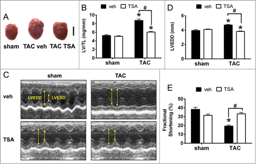
Echocardiography tests of cardiac performance clearly show that TAC veh mice have dilated hearts and TSA administration with TAC surgery prevented left ventricle (LV) dilation (). Increased wall thickness (25% higher in left ventricular end diastolic dimensions, LVEDD) induced by TAC was blunted by TSA (). Decline in fractional shortening (FS) (P < 0.05) induced by pressure overload was also abrogated by TSA (). Increased fetal cardiac expression and suppression of adult genes accompany pathological cardiac hypertrophy.Citation32,33 We observed increased Atrial natriuretic peptide (Anp), Brain natriuretic peptide (Bnp) and Beta myosin heavy chain (Myh7) genes induced by pressure overload were blunted by TSA (Fig. S1A). Reduced expression of the Sarcoplasmic reticulum ATPase (Serca2a) and Alpha myosin heavy chain (Myh6) adult genes by TAC was reversed by HDAC inhibition (Fig. S1B). Taken together, these data demonstrate that HDAC inhibition with TSA attenuates hypertrophic gene response and preserves cardiac function and wall thickness in load-stressed myocardium.
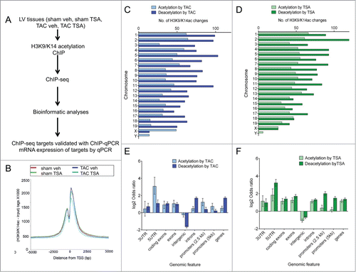
Massive parallel sequencing of H3K9/K14ac in the heart
Recent observations based on genome-wide analysis indicate that HDAC inhibition confers acetylation and deacetylation of H3K9/K14 at gene promoters.Citation27 To test the hypothesis that the myocardium is subject to TSA mediated changes we used ChIP coupled with massive parallel sequencing (ChIP-seq) to map genome-wide histone acetylation patterns in vivo ().Citation34,35 Sequencing generated a total of 134 million single-end reads (on average 32 million 36 bp reads per lane) with 98.27% of reads aligned to the mm9 mouse genome (Table S1). The sequencing depth for all samples was close to the maximum number of peaks generated and close to saturation (Fig. S2A). H3K9/K14ac peaks are observed at transcription start sites (TSS) (Fig. S2B). Characteristic nucleosome free regions Citation36-38 are subject to reduced H3K9/K14ac directly at start sites of genes (). Interestingly, we observed reduced H3K9/K14ac read intensity on promoters in mice subject to TSA attenuation of pathological hypertrophy ().
Figure 2. Dynamic H3K9/K14 acetylation and deacetylation in the heart. (A) Genome-wide changes of TAC and TSA were investigated using H3K9/K14ac ChIP coupled with massive parallel sequencing (ChIP-seq). The targets identified from ChIP-seq were validated with ChIP-qPCR. Expression of corresponding genes was validated by qPCR. (B) H3K9/K14ac ChIP-seq profiles on the transcription start sites of promoters are shown. (C-F) MACS peak calling was used to identify differential H3K9/K14ac in response to TAC (comparison between TAC veh and sham veh) and TSA (TAC TSA vs. TAC veh). (C) Ideogram showing differential H3K9/K14ac regions on mouse (mm9) genome of TAC and (D) TSA group. (E) Genome-wide distribution of H3K9/K14 acetylation and deacetylation for TAC and (F) TSA shown by genomic feature using Fisher's exact test, represented as the log2 odds ratio. Error bars represent 95% confidence intervals. Feature annotation is described in the methods.
Extensive deacetylation by HDAC inhibition
H3K9/K14ac patterns were examined in mice hearts subject to pressure overload (TAC veh vs. sham veh) and attenuation of cardiac hypertrophy with TSA (TAC TSA vs. TAC veh). We also examined acetylation changes mediated by HDAC inhibition in healthy C57BL/6 animals (sham TSA vs. sham veh). To do this, MACS peak calling was used to identify genome-wide differential H3K9/K14ac changes with P = 10−4 cut-off.Citation39 We defined these comparisons as (1) TAC (TAC veh vs. sham veh), (2) TSA (TAC TSA vs. TAC veh), (3) sham controls (sham TSA vs. sham veh). We observed broad increases and decreases in H3K9/K14ac across all comparisons (Table S2). Whereas 24% of peaks showed acetylation, 76% of peaks were deacetylated after pressure overload mediated by TAC (Table S2). HDAC inhibition with TSA induced broad histone deacetylation in mice subject to pressure overload (H3K9/K14 deacetylation 61% in TSA) and healthy mice (H3K9/K14 deacetylation 55% in sham controls) (Table S2). Histone acetylation peaks were distributed uniformly across the genome except for the sex chromosomes in response to TAC () and TSA (). The distribution of H3K9/K14ac peaks at specific regions of the mouse genome were examined, including promoters (5 kb and 2.5 kb up- and down-stream of TSS), coding exons, exons, introns, 3′ untranslated region (UTR) and 5′ UTR, intergenic, and gene body. These regions were annotated using UCSC genome browser.Citation40 Fisher's exact test was used to determine H3K9/K14 acetylation and deacetylation at these genomic sites. The results are expressed as the log2 odds ratios with 95% confidence interval of the observed differences compared to randomly generated peaks. Histone acetylation and deacetylation changes were more likely to occur within a gene than in intergenic regions in both comparisons: TAC () and TSA (). In TAC hearts, gene promoters were preferentially acetylated (). In contrast, gene promoters were deacetylated in TSA group (), suggesting that HDAC inhibition can reverse TAC-induced histone acetylation changes on gene promoters. Together, these data show that pressure overload and HDAC inhibition confers broad H3K9/K14 acetylation and deacetylation at gene promoters in the myocardium.
TSA reverses histone acetylation changes conferred by pathological hypertrophy
H3K9/K14ac is functionally associated with gene promoters and can influence mRNA expression.Citation13,15 We examined promoter specific H3K9/K14ac changes on regions 2.5 kb up- and down-stream of the TSS. The top 20 genes associated with H3K9/K14ac changes at gene promoters in response to TAC and TSA are summarized in Table S3. Pharmacological HDAC inhibition in the TAC group confers promoter H3K9/K14ac changes on genes involved in pathological processes of cardiac disease such as cardiac contractile function, metabolic processes, calcium signaling and inflammation (Table S3).
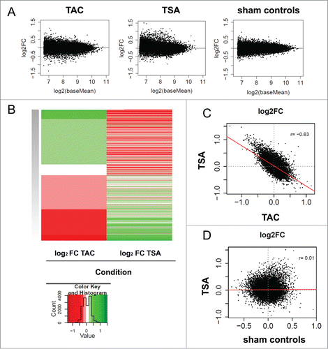
MA plots visualize differential H3K9/K14ac in the heart showing greatest change in mice subject to pressure overload suppressed by TSA (). The heatmap clearly shows 2 clusters for gene promoters: (1) increased acetylation by pressure overload and deacetylation by HDAC inhibition with TSA and (2) deacetylation by TAC and increased acetylation with TSA (). Changes in histone acetylation in TAC are inversely correlated with histone acetylation changes in TSA (). These changes are disease specific; no correlation between TSA and sham controls was observed (). The opposing effects of histone acetylation in TAC and TSA suggest HDAC inhibition reverses promoter specific histone acetylation changes induced by pathological cardiac remodeling ().
Figure 3. HDAC inhibition with TSA reverses pressure overload-induced changes in histone acetylation and deacetylation. Three comparisons are investigated: 1) TAC (TAC veh vs. sham veh), 2) TSA (TAC TSA vs. TAC veh), and 3) sham controls (sham TSA vs. sham veh) (A) MA plots are shown to visualize the concentration of read counts for differential H3K9/K14ac changes in TAC, TSA and sham controls. The log2 (base mean) is a measure of the mean read concentration for each gene. (B) Heat map showing inverse fold changes in H3K9/K14ac between TAC and TSA. Red indicates increases in histone acetylation and green corresponding decreases in histone acetylation (C) Correlation of fold changes between TSA with TAC as well as (D) TSA with sham controls. Correlation was determined using Pearson's correlation (r). Red line represents the linear model. Only promoter (±2.5 kb from TSS) specific changes in H3K9/K14ac are shown for all plots.
TSA suppresses pro-inflammatory NFκB target genes induced by pressure overload
Gene Set Enrichment Analyses (GSEA) was used to explore potential mechanisms of histone acetylation mediated gene expression.Citation41 GSEA of ChIP-seq datasets identified major pathways altered with pressure overload and TSA (). Gene sets related to the extracellular matrix, inflammation, heart contraction, and TCA cycle were associated with histone acetylation in hypertrophied hearts (Table S4). HDAC inhibition in mice subjected to pressure overload altered gene sets related to heart contraction, potassium channel, and inflammation (Table S4). We identified opposing gene sets in TAC and TSA groups (). Gene sets related to metabolism and cardiac contraction are associated with promoter deacetylation in cardiac hypertrophy and gain of acetylation with HDAC inhibition (Table S5). In contrast, collagen deposition and inflammation pathways are associated with acetylation following TAC but deacetylation with TSA (Table S5).
Figure 4. HDAC inhibition with TSA prevents histone acetylation of NFκB target genes. Two comparisons were investigated TAC (TAC veh vs. sham veh) and TSA (differences between TAC TSA and TAC veh). (A) Bar plot of number of GSEA pathways identified with histone acetylation and deacetylation changes for each condition. (B) Venn diagram showing the overlap for GSEA pathways between TAC and TSA. All gene sets are FDR q val < 0.05 according to standard GSEA output. (C) GSEA was used to identify H3K9/K14ac at promoters associated with transcription factor binding using the ENCODE ChIP-seq collection of transcription and co-regulatory factors and chromatin-associated proteins (TFBS). Bar plot of the top 10 TFBS gene sets by deacetylation with TSA and its corresponding association in response to TAC. Numbers following the protein name represent the cell line and have been defined in the methods. Gene sets for TSA are FDR q val < 0.05 according to standard GSEA output. A negative normalized enrichment score (NES) shows deacetylated gene sets, while a positive NES score indicates gene sets associated with histone acetylation. (D) GSEA plot showing an association of NFκB bound genes associated with histone acetylation in response to TAC and corresponding deacetylation with HDAC inhibitor TSA. Genes are ranked by changes in H3K9/K14ac.
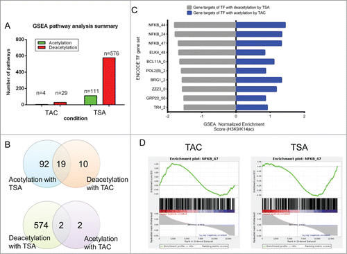
Next, we examined H3K9/K14ac profiles with ENCODE Citation42 to identify relevant transcription factor binding sites that might be associated with pharmacological HDAC inhibition. GSEA revealed strong associations for multiple transcription factors on promoters with increased H3K9/K14 acetylation in pressure overload mice, while these same promoters were deacetylated by HDAC inhibition with TSA (). Pathological cardiac remodeling is associated with inflammation, and novel anti-inflammatory therapies have shown promise in heart failure patients.Citation43,44 In addition to attenuating cardiac hypertrophy, compounds that inhibit HDAC activity have potent anti-inflammatory actions.Citation45 NFκB is a central player in regulation of the immune and inflammatory response and we observed a strong association of NFκB in our data set with pressure overload and HDAC inhibition ().
Since pharmacological HDAC inhibition attenuates pro-inflammatory gene expression, we hypothesized that histone deacetylation may play a role in the transcription of NFκB targets genes in pathological cardiac remodeling. To visualize differential acetylation patterns, we examined 3 NFκB target genes, Icam1, Cxcl10, and Il6ra, implicated in vascular and cardiac inflammation. Increased promoter acetylation by load-induced hypertrophy was reversed by HDAC inhibition with TSA ().
Figure 5. TAC confers histone acetylation and HDAC inhibition attenuates the increase in histone acetylation and gene expression on NFκB target genes. (A) Increased promoter acetylation of Icam1, Cxcl10, and Il6r genes (blue line, blue arrow) are attenuated by HDAC inhibition with TSA (red line, red arrow). (B) Chromatin immunopurification of acetylated H3K9/K14 histones in the LV tissues assessed by real time qPCR on NFκB target genes. N = 4–7/group, TAC veh vs. sham veh: * P < 0.05; ** P < 0.01, *** P < 0.001; TAC TSA vs. TAC veh: # P < 0.05; ## P < 0.01; ### P < 0.001. (C) Gene expression changes were validated using qPCR of NFKB target genes. n = 4-5/group, TAC veh vs. sham veh: * P < 0.05, ** P < 0.01; *** P < 0.001; TAC TSA vs. TAC veh: # P < 0.05; ## P < 0.01; ### P < 0.001. Cxcl10, chemokine (C-X-C Motif) ligand 10; Il6ra, interleukin-6 receptor; Il21r, interleukin-21 receptor; Icam1, intercellular adhesion molecule-1; Ticam2, toll-like receptor adaptor molecule 2; Traf3, TNF receptor-associated factor 3; Vcam1, vascular cell adhesion molecule 1.
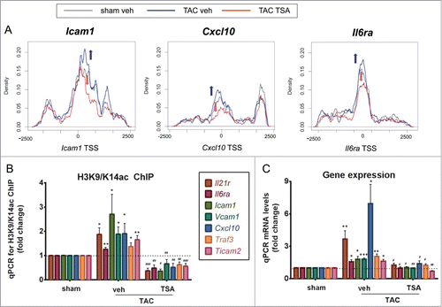
Next we validated ChIP-seq signals for histone acetylation using qPCR. NFκB target genes IL21r, Il6ra, Icam1, Vcam1, Cxcl10, Traf3, and Ticam2 were subject to histone acetylation in response to pressure overload and were reversed by HDAC inhibition (). We sought to identify if histone acetylation changes identified by massive parallel sequencing were closely associated with gene expression. We show a tight correspondence between gene activation and histone acetylation at these promoters in TAC as well as gene suppression and histone deacetylation with TSA treatment (). HDAC inhibition with TSA reversed inflammatory gene expression changes during cardiac remodeling. Our results suggest that changes in histone deacetylation may influence the expression of NFκB target genes mediated by load-induced pathological hypertrophy. HDAC inhibition with TSA attenuates gene expression of NFκB target genes by histone deacetylation and the anti-inflammatory response in cardiac hypertrophy.
Discussion
HDAC inhibitors are efficacious in preclinical models of heart failure.Citation22-24 These pharmacological compounds block pathological processes such as cardiac hypertrophy, inflammation, autophagy, and fibrosis,Citation45 but the mechanism underpinning these events are not well understood. To elucidate the impact of HDAC inhibition in the heart, we used unbiased next generation sequencing approach to explore histone acetylation by ChIP-seq in a model of pressure overload induced hypertrophy using the prototypical inhibitor of HDAC activity, TSA. To our knowledge, this is the first report of genome-wide maps determined for histone acetylation in hypertrophied heart attenuated by pharmacological HDAC inhibition. This study is particularly novel for several reasons. First, HDAC inhibition by TSA induced broad acetylation and deacetylation on gene promoters in the myocardium. Second, histone acetylation mediated by load-induced hypertrophy was blunted by HDAC inhibition at genes implicated in inflammation, cardiac contraction, and extracellular matrix. Third, transcription factor analysis revealed a role for NFκB in the regulation of a subset of pro-inflammatory genes. Taken together, this data reveals a novel mechanism of TSA mediated cardioprotection by deacetylation of NFκB target genes implicated in inflammation.
Although systemic pharmacological compounds that inhibit HDAC activity are strongly predicted to increase histone acetylation, our report and its findings appear paradoxical in that the actions of TSA attenuating cardiac hypertrophy include histone deacetylation. Whereas, the paradigmatic view of HDAC inhibition involves increased acetylation of total histones, the results presented here are consistent with very recent genome-wide studies.Citation27 Indeed, the expression of NFκB gene targets, such as Icam1, Vcam1, Il21r, Il6ra, Ticam2, and Cxcl10, closely correlate with histone deacetylation. Furthermore, pressure overload increased the expression of pro-inflammatory genes that were closely associated with histone acetylation status. While there is no precedent for blunting hypertrophic-induced inflammation by histone deacetylation, some genes are sheltered from hyperacetylation by HDAC inhibition.Citation14 We cannot exclude a role for other histone modifications. N-terminal histone tails are targets for enzymes that mediate specific modifications, particularly on lysine and arginine residues, which, together, form a “histone code” that coordinates the transcriptional events that precisely regulate chromatin structure and gene function.Citation11,12 The functional state of cardiac chromatin can be altered by four regulatory mechanisms: covalent modification of histones,Citation19,20 differential association of non-histone proteins,Citation9 ATP-dependent chromatin remodeling,Citation46,47 and gene regulation under the control of non-coding RNAs.Citation48 Recent studies from our laboratory have also indicated that the cooperation between non-coding RNAs integrating chromatin modifications and epigenetic changes regulate hypertrophic gene activity.Citation49
Using GSEA to intersect our data against ENCODE transcription factor database, we identified a role for NFκB in regulating hypertrophic gene expression. NFκB is an inducible transcription factor and a master regulator of inflammation. With five members of mammalian NFκB family, the most widely studied and abundant form is the p50 and p65 heterodimer. In the heart, the importance of NFκB is highlighted in preclinical models of heart failure. NFκB activation is required for adaptive cardiac hypertrophy.Citation50,51 We also observed changes in acetylation in genes functionally associated with cardiac contractile and extracellular matrix regulation; and these are the focus of future studies.Citation50,51 Conversely, other studies show that NFKB promotes adverse cardiac remodeling.52,53 It is postulated that the microenviroment, intensity and chronicity of NFKB stimulus dictate its multifaceted outcomes in the heart.Citation54 In addition, TSA improves cardiac function in an NFκB dependent manner.Citation55 In a Langendorff hanging heart model of ischemia-reperfusion injury, HDAC inhibition reduced infarct size and improved cardiac function, and TSA-induced cardioprotection was absent with genetic deletion of NFκB p50 subunit.Citation55 Whereas the anti-inflammatory and anti-hypertrophic role of NFκB in the heart is documented, its mechanism of action remains unclear. One study suggests a role for protein acetylation in the heart with HDAC inhibition.Citation55 Histone proteins are substrates for acetylation and deacetylation.Citation56 Treatment with the TSA in the myocardium mediates p50 protein acetylation and enhanced NFκB DNA binding activity in the heart.Citation55
Heart failure represents complicated pathophysiology typified as systolic or diastolic with the activation of multiple pro-inflammatory pathways. Preclinical studies indicate potential to develop small molecule HDAC inhibitors to block cardiac remodeling. The US FDA has approved 2 compounds that inhibit HDAC activity for the treatment of cancer, vorinostat (suberoylanilide hydroxamic acid, Zolinza) and depsipeptide (romidepsin, Istodax).Citation57 The protective activity of HDAC inhibitors is related to the efficacy of these compounds in preclinical models of heart failure. For example, cardiac hypertrophy is blocked for 2 weeks by the hydroxamate, TSA, or the aliphatic acid valproate, indicating that these compounds are well tolerated.Citation58 In addition to TSA, the pan-HDAC inhibitor scriptaid attenuated cardiac hypertrophy in both short-term (3 week) and long-term (9 week) studies in mice models, and was well tolerated with chronic administration not adversely impacting survival.Citation59 There is a tremendous push for isoform-selective small molecule HDAC inhibitors to further improve safety profiles based on the identification of HDAC isoforms implicated in heart failure.Citation60 Before preclinical safety and efficacy studies can begin to evaluate isoform selective inhibitors it will be important to identify which HDACs promote pathological cardiac remodeling.
There are several considerations that are beyond the scope of this report but relevant to future studies. Firstly, the histone cross-talk hypothesis suggests that different inter-related post-translational histone modifications are important in regulating gene expression.Citation61,62 For example, histone H3 acetylation and H3K4m3 are functionally linked with the inhibition of HDAC activity.Citation63 Comprehensive mapping of chromatin modifications in vascular cells have shown that transcriptional events are coordinated by multiple post-translational changes to histone residues.Citation27 This is evident by the strong correlation between increased gene expression and the co-occurrence of H3K9/K14ac and H3K4m3. Interestingly, several histone methyltransferases are downregulated in primary vascular cells stimulated by compounds that inhibit HDAC activity. Although beyond the scope of this study, it will be interesting to characterize histone code changes of attenuation of cardiac hypertrophy with HDAC inhibition.
While preclinical studies justify the potential use of HDAC inhibitors blunting pathological remodeling, studies such as ours are designed to prevent rather than reverse pre-existing cardiac pathology. In this context, it would be important to determine whether histone deacetylation of target genes mediated by HDAC inhibition reverse pro-inflammatory gene expression patterns in models of load-induced hypertrophy. Indeed, such studies would closely correspond to patients with heart failure and therapies targeted to reduce pro-inflammatory expression once symptoms are present or after a stress event. Finally, this report focused on gene targets implicated in cardiac inflammation based on unbiased genome-wide mapping. We also observed changes in acetylation status associated with cardiac contractile and extracellular matrix regulation and the focus of future studies. In conclusion, our results demonstrate that HDAC inhibition with TSA attenuates cardiac hypertrophy by mediating acetylation as well as histone deacetylation on gene promoters. We describe a potential mechanism of the anti-inflammatory effects of HDAC inhibitors on NFκB target genes and re-examination of cardiac deacetylation by HDAC inhibition.
Materials and Methods
TAC surgery
This study used a previously established model of pathological cardiac hypertrophy, induced by transverse aortic constriction (TAC) and the attenuation of cardiac hypertrophy by treatment with the HDAC inhibitor Trichostatin A (TSA, Sigma, Cat-T8552).Citation46,49 Briefly, C57/BL6 male mice at 10-12 weeks old were anesthetized with a dose mixture of ketamine/xylazine/atropine (50, 10, and 1.2 mg/kg, respectively). The aorta constricted at the mid aortic arch level with a 5/0 braided silk suture using a blunted 26/27-gauge needle as a calibrator, producing approximately 60% reduction in luminal diameter. Sham control mice underwent the same procedure but without the aortic constriction. Mice were then randomized to receive either vehicle (DMSO) or TSA treatment. DMSO or TSA (dissolved in 50% DMSO) were injected subcutaneously twice daily at 0.6 mg/kg/day for a period of 4 weeks. At the end of the 4-week treatment period, establishment of cardiac hypertrophy and attenuation was confirmed by echocardiography. 2-D guided M-mode traces were acquired using a Phillip iE33 ultrasound machine and 15 MHz linear-transducer. LV dimensions at diastole (LVDd), systole (LVDs) and wall thickness were measured. During autopsy, the left ventricles were dissected, weighed, and snapped frozen in liquid nitrogen for downstream molecular analysis.
Total RNA isolation and cDNA synthesis
LV tissue was homogenized in TRIzol® reagent (Invitrogen). Phenol chloroform extraction was performed to obtain aqueous containing RNA phase and RNA was purified with RNeasy Mini Kit spin columns (Qiagen, Cat-74106) following manufacturer's instructions. Genomic DNA contamination was removed by on column DNase treatment (Qiagen). RNA quality was measured with MultiNA and Nanodrop. cDNA synthesis was performed using High Capacity cDNA Reverse Transcription Kit (Applied Biosystems, Cat-4368813) according to manufacturer's instructions.
Chromatin immunoprecipitation
LV tissue was minced then cross-linked with 1% formaldehyde (in phosphate buffered saline without calcium and magnesium) by rotation for 10 min at room temperature. Quenching for excess formaldehyde was performed with 0.125 M glycine for 10 min. Tissue was then resuspended in SDS lysis buffer containing 1% SDS, 10 mM EDTA, 50 mM Tris-HCl pH8.1 supplemented with a protease inhibitor cocktail (Roche). Tissue was then homogenized to a clear solution in SDS lysis buffer and placed on ice to complete cell lysis. Chromatin shearing was performed by sonication with a Diagnode bioruptor for 35 min with 0.5 minute on and off intervals. Sheared soluble chromatin was size fractionated with the MultiNA (Shimadzu) to ensure proper sonication of chromatin (range 75-300 bp). One quarter of the sheared chromatin was kept as input material. Sheared chromatin was diluted 10 times in ChIP dilution buffer (0.01% SDS, 1% Triton-X, 1.2 mM EDTA, 16.7 mM Tris-HCl pH 8.0, 167 mM NaCl, and protease inhibitor) and incubated with antibody to acetylated H3K9/K14 (Upstate, Cat-06-599) overnight at 4°C. A no antibody ChIP was used as a negative control for non-specific background enrichment. Antibody-bound chromatin complex was captured with Dynabeads® Protein A magnetic beads (Invitrogen, Cat-100.01D) and given a sequence of 5 washes beginning with low salt, high salt, lithium chloride, TE buffer (pH 8.0) followed with TE + 0.01% SDS. Bound DNA was eluted with ChIP elution buffer (20 mM Tris-HCl pH7.5, 5 mM EDTA, 50 mM NaCl, 1% SDS) and reverse cross-linked for 2 h at 62°C on a thermomixer (Eppendorf) set to 1,400 rpm. DNA was recovered and purified with NucleoSpin® PCR Clean-up Columns and Reagents (Macharey-Nagel) following manufacturer's instructions. Percentage input (% input) was calculated for each ChIP experiment, and results are expressed as relative fold enrichment/ratio for the target sequences compared between the treated versus control groups.
Quantitative real-time PCR (qPCR)
SYBR Green quantitative PCR was performed with Applied Biosystems 7500 Fast Real-Time PCR System. 5 pmoles of forward and reverse primer, cDNA template/ChIP material and FAST SYBR® Green Master Mix (Roche) were mixed to a final volume of 20 µl. Reactions were incubated at 95°C for 10 min, followed by 40 cycles of 95°C for 10 sec and 60°C for 30 sec.
Primers used for gene expression validation:
Amhc, F:CCACCTGGGCAAGTCTAACAA, R:TGTAGTCCACGGTGCCAGC;
Anp, F: ACAGCCAAGGAGGAAAAGGC, R: CCACAGTGGCAATGTGACCA;
Bmhc, F: GATGTTTTTGTGCCCGATGA, R:ACCGTCTTGCCATTCTCCG;
Bnp, F: TCCAGAGCAATTCAAGATGCA, R:CTTTTGTGAGGCCTTGGTCC;
Cxcl10, F: TCTGAGTGGGACTCAAGGGAT, R: AGGCTCGCAGGGATGATTTC;
Gapdh, F: TGAAGCAGGCATCTGAGGG, R: CGAAGGTGGAAGAGTGGGAG;
Icam1, F: GCTCCGCTGCTACCTGCACT, R: CCAGGCCCAGGGATCACAACG;
Il6ra, F: TGCGTCATCCATGATGCCTT, R: CCTGGGCTCTGCTATCCAAG;
Il21r, F: CACCTGACTGAACTCCTGCC, R: TGCTGTGTCCCAGACCTACT;
Serca2a, F: CCCCCTGGGAGAATATCTGG, R:GATCTGGAAAATGAGCGGCA;
Ticam2, F: GCAGTACCACTTCCCAGCTAA, R: ACTCATTTGACACTGGGCTCT;
Traf3, F: GCCTCGGGAATTTCAGTTTCCT, R: CCTTGTAGCCTCCTTGCTCC;
Vcam1, F: AAGTCTGTGGATGGCTCGTACA, R: TCAGTCTTAGATTCACACTCG TATATGC.
Primers used for H3K9/K14 acetylation ChIP:
Cxcl10, F: TCAGAACAGAAGCCGGAAGT, R: GGTGCTGTGCAGAGTGACAT;
Icam1, F: CTTCTCTCCGGACTCACCTG, R: GGTATTTCCGGGTGGAGACT;
Il6ra, F: GCAAGATGCAAATGATGTGG, R: TGAGTTCTGAGGCATTGCAC;
Il21r, F: TGAGTCTCGGAGGCAGATTT, R: TCACCATTCCTGTCAAACCA;
Ticam2, F: GTTTGGGTCAGCATTCTGGT, R: TCACACCTCGGGGTTTAGAG;
Traf3, F: TCGGGTGAATCCTCTGGTAG,R: AGCTCAGTGCCTGGTATGCT;
Vcam1, F: TGAAAGGACGTTGATGCAGA, R: ATGCAGCCAAAGAAATCCAC.
Data analysis and statistics
For qPCR data analysis, threshold cycle numbers (Ct) were measured in the exponential phase for all samples. For gene expression, analyzed genes were normalized to the level of Gapdh. For chromatin immunoprecipitation, Ct values were converted to final relative values of % input enrichment. To compare between ChIP enrichment in treated compared to control samples, enrichment were calculated against sham veh; for example, % input TAC veh/% input sham veh and % input TAC TSA/% input sham veh. Data is shown as mean ± standard error of the mean (SEM). Statistical significance and P-values were calculated by 2-tailed, unpaired Student's t-tests (Graphpad Prism) by comparing groups TAC veh vs. sham veh and TAC veh versus TAC TSA. A P-value < 0.05 was considered statistically significant.
ChIP sequencing
Purified ChIP DNA sequences and their input controls were sequenced on the Illumina GAIIx Next Generation Sequencer (GAIIx) as described previously.Citation27 ChIP-seq libraries were prepared according to the protocols described in the Illumina ChIP-seq library preparation kit (Illumina, Cat-IP-102-1001). Briefly, 10 ng of immunopurified DNA or genomic DNA from an input sample was end-repaired, followed by the 3′ addition of an adenosine nucleotide and ligated to universal library adaptors. Ligated material was separated on a 2.0% agarose gel, and fragments in the range of 250–350 bp were excised and column-purified (Qiagen). A DNA library was obtained by an 18-cycle PCR amplification using Phusion DNA polymerase (10 sec 98°C, 30 sec 65°C, 30 sec 72°C) with oligonucleotides complementary to Illumina sequencing adapters. Column- purified libraries were quantified fluorometrically (Qubit) and visualized by MultiNA capillary electrophoresis (DNA 500 kit) for quality assurance. Libraries were diluted to 10 nM and stored at −20°C prior to cluster generation at a final DNA concentration of 6 pM. Sequencing of the first 36 nucleotides was performed on the Genome Analyzer IIx instrument (Illumina) according to the manufacturer's protocols. Short-read sequences of 36 bp single ended reads were generated using Illumina Pipeline 1.4.
Analysis of ChIP-seq data
ChIP-seq 36bp single-ended reads were aligned to the mouse genome (NCBI37/mm9) using the Burrows-Wheeler Aligner (BWA, version 0.6.1), with default settings.Citation64 Uniquely mapped reads with no more than one mismatch were used for binding peak detection.
Distribution of peaks across genome features
To identify peaks, Model-based Analyis of ChIP-seq (MACS) algorithm was used with default parameters with a cut off of P = 10−4.Citation39 Binding peaks were detected for H3K9/K14ac ChIP in 2 comparisons; TAC (TAC veh to sham veh) and TSA (TAC TSA to TAC veh). A peak is defined as discrete, non-overlapping region in which a distinct H3K9/K14ac enrichment pattern is observed when compared between 2 samples. All reference BED format files corresponding to the coordinate of common genomic features such as intergenic regions, introns, exons, RefSeq genes, coding exons, 3′ UTR, and 5′UTR were acquired from the UCSC Genome Browser.Citation65 To determine whether increases or decreases of histone acetylation are enriched at specific features of the genome, the data was divided into differential histone acetylation and a randomly generated bed file of peaks, generated using bedtools shuffle. The number of histone acetylation at each feature was counted and the Fisher's exact test was used to compare the enrichment of differential vs. random peaks. The results are reported as the odds ratio as a log to the base of 2 ( and ).
Promoter-specific H3K9/K14ac and DESeq
In addition to peak calling using MACS, promoter-specific changes to H3K9/K14ac were determined (, Table S3, Table S4). The raw number of reads were counted using BEDTools multicovCitation66 for H3K9/K14ac at gene promoters. Promoter regions are defined as 2.5 kb either side of the TSS (transcription start site).
DESeq was used to determine changes in histone acetylation with the following parameters; (parametric fit for dispersion estimation, contigs were ranked by number of reads and the bottom 30% of contigs with the least reads were removed before further analysis, FDR method=Benjamini-Hochberg). Fold changes were reported as the log2 of the fold change (log2FC). DESeq also reports the base mean, a measure of average read abundance across all samples for each promoter (ChIP-seq). A smaller number correlates with lower read abundance for that promoter.
MA plots, correlation plots, and heat map generation
Promoter specific H3K9/K14ac log2FC from DESeq output were used for MA plots, correlation plots and heat maps. Three comparisons were of interest: TAC (TAC veh compared to sham veh), sham controls (sham TSA compared to sham veh) and TSA (TAC TSA compared to TAC veh). MA plots were generated by plotting the log2FC of each comparison versus the log2 of base mean (a measure of relative read abundance at each promoter). Correlation plots were determined comparing the log2FC from one condition compared to another. Pearsons correlation coefficient (r) was also reported. Heat maps were generated in R using the tool heatmap.2 from the gplots package (http://CRAN.R-project.org/package=gplots). The heat map was generated including only the top sixth most acetylated and sixth most deacetylated promoters in the TAC condition (based on log2FC).
Vizualization of changes in acetylation at the promoter region
Samtools view was used to determine changes in acetylation at the promoter region (2.5 kb either side of TSS) of selected genes by counting reads directly from the bam file.Citation64 In R, localization of the reads was adjusted by shifting the position of the read by 100 bp toward the center of the read, based on the average read size of 200 bp. The number of reads was then normalized to the total number of reads in each bam file. Rectangular kernel density plots were then generated in R using a bandwidth of 200.
Gene set enrichment analysis
Pathway analysis was performed using Gene Set Enrichment Analysis (GSEA) using annotated gene sets from MSigDB default parameters Citation41 (Table S4). GSEA was used to determine enrichment of histone acetylation or deacetylation at the promoters of genes associated with transcription factor and chromatin associated protein binding. ChIP-seq data of transcription factor and chromatin associated proteins in diverse cell lines were accessed from ENCODE source: ftp://hgdownload.cse.ucsc.edu/apache/htdocs/goldenpath/hg19/encodeDCC/wgEncodeRegTfbsClustered/. Mouse genes were converted to hg19 human homologues using BioMart to enable comparsion to human ENCODE transcription factor binding site.Citation42 A series of gene sets were created for each ENCODE ChIP-seq data set that contained genes bound by the precipitated protein within 3 kb of its transcription start site. These gene sets were compared to the ranks of acetylation via GSEA. Ranks of changes of acetylation at the TSS were generated from product of the log of the adjusted p value and sign of the fold change based on DESeq output. Default GSEA parameters were used, except for the number of permutations (1000). An association was deemed significant if it had an FDR q value <0.05 as determined by GSEA. Only ENCODE ChIP-seq gene sets containing 15-1000 genes were included for further analysis. Cell lines are presented as numbers in followed by the name of the transcription or co-regulatory factor.
Data access
ChIP-seq data used for this research have been submitted to the NCBI Gene Expression Omnibus (GEO) under accession number GSE63590. The data is available at the following link: http:/www.ncbi.nlm/nih.gov/geo/ under accession number GSE63590.
Disclosure of Potential Conflicts of Interest
No potential conflicts of interest were disclosed.
Supplemental_Material.zip
Download Zip (1.6 MB)Acknowledgments
The authors thank Ross Lazarus and Antony Kaspi for bioinformatics expertise and technical support.
Funding
The authors acknowledge grant and fellowship support from the National Health and Medical Research Council (NHMRC). A E-O. is an NHMRC Senior Research Fellow. Supported in part by the Victorian Government's Operational Infrastructure Support program.
References
- Hunter JJ, Chien KR. Signaling pathways for cardiac hypertrophy and failure. N Engl J Med 1999; 341: 1276-83; PMID:10528039; http://dx.doi.org/10.1056/NEJM199910213411706
- Bernardo BC, Weeks KL, Pretorius L, McMullen JR. Molecular distinction between physiological and pathological cardiac hypertrophy: experimental findings and therapeutic strategies. Pharmacol Ther 2010; 128: 191–227; PMID:20438756; http://dx.doi.org/10.1016/j.pharmthera.2010.04.005
- Bozkurt B, Mann DL, Deswal A. Biomarkers of inflammation in heart failure. Heart failure reviews 2010; 15: 331–41; PMID:19363700; http://dx.doi.org/10.1007/s10741-009-9140-3
- Imamura S, Matsuoka R, Hiratsuka E, Kimura M, Nakanishi T, Nishikawa T, Furutani Y, Takao A. Adaptational changes of MHC gene expression and isozyme transition in cardiac overloading. Am J Physiol 1991; 260: H73–9; PMID:1825154
- Mayer Y, Czosnek H, Zeelon PE, Yaffe D, Nudel U. Expression of the genes coding for the skeletal muscle and cardiac actions in the heart. Nucleic Acids Res 1984; 12: 1087–100; PMID:6546444; http://dx.doi.org/10.1093/nar/12.2.1087
- McKinsey TA, Olson EN. Cardiac histone acetylation–therapeutic opportunities abound. Trends Genet 2004; 20: 206–13; PMID:15041175; http://dx.doi.org/10.1016/j.tig.2004.02.002
- McKinsey TA, Olson EN. Dual roles of histone deacetylases in the control of cardiac growth. Novart Found Symp 2004; 259: 132–41; discussion 41-5, 63-9; PMID:15171251; http://dx.doi.org/10.1002/0470862637.ch9
- McKinsey TA, Olson EN. Toward transcriptional therapies for the failing heart: chemical screens to modulate genes. J Clin Invest 2005; 115: 538–46; PMID:15765135; http://dx.doi.org/10.1172/JCI24144
- McKinsey TA, Zhang CL, Olson EN. Signaling chromatin to make muscle. Curr Opin Cell Biol 2002; 14: 763–72; PMID:12473352; http://dx.doi.org/10.1016/S0955-0674(02)00389-7
- Olson EN, Backs J, McKinsey TA. Control of cardiac hypertrophy and heart failure by histone acetylation/deacetylation. Novart Found Symp 2006; 274: 3–12; discussion 3-9, 152-5, 272-6; PMID:17019803; http://dx.doi.org/10.1002/0470029331.ch2
- Luger K, Mader AW, Richmond RK, Sargent DF, Richmond TJ. Crystal structure of the nucleosome core particle at 2.8 A resolution. Nature 1997; 389: 251–60; PMID:9305837; http://dx.doi.org/10.1038/38444
- Jenuwein T, Allis CD. Translating the histone code. Science 2001; 293: 1074–80; PMID:11498575; http://dx.doi.org/10.1126/science.1063127
- Pokholok DK, Harbison CT, Levine S, Cole M, Hannett NM, Lee TI, Bell GW, Walker K, Rolfe PA, Herbolsheimer E, et al. Genome-wide map of nucleosome acetylation and methylation in yeast. Cell 2005; 122: 517–27; PMID:16122420; http://dx.doi.org/10.1016/j.cell.2005.06.026
- Wang Z, Zang C, Rosenfeld JA, Schones DE, Barski A, Cuddapah S, Cui K, Roh TY, Peng W, Zhang MQ, et al. Combinatorial patterns of histone acetylations and methylations in the human genome. Nat Genet 2008; 40: 897–903; PMID:18552846; http://dx.doi.org/10.1038/ng.154
- Liang G, Lin JC, Wei V, Yoo C, Cheng JC, Nguyen CT, Weisenberger DJ, Egger G, Takai D, Gonzales FA, et al. Distinct localization of histone H3 acetylation and H3-K4 methylation to the transcription start sites in the human genome. Proc Natl Acad Sci U S A 2004; 101: 7357–62.
- Zhang CL, McKinsey TA, Chang S, Antos CL, Hill JA, Olson EN. Class II histone deacetylases act as signal-responsive repressors of cardiac hypertrophy. Cell 2002a; 110: 479–88; http://dx.doi.org/10.1016/S0092-8674(02)00861-9
- Trivedi CM, Luo Y, Yin Z, Zhang M, Zhu W, Wang T, Floss T, Goettlicher M, Noppinger PR, Wurst W, et al. Hdac2 regulates the cardiac hypertrophic response by modulating Gsk3 beta activity. Nat Med 2007; 13: 324–31; PMID:17322895; http://dx.doi.org/10.1038/nm1552
- McKinsey TA. The biology and therapeutic implications of HDACs in the heart. Handbook Exp Pharmacol 2011; 206: 57–78; PMID:21879446; http://dx.doi.org/10.1007/978-3-642-21631-2_4
- Papait R, Cattaneo P, Kunderfranco P, Greco C, Carullo P, Guffanti A, Vigano V, Stirparo GG, Latronico MV, Hasenfuss G, et al. Genome-wide analysis of histone marks identifying an epigenetic signature of promoters and enhancers underlying cardiac hypertrophy. Proc Natl Acad Sci U S A 2013; 110: 20164–9; PMID:24284169; http://dx.doi.org/10.1073/pnas.1315155110
- Sayed D, He M, Yang Z, Lin L, Abdellatif M. Transcriptional regulation patterns revealed by high resolution chromatin immunoprecipitation during cardiac hypertrophy. J Biol Chem 2013; 288: 2546–58; PMID:23229551; http://dx.doi.org/10.1074/jbc.M112.429449
- Thaler F, Minucci S. Next generation histone deacetylase inhibitors: the answer to the search for optimized epigenetic therapies? Expert Opin Drug Disc 2011; 6: 393–404; PMID:22646017; http://dx.doi.org/10.1517/17460441.2011.557660
- Antos CL, McKinsey TA, Dreitz M, Hollingsworth LM, Zhang CL, Schreiber K, Rindt H, Gorczynski RJ, Olson EN. Dose-dependent blockade to cardiomyocyte hypertrophy by histone deacetylase inhibitors. J Biol Chem 2003; 278: 28930–7; PMID:12761226; http://dx.doi.org/10.1074/jbc.M303113200
- Gallo P, Latronico MV, Grimaldi S, Borgia F, Todaro M, Jones P, Gallinari P, De Francesco R, Ciliberto G, Steinkuhler C, et al. Inhibition of class I histone deacetylase with an apicidin derivative prevents cardiac hypertrophy and failure. Cardiov Res 2008; 80: 416–24; PMID:18697792; http://dx.doi.org/10.1093/cvr/cvn215
- Kee HJ, Sohn IS, Nam KI, Park JE, Qian YR, Yin Z, Ahn Y, Jeong MH, Bang YJ, Kim N, et al. Inhibition of histone deacetylation blocks cardiac hypertrophy induced by angiotensin II infusion and aortic banding. Circulation 2006; 113: 51–9; PMID:16380549; http://dx.doi.org/10.1161/CIRCULATIONAHA.105.559724
- Halsall J, Gupta V, O'Neill LP, Turner BM, Nightingale KP. Genes are often sheltered from the global histone hyperacetylation induced by HDAC inhibitors. PloS One 2012; 7: e33453; PMID:22479401; http://dx.doi.org/10.1371/journal.pone.0033453
- Rada-Iglesias A, Enroth S, Ameur A, Koch CM, Clelland GK, Respuela-Alonso P, Wilcox S, Dovey OM, Ellis PD, Langford CF, et al. Butyrate mediates decrease of histone acetylation centered on transcription start sites and down-regulation of associated genes. Genome Res 2007; 17: 708–19; PMID:17567991; http://dx.doi.org/10.1101/gr.5540007
- Rafehi H, Balcerczyk A, Lunke S, Kaspi A, Ziemann M, Kn H, Okabe J, Khurana I, Ooi J, Khan AW, et al. Vascular histone deacetylation by pharmacological HDAC inhibition. Genome Res 2014; 24: 1271–84; PMID:24732587; http://dx.doi.org/10.1101/gr.168781.113
- Cao DJ, Wang ZV, Battiprolu PK, Jiang N, Morales CR, Kong Y, Rothermel BA, Gillette TG, Hill JA. Histone deacetylase (HDAC) inhibitors attenuate cardiac hypertrophy by suppressing autophagy. Proc Natl Acad Sci U S A 2011; 108: 4123–8; PMID:21367693; http://dx.doi.org/10.1073/pnas.1015081108
- Cardinale JP, Sriramula S, Pariaut R, Guggilam A, Mariappan N, Elks CM, Francis J. HDAC inhibition attenuates inflammatory, hypertrophic, and hypertensive responses in spontaneously hypertensive rats. Hypertension 2010; 56: 437–44; PMID:20679181; http://dx.doi.org/10.1161/HYPERTENSIONAHA.110.154567
- Iyer A, Fenning A, Lim J, Le GT, Reid RC, Halili MA, Fairlie DP, Brown L. Antifibrotic activity of an inhibitor of histone deacetylases in DOCA-salt hypertensive rats. Brit J Pharmacol 2010; 159: 1408–17; PMID:20180942; http://dx.doi.org/10.1111/j.1476-5381.2010.00637.x
- Du XJ, Fang L, Gao XM, Kiriazis H, Feng X, Hotchkin E, Finch AM, Chaulet H, Graham RM. Genetic enhancement of ventricular contractility protects against pressure-overload-induced cardiac dysfunction. J Mol Cell Cardiol 2004; 37: 979–87; PMID:15522275; http://dx.doi.org/10.1016/j.yjmcc.2004.07.010
- Nakao K, Minobe W, Roden R, Bristow MR, Leinwand LA. Myosin heavy chain gene expression in human heart failure. J Clin Invest 1997; 100: 2362–70; PMID:9410916; http://dx.doi.org/10.1172/JCI119776
- Tardiff JC, Hewett TE, Factor SM, Vikstrom KL, Robbins J, Leinwand LA. Expression of the beta (slow)-isoform of MHC in the adult mouse heart causes dominant-negative functional effects. Am J Physiol Heart Circ Physiol 2000; 278: H412–9; PMID:10666070
- Mardis ER. The impact of next-generation sequencing technology on genetics. Trends Genet: TIG 2008; 24: 133–41; PMID:18262675; http://dx.doi.org/10.1016/j.tig.2007.12.007
- Park PJ. ChIP-seq: advantages and challenges of a maturing technology. Nat Rev Genet 2009; 10: 669–80; PMID:19736561; http://dx.doi.org/10.1038/nrg2641
- Heintzman ND, Stuart RK, Hon G, Fu Y, Ching CW, Hawkins RD, Barrera LO, Van Calcar S, Qu C, Ching KA, et al. Distinct and predictive chromatin signatures of transcriptional promoters and enhancers in the human genome. Nat Genet 2007; 39: 311–8; PMID:17277777; http://dx.doi.org/10.1038/ng1966
- Lee CK, Shibata Y, Rao B, Strahl BD, Lieb JD. Evidence for nucleosome depletion at active regulatory regions genome-wide. Nat Genet 2004; 36: 900–5; PMID:15247917; http://dx.doi.org/10.1038/ng1400
- Sekinger EA, Moqtaderi Z, Struhl K. Intrinsic histone-DNA interactions and low nucleosome density are important for preferential accessibility of promoter regions in yeast. Mol Cell 2005; 18: 735–48; PMID:15949447; http://dx.doi.org/10.1016/j.molcel.2005.05.003
- Zhang Y, Liu T, Meyer CA, Eeckhoute J, Johnson DS, Bernstein BE, Nusbaum C, Myers RM, Brown M, Li W, et al. Model-based analysis of ChIP-Seq (MACS). Genome Biol 2008; 9: R137
- Kent WJ, Sugnet CW, Furey TS, Roskin KM, Pringle TH, Zahler AM, Haussler D. The human genome browser at UCSC. Genome Res 2002; 12: 996–1006; PMID:12045153; http://dx.doi.org/10.1101/gr.229102
- Subramanian A, Tamayo P, Mootha VK, Mukherjee S, Ebert BL, Gillette MA, Paulovich A, Pomeroy SL, Golub TR, Lander ES, et al. Gene set enrichment analysis: a knowledge-based approach for interpreting genome-wide expression profiles. Proc Natl Acad Sci U S A 2005; 102: 15545–50; PMID:16199517; http://dx.doi.org/10.1073/pnas.0506580102
- Raney BJ, Cline MS, Rosenbloom KR, Dreszer TR, Learned K, Barber GP, Meyer LR, Sloan CA, Malladi VS, Roskin KM, et al. ENCODE whole-genome data in the UCSC genome browser (2011 update). Nucleic Acids Res 2011; 39: D871–5; PMID:21037257; http://dx.doi.org/10.1093/nar/gkq1017
- Bozkurt B, Torre-Amione G, Warren MS, Whitmore J, Soran OZ, Feldman AM, Mann DL. Results of targeted anti-tumor necrosis factor therapy with etanercept (ENBREL) in patients with advanced heart failure. Circulation 2001; 103: 1044–7; PMID:11222463; http://dx.doi.org/10.1161/01.CIR.103.8.1044
- Deswal A, Bozkurt B, Seta Y, Parilti-Eiswirth S, Hayes FA, Blosch C, Mann DL. Safety and efficacy of a soluble P75 tumor necrosis factor receptor (Enbrel, Etanercept) in patients with advanced heart failure. Circulation 1999; 99: 3224–6; PMID:10385494; http://dx.doi.org/10.1161/01.CIR.99.25.3224
- McKinsey TA. Targeting inflammation in heart failure with histone deacetylase inhibitors. Mol Med 2011; 17: 434–41; PMID:21267510; http://dx.doi.org/10.2119/molmed.2011.00022
- Chang L, Kiriazis H, Gao XM, Du XJ, El-Osta A. Cardiac genes show contextual SWI/SNF interactions with distinguishable gene activities. Epigenetics: Off Jo DNA Methylation Soc 2011; 6: 760–8; PMID:21586902; http://dx.doi.org/10.4161/epi.6.6.16007
- Hang CT, Yang J, Han P, Cheng HL, Shang C, Ashley E, Zhou B, Chang CP. Chromatin regulation by Brg1 underlies heart muscle development and disease. Nature 2010; 466: 62–7; PMID:20596014; http://dx.doi.org/10.1038/nature09130
- Haddad F, Qin AX, Bodell PW, Zhang LY, Guo H, Giger JM, Baldwin KM. Regulation of antisense RNA expression during cardiac MHC gene switching in response to pressure overload. Am J Physiol Heart Circ Physiol 2006; 290: H2351–61; PMID:16415074; http://dx.doi.org/10.1152/ajpheart.01111.2005
- Mathiyalagan P, Okabe J, Chang L, Su Y, Du XJ, El-Osta A. The primary microRNA-208b interacts with Polycomb-group protein, Ezh2, to regulate gene expression in the heart. Nucleic Acids Res 2014; 42: 790–803; PMID:24137001; http://dx.doi.org/10.1093/nar/gkt896
- Hamid T, Guo SZ, Kingery JR, Xiang X, Dawn B, Prabhu SD. Cardiomyocyte NF-kappaB p65 promotes adverse remodelling, apoptosis, and endoplasmic reticulum stress in heart failure. Cardiovasc Res 2011; 89: 129–38; PMID:20797985; http://dx.doi.org/10.1093/cvr/cvq274
- Zelarayan L, Renger A, Noack C, Zafiriou MP, Gehrke C, van der Nagel R, Dietz R, de Windt L, Bergmann MW. NF-kappaB activation is required for adaptive cardiac hypertrophy. Cardiovasc Res 2009; 84: 416–24; PMID:19620128; http://dx.doi.org/10.1093/cvr/cvp237
- Gupta S, Young D, Maitra RK, Gupta A, Popovic ZB, Yong SL, Mahajan A, Wang Q, Sen S. Prevention of cardiac hypertrophy and heart failure by silencing of NF-κB. J Mol Biol. 2008; 375:637–49; doi:10.1016/j.jmb.2007.10.006
- Hamid T, Guo SZ, Kingery JR, Xiang X, Dawn B, Prabhu SD. Cardiomyocyte NF-κB p65 promotes adverse remodelling, apoptosis, and endoplasmic reticulum stress in heart failure. Cardiovasc Res. 2011; 89:129–38; doi:10.1093/cvr/cvq274
- Gordon JW, Shaw JA, Kirshenbaum LA. Multiple facets of NF-κB in the heart: to be or not to NF-κB. Circ Res. 2011; 108:1122–32; doi:10.1161/CIRCRESAHA.110.226928
- Zhang LX, Zhao Y, Cheng G, Guo TL, Chin YE, Liu PY, Zhao TC. Targeted deletion of NF-kappaB p50 diminishes the cardioprotection of histone deacetylase inhibition. Am J Physiol Heart Circ Physiol 2010; 298: H2154–63; PMID:20382965; http://dx.doi.org/10.1152/ajpheart.01015.2009
- Choudhary C, Kumar C, Gnad F, Nielsen ML, Rehman M, Walther TC, Olsen JV, Mann M. Lysine acetylation targets protein complexes and co-regulates major cellular functions. Science 2009; 325: 834–40; PMID:19608861; http://dx.doi.org/10.1126/science.1175371
- Marks PA. The clinical development of histone deacetylase inhibitors as targeted anticancer drugs. Expert Opin Invest Drugs 2010; 19: 1049–66; PMID:20687783; http://dx.doi.org/10.1517/13543784.2010.510514
- Kook H, Lepore JJ, Gitler AD, Lu MM, Wing-Man Yung W, Mackay J, Zhou R, Ferrari V, Gruber P, Epstein JA. Cardiac hypertrophy and histone deacetylase-dependent transcriptional repression mediated by the atypical homeodomain protein Hop. J Clin Invest 2003; 112: 863–71; PMID:12975471; http://dx.doi.org/10.1172/JCI19137
- Kong Y, Tannous P, Lu G, Berenji K, Rothermel BA, Olson EN, Hill JA. Suppression of class I and II histone deacetylases blunts pressure-overload cardiac hypertrophy. Circulation 2006; 113: 2579–88; PMID:16735673; http://dx.doi.org/10.1161/CIRCULATIONAHA.106.625467
- McKinsey TA. Isoform-selective HDAC inhibitors: closing in on translational medicine for the heart. J Mol Cell Cardiol 2011; 51: 491–6; PMID:21108947; http://dx.doi.org/10.1016/j.yjmcc.2010.11.009
- Nightingale KP, O'Neill LP, Turner BM. Histone modifications: signalling receptors and potential elements of a heritable epigenetic code. Curr Opin Genet Dev 2006; 16: 125–36; PMID:16503131; http://dx.doi.org/10.1016/j.gde.2006.02.015
- Fischle W, Wang Y, Allis CD. Histone and chromatin cross-talk. Curr Opin Cell Biol 2003; 15: 172–83; PMID:12648673; http://dx.doi.org/10.1016/S0955-0674(03)00013-9
- Nightingale KP, Gendreizig S, White DA, Bradbury C, Hollfelder F, Turner BM. Cross-talk between histone modifications in response to histone deacetylase inhibitors: MLL4 links histone H3 acetylation and histone H3K4 methylation. J Biol Chem 2007; 282: 4408–16; PMID:17166833; http://dx.doi.org/10.1074/jbc.M606773200
- Li H, Durbin R. Fast and accurate short read alignment with Burrows-Wheeler transform. Bioinformatics 2009; 25: 1754–60; PMID:19451168; http://dx.doi.org/10.1093/bioinformatics/btp324
- Kuhn RM, Haussler D, Kent WJ. The UCSC genome browser and associated tools. Briefings Bioinformatics 2012; 14: 144–61; PMID:22908213
- Quinlan AR, Hall IM. BEDTools: a flexible suite of utilities for comparing genomic features. Bioinformatics 2010; 26: 841–2; PMID:20110278; http://dx.doi.org/10.1093/bioinformatics/btq033