
 ?Mathematical formulae have been encoded as MathML and are displayed in this HTML version using MathJax in order to improve their display. Uncheck the box to turn MathJax off. This feature requires Javascript. Click on a formula to zoom.
ABSTRACT
Objectives: Aplastic anemia (AA) is one of the immune-mediated bone marrow failure disorders caused by multiple factors, including the inability of CD4 + CD25 + regulatory T cells (Tregs) to negatively regulate cytotoxic T lymphocytes (CTLs). Dioscin is a natural steroid saponin that has a similar structure to steroid hormones. The purpose of this study is to look into the effect of Dioscin on the functions of CD4 + CD25+ Tregs in the AA mouse model and explore its underlying mechanism.
Methods: To begin with, bone marrow failure was induced through total body irradiation and allogeneic lymphocyte infusion using male Balb/c mice. After 14 consecutive days of Dioscin orally administrated, the AA mouse model was tested for complete blood counts, HE Staining of the femur, Foxp3, IL-10 and TGF-β. Then CD4 + CD25+ Tregs were isolated from splenic lymphocytes of the AA mouse model, Tregs and the biomarkers and cytokines of Tregs were measured after 24 h of Dioscin intervention treatment in vitro.
Results: Dioscin promotes the expression of Foxp3, IL-10, IL-35 and TGF-β, indicating its Tregs-promoting properties. Mechanistically, the administration of Dioscin resulted in the alteration of CD152, CD357, Perforin and CD73 on the surface of Tregs, and restored the expression of Foxp3.
Conclusion: Dioscin markedly attenuated bone marrow failure, and promoted Tregs differentiation, suggesting the maintenance of theimmune balance effect of Dioscin. Dioscin attenuates pancytopenia and bone marrow failure via its Tregs promotion properties.
Introduction
Aplastic anemia (AA) is a hematological immune dysfunction characterized by pancytopenia and bone marrow failure [Citation1]. Based on the pathogenesis, AA can be divided into primary and acquired forms. Cases of AA can also be categorized into severe aplastic anemia (SAA) and non-severe aplastic anemia (NSAA) by the severity of the disease. Patients with SAA are more vulnerable to life-threatening infections or bleeding due to a lack of neutrophils or platelets [Citation2], and treatment is less effective [Citation3]. In addition, patients with NSAA receive long-term medication therapy with a lower quality of life [Citation4]. It is generally believed that reduced quantity and impaired function of CD4 + CD25 + regulatory T cells (Tregs) in most AA patients, which causes Tregs unable to inhibit cytotoxic T lymphocytes (CTLs). The excessive CTL activation that follows results in the release of pro-inflammatory cytokines, which damages hematopoietic stem cells [Citation5].
Tregs, a type of CD4+ T cells, are critical for maintaining immunological tolerance [Citation6]. To maintain immunity homeostasis, Tregs suppress immune responses mediated by cytotoxic T lymphocytes (CTLs). It is now known that Tregs with high levels of the transcription factor Foxp3 expression are required for the maintenance of immune self-tolerance and immunosuppression [Citation7]. Additionally, mounting data suggests that the development of AA may be influenced by the absence of Tregs and their dysfunction [Citation8]. By the release of anti-inflammatory cytokines including IL-10, IL-35, and TGF-β, as well as cell contact inhibition mechanisms like perforin, Tregs induce immunological tolerance and restrain the progress of CTLs in physiology [Citation9]. In addition, Tregs express various cell surface molecules, some of which are more closely related to their functions, such as CD152, CD357, CD39 and CD73.
Analogous to the costimulatory signal receptor CD28, which is expressed by Tregs, CD152 is more receptive to CD80 and CD86 on antigen-presenting cells (APCs) than CD28. Combining CD152 with CD80/86 disables APCs and significantly limits T-cell activation [Citation10]. Contrarily, Tregs and effector T cells(Teffs) express CD357, a positive regulator of T cell activation [Citation11]. CD357 is activated by its ligand (GITRL) on APCs, Teffs multiply and Tregs are unable to block CTLs. The absence of CD357 can increase one's susceptibility to activation-induced cell death (AICD). NF-b and MAPK signaling pathways are also activated by CD357 to control the immunological response [Citation12]. CD39, as well as ectonucleoside triphosphate diphosphohydrolase-1, is a transmembrane protein encoded by the ENTPD1 gene. CD73, an ecto-5′-nucleotidase encoded by the NT5E gene. CD39 converts ATP and ADP to immunosuppressive adenosine (ADO) through a cascade reaction in concert with CD73 [Citation13]. CD39 is involved in the transition from ATP/ADP to AMP and CD73 is associated with the transition from AMP to ADO. The combination of CD39 and CD73 avoids the accumulation of ATP at sites of chronic inflammation [Citation14].
Hematopoietic stem cell transplantation (HSCT), immunosuppressive therapy (IST), and other replacement therapies are the key components of AA treatment. While IST is more frequently used on patients, HSCT is thought to be the sole effective treatment for AA. Nevertheless, due to their adverse effects and age restrictions, these interventions are limited [Citation3]. Finding current and potent medications for the treatment of AA is therefore important. Dioscin (C45H72O16), a dominant steroidal saponin derived from the traditional medicinal herb Dioscorea nipponica Makino (TCDN), is structurally similar to steroid hormones, with clear effects in hypolipidemic [Citation15], antioxidant [Citation16], anti-inflammatory [Citation17] and antitumor [Citation18], has become a common drug for the treatment of coronary heart disease [Citation19].
In our previous studies, Dioscin was beneficial in ameliorating pancytopenia, modulating immunity and inhibiting apoptosis of hematopoietic stem cells (HSCs) in AA mice [Citation20]. In this paper, we found that Dioscin significantly improved the symptoms of AA mice by promoting Tregs differentiation. The effects of Dioscin on Tregs were observed in AA mice in vivo and in vitro. These findings helped to summarize the therapeutic effects of Dioscin on aplastic anemia.
Materials and methods
Chemicals and materials
Dioscin (purity ≥ 98%; lot number: B21176) was purchased from Shanghai Yanye Bio-Technology Co., Ltd (Shanghai, China). Mouse IL-10, IL-35 and TGF-β ELISA KIT were purchased from Sangon Biotech (Shanghai, China). CD4 + CD25+ Regulatory T Cell Isolation Kit was purchased from Miltenyi Biotec (Bergisch Gladbach, Germany). The fluorophore-conjugated monoclonal antibodies: CD4-FITC, CD25-PE, Foxp3-APC, CD127-APC, CD152-APC, CD357-PerCP, CD39-PerCP, CD73-FITC and Perforin-APC were from Biolegend (San Diego, CA, USA).
Animals
Male Balb/c mice and DBA/2 mice (specifically pathogen-free, 4–6 weeks old) were purchased from Beijing Huafukang Biotechnology Co., Ltd. and raised in the Institute of Radiation Medicine’s laboratory animal facility. Laboratory animals were used according to the guidelines of the Chinese National Institute of Health.
Induction of bone marrow failure
DBA/2 donor mice’s spleens and thymuses were removed under aseptic conditions, homogenized, and washed with PBS. After that, the tissues were filtered through 70 μm cell strainers, separated using a Mouse Lymphocytes Separation Kit, and then counted, respectively. Then, to make the mixture of cell suspension, the splenic lymphocytes and the thymus lymphocytes were combined at a ratio of 2:1. Within four hours of receiving a dose of 5Gy total body radiation (TBI) (137Cs source), Balb/c mice got a tail vein injection with 5 × 10⁶ DBA/2 mixed lymphocytes [Citation21].
Accessment of AA mouse model
White blood cells, neutrophils, red blood cells, platelets and reticulocytes were analyzed using an automated hematology analyzer for animals (BC-5000Vet; Mindray bio-medical electronics Co., Ltd., Shenzhen, China). The number of bone marrow nucleated cells (BMNC) were obtained by flushing marrow from femora each mice; cells were counted by hemocytometer, and blood cell subset counts were determined by multiplying cell counts by percentage of individual leukocyte subsets.
Animal treatment
According to the equivalence calculation method of converting human and animal body surface area in the Method of Pharmacological Study of Chinese Medicine, the dose of Dioscin is 74.88 mg/kg/d (equivalent to 9.60 mg/kg/d for adults).
After receiving TBI and allogeneic lymphocyte infusion, AA model mice were randomly divided into the AA group and AA + Dioscin group. Healthy Balb/c mice as a healthy control (Control group), for a total of three groups. The Dioscin treatment group was administered by gavage, while the control and AA models were given an equal volume of distilled water once daily for 14 days. Each groups’ venous blood was collected from a face vein on day 15 and put into tubes containing anticoagulants. Using a veterinarian auto hematology analyzer, complete blood counts were performed. The mice were then slaughtered via cervical dislocation to obtain spleens and thighbones. Thighbones were decalcified with EDTA solution, embedded in paraffin, and preserved with 4% paraformaldehyde, respectively. HE Staining Kit was used to stain bone sections, and an inverted microscope (Nikon Eclipse Ti2, Tokyo, Japan) was used for observation.
Level of Foxp3, IL-10, IL-35 and TGF-β
Spleen lymphocyte suspension was obtained from model mice in each group on day 15, then divided into two groups. One group was stained for 30 min at room temperature with CD4-FITC, CD25-PE, and Foxp3-APC before being washed with PBS. Finally, the NovoCyte Flow Cytometer (NovoCyte 2000, Agilent Technologies, CA, USA) filtered the cells for examination. The other group was centrifuged at 4°C for 10 min at 5000×g, and the supernatant was taken for assay. According to the instructions of the IL-10, IL-35 and TGF-β ELISA kit, the diluted sample was added to the coated plate and the enzyme-labeled antibody was added after 90 min incubation.
The substrate for color development was applied after four washes, and the absorbance was measured at 450 nm with a microplate reader (Becton Dickinson, New Jersey, USA).
CD4 + CD25+ Tregs isolation and stimulation
After receiving TBI and allogeneic lymphocyte infusion, the mice were sacrificed by decortication, and then the spleens were taken out by aseptic manipulation. Following the manufacturer's instructions, a CD4 + CD25+ Regulatory T Cell Isolation Kit was used to separate Tregs from splenic lymphocytes. The cells were then gathered, and stained with CD4-FITC, CD25-PE, and CD127-APC, and then PBS was added to the mix. Finally, a NovoCyte Flow Cytometer was used to identify the cells. CD4 + CD25+ Tregs were tagged with CFDA-SE, seeded into 96-well plates at a density of 4 × 10⁵ cells per well, and treated for 5 days with anti-mouse CD3 antibody (5 μg/ml), anti-mouse CD28 antibody (2 μg/ml), and IL-2 protein (20 ng/ml) to examine the proliferation.
CCK-8 assays
At a density of 4 × 10⁵ cells per well, CD4 + CD25+ Tregs were plated in 96-well plates. They received different doses of dioscin (1.25, 2.5, 3.75, and 5 μM) for 24, 48, and 72 h, respectively. The CCK-8 reagent was added, and the plates were then incubated for 4 h at 37°C. Then, a microplate reader was used to measure the absorbance value at 450 nm. The cell viability calculation using the CCK-8 reagent validated the effects of Dioscin:
(1)
(1)
Level of biomarkers on Tregs
Sorted CD4 + CD25+ Tregs from healthy control mice and AA mice were grown in anti-mouse CD3 antibody-coated well plates and cultured with medium containing anti-mouse CD28 antibody and IL-2 protein, respectively. Then they were treated with culture mediums as follows at 37°C for 24 h. (1) In the control group, the cells were cultured with the medium. (2) In the AA group, the cells were cultured with the medium. (3) In the AA + Dioscin group, the cells were exposed to the medium with 2.5μM Dioscin. Following the collection of cells from each group, they were stained with CD152-APC, CD357-PerCP, Perforin-APC, CD39-PerCP, and CD73-FITC before being washed with PBS. The NovoCyte Flow Cytometer then filtered the cells for examination.
Statistical analysis
Data that followed a normal distribution were analyzed as mean ± standard deviation using the software Graphpad. A t-test or one-way analysis of variance was used to compare the characteristics of the various groups, and P < 0.05 was regarded as statistically significant.
Results
Peripheral blood data, weight and the amount of BMNC in AA model mice
In the control group, the white blood cell (WBC), red blood cell (RBC), neutrophils (NET), hemoglobin (HGB), platelet (PLT) and reticulocyte (RET) concentrations of the group normal control were 6.20 × 10⁹/L, 8.70 × 10¹²/L, 0.70 × 10⁹/L, 143.0 g/L, 1020.4 × 109/L and 7.93%, respectively. Comparing with the control group, the level of the WBC, RBC,NET, HGB, PLT and RET in the AA group decreased which is shown in (P<0.01). In addition, we observed the weight and the BMNC in the Control group and AA group. We found that the weight in the AA mice model was lower than control group (P<0.01). The number of the BMNC in the Control group was 22.85 × 10⁶/femur, compared with the control group, the amount of the BMNC in the AA group was obviously decreased which show in the (P<0.01). Taking 50 days as the observation node, all the mice in the control group survived, the median survival time of the model group was 21.7 days, and the 50-day survival rate was 0. The median survival time of the treatment group was 34.1 days, and the 50-day survival rate was 0. The Kaplan-Meier survival curve of each group was shown in (A), and the difference in survival curve between the control group and the model group was extremely significant (Log Rank test = 7.35, p = 0.03).
Figure 1. The pancytopenia of AA mice model and Effects of Dioscin in mice.(A) The probability of survavial (n = 3). (B) The molecular structure of dioscin (n = 3). (C) Effects of Dioscin on pancytopenia in AA model mouse. Data were expressed as mean ± SD (n = 3). (D) Bone marrow biopsies were stained with HE in the Control, AA and Dioscin groups (200X) (n = 4). (E) IL-10, IL-35 and TGF-β in the Control, AA and Dioscin groups (n = 3). (a. Control group. b. AA group. c. AA + Dioscin group. **P<0.01 vs. control group. ##P<0.01 vs. AA group).
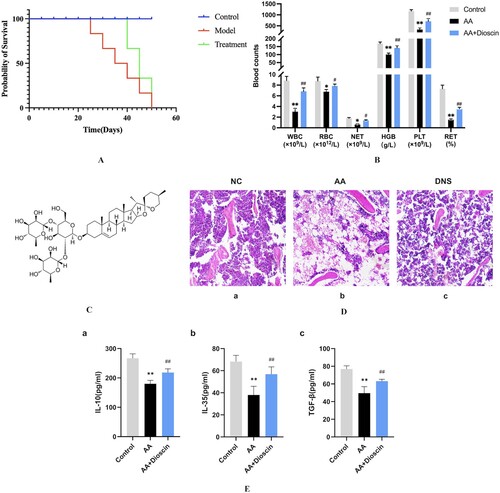
Table 1. Peripheral blood cell count in mice (n = 10).
Table 2. Weight and BMNC in mice (n = 10).
Dioscin ameliorated pancytopenia and bone marrow failure symptoms
To begin with, we investigated whether Dioscin could improve bone marrow failure during aplastic anemia. The molecular structure of dioscin is shown in (C). Blood counts and bone marrow biopsies are two widely used indicators for assessing hematopoietic function. After TBI and allogeneic lymphocyte infusion in Balb/c mice, we observed decreased levels of WBC, RBC, NET, HGB, PLT and RET compared with the Control group, which confirmed the successful establishment of the AA model. Importantly, the WBC, RBC, NET, HGB, PLT and RET levels in the AA + Dioscin group were significantly higher than in the AA model group, which is shown in (P<0.01). In addition, we observed the DNS treatment obviously increased the level of PLT and HGB compared with AA model group. After DNS treatment, the weight of the mice and the number of BMNC increased compared with the AA group, the differences are statistically significant (, (B)). Then we observed the different histopathological alterations among the various groups. We stained the bone marrow biopsies with HE. In the healthy control group, we observed densely packed hematopoietic cell distribution. We observed a marked reduction in bone marrow hematopoietic tissue and an increase in adipose tissue in the femur sections of the AA group. However, the AA + Dioscin group showed slight adipose vacuolated tissue accompanied by active bone marrow hematopoietic cell proliferation ((D)). These data showed that Dioscin significantly alleviated pancytopenia symptoms in AA mice.
Immunosuppressive effect of Dioscin in AA
Aplastic anemia has immune homeostasis imbalance, including abnormal activation of CD8 + T and impaired immune tolerance of Treg [Citation22]. We sought to determine whether dioscin recovered hematopoiesis by regulating Treg function. The levels of Foxp3 in splenic lymphocytes were measured using flow cytometry, and the results are shown in . In comparison to the healthy control group, CD4 + Foxp3 + Tregs were considerably lower in the AA model group (P<0.01). In contrast, treatment with Dioscin significantly increased the expression of Treg (P<0.05). In addition, we tried to evaluate the effect of Dioscin on inhibitory cytokines and measured the levels in spleens by ELISA. TBI and allogeneic lymphocyte infusion significantly reduced the expression of IL-10, IL-35 and TGF-β in AA mice (P<0.01). The level of inhibitory cytokines was increased in the AA + Dioscin group compared with the AA group (P<0.01) ((E)). These data indicate that Dioscin increased the number of Treg and promoted the secretion of immunosuppressive cytokine.
Dioscin modulates the function of Treg in AA
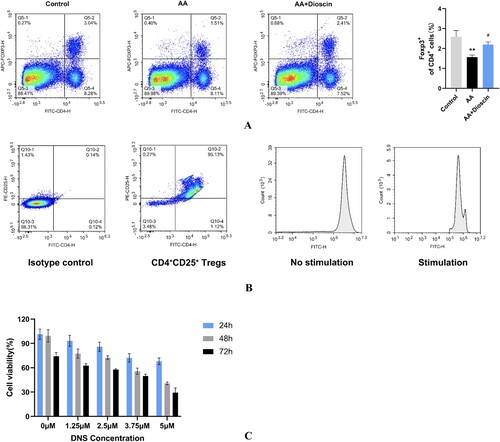
Besides the number of Treg, its function was crucial to immune tolerance and represented as immunophenotype and secretory capacity [Citation23]. To determine the targeting effect of Dioscin on Tregs in AA, we examined the surface molecules of Tregs in vitro by flow cytometry. First, we sorted Tregs with magnetic beads, which showed a positive rate of over 90% and a low expression of CD127((A)). Theoretically, Tregs perform their role of suppressing CTLs in vitro after stimulation with anti-CD3 and CD28 antibodies and IL-2 protein. Therefore, we stimulated the sorted Tregs, which exhibited proliferation after stimulation ((B)), which could be used for subsequent experiments. In addition, we tested the optimal concentration and timing of Dioscin administration using the CCK8 assay. As shown in (C), Dioscin at 1.25 and 2.5 μM concentrations showed cell survival rates above 80% after 24 h of intervention. All concentration groups exhibited very low cell survival after 48 and 72 h of intervention. Thus, we chose 2.5 μm as the administration concentration and the intervention time was 24 h.
Figure 2. Effects of Dioscin on Foxp3 in splenic lymphocytes of AA mice. (A) Purification of CD4+ CD25+ Tregs (n = 3). (B) The proliferation of Tregs (n = 3). (C) The cell viability of Tregs treated with Dioscin at 24 h, 48 and 72 h (n = 3). Data were mean ± SD (*P < 0.05 vs. NC group, #P < 0.05 vs. AA model group, **P<0.01 vs. control group. ##P<0.01 vs. AA group).
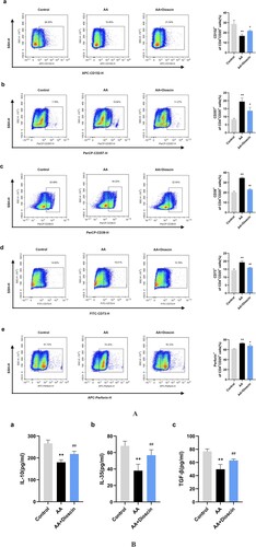
The changes of surface molecules on Tregs are an essential part of the development of AA, as they can lead to damage to Tregs. Therefore, we performed the flow cytometry technique to evaluate the levels of CD152, CD357, CD39, CD73 and perforin ((A)). Compared to those in the control group, the levels of CD357, CD39, CD73 and perforin were increased and CD152 was decreased in the AA group, implying the alteration of Tregs (P<0.01). In the AA + Dioscin group, the levels of CD357, CD39, CD73 and perforin were significantly decreased and CD152 expression was upregulated (P<0.05). These data suggested that Dioscin activated the immune tolerance function of Tregs in AA. At the same time, the supernatant of the Treg was collected for testing cytokine secretion. We found that the level of TGF-β, IL-10 and IL-35 was obviously decrease in the AA group (P<0.01). However, compared with the AA group, the level of anti-inflammatory factors TGF-β, IL-10 and IL-35 secreted by Treg cells were increased, and the differences were statistically significant (P<0.05) ((B)).
Figure 3. Effects of Dioscin on surface molecules of Tregs. (A): (a) The proportion of CD152 on Tregs among groups (n = 3). (b) The proportion of CD357 on Tregs among groups (n = 3). (c) The proportion of CD39 on Tregs among groups (n = 3). (d) The proportion of CD73 on Tregs among groups (n = 3). (e) The proportion of perforin on Tregs among groups (n = 3). (B) Level of IL-10, IL-35 and TGF-β in the Control, AA and Dioscin groups secreted by Tregs (n = 3). Data were mean ± SD (*P < 0.05 vs. NC group, #P < 0.05 vs. AA model group, **P<0.01 vs. control group. ##P<0.01 vs. AA group).
Discussion
AA, a refractory blood disorder, has numerous pathogenic processes and complex disease causes. According to an increasing number of studies, AA is an immunological-mediated disease caused by the dysregulation of immune cell subsets, particularly T lymphocytes. The primary mechanism is that bone marrow failure is brought on by HSC destruction caused by excessive CTLs activity. Tregs serve a crucial function in maintaining immunological tolerance by blocking the cytotoxic effect of activated CTLs. Patients with AA have much lower Treg ratios in their peripheral blood and bone marrow, and their Tregs’ capacity to suppress CTLs is compromised [Citation7]. A higher proportion of Tregs indicates a greater response to IST and is correlated with disease severity in AA. Our previous study found that Dioscin recovered the ameliorating pancytopenia by modulating immunity and inhibiting apoptosis of hematopoietic stem cells (HSCs) in AA mice [Citation24]. These reports support our hypothesis that Dioscin may regulate the function of Treg in AA.
We established the immune mediated AA model according to our previous method [Citation21]. The AA mice showed peripheral hemopenia and bone marrow failure, which is according with our previous study. Here, we demonstrated that Dioscin prolonged the overall survival and recovered peripheral hemogram and hematopoiesis.
To regulate the function of Treg, we performed the Treg cell studies with Dioscin in vitro and animal studies in vivo. Foxp3, which is essential for the developmental maturity and functional amplification of Tregs, is selectively expressed by CD4 + CD25 + Tregs [Citation25]. And CD4 + CD25 + Tregs lowly express CD127, which contributes to the experimental identification of Tregs [Citation26]. IL-10, IL-35 and TGF-β are the main cytokines secreted by Tregs, inhibiting the immune response of CTLs, suppressing the secretion of inflammatory factors, and contributing to the body's autoimmune tolerance [Citation27,Citation28]. The results of this investigation supported the findings above by demonstrating that the level of Foxp3, IL-10, IL-35, and TGF-β in the spleen of AA mice was much lower than that of the control group. The levels of those also rose with Dioscin therapy, indicating that upregulating Treg-targeted therapy may improve the outcomes for AA patients.
In Hashimoto's thyroiditis, dioscin increases the proportions of CD4 + CD25 + Foxp3 + Tregs and upregulates the SUMO-specific protease 1 [Citation29]. To determine whether Dioscin promotes Tregs differentiation by regulating CD152, CD357, CD39, CD73 and perforin, we detected the level of those in Tregs. Our results showed that the levels of CD357, CD39, CD73 and perforin were increased and CD152 was decreased in the AA group. Treatment with Dioscin resulted in elevated expression of CD152 and decreased expression of CD357, CD39, CD73 and perforin, suggesting that Dioscin exerted immunomodulatory effects by affecting the expression of surface molecules on Tregs. We detected a consistent trend in the levels of cytokines secreted by Tregs that we had previously detected in splenic tissue. DNS increased the level of anti-inflammatory cytokines.
Our foundings indicated that Dioscin could recover hematopoietic function and prolong survival in AA mice. Specifically, Dioscin increased the cell number, regulated the founctional surface molecules and influenced the secretion of cytokines in Treg. Building on our findings, we suggested that Dioscin may be a prospective candidate for treating AA by regulating immune torerance.
Ethics approval
According to the guidelines of the Helsinki Declaration, this study was conducted. The Animal Management and Ethics Committee of Tianjin Medical University gave its approval.
Disclosure statement
No potential conflict of interest was reported by the author(s).
Additional information
Funding
References
- Young NS, Scheinberg P, Calado RT. Aplastic anemia. Curr Opin Hematol. 2008;15(3):162–168. doi:10.1097/MOH.0b013e3282fa7470
- Park YB, Lee JW, Cho BS, et al. Incidence and etiology of overt gastrointestinal bleeding in adult patients with aplastic anemia. Dig Dis Sci. 2010;55(1):73–81. doi:10.1007/s10620-008-0702-3
- Dezern AE, Brodsky RA. Clinical management of aplastic anemia. Expert Rev Hematol. 2011;4(2):221–230. doi:10.1586/ehm.11.11
- Chen FF, Guo ZW, Zhang LN, et al. The change of quality of life in 52 patients with non-severe aplastic anemia after cyclosporine A therapy]. Zhonghua Xue Ye Xue Za Zhi. 2020;41(10):806–810. doi:10.3760/cma.j.issn.0253-2727.2020.10.003
- Sloand E, Kim S, Maciejewski JP, et al. Intracellular interferon-γ in circulating and marrow T cells detected by flow cytometry and the response to immunosuppressive therapy in patients with aplastic anemia. Blood. 2002;100(4):1185–1191. doi:10.1182/blood-2002-01-0035
- Schlöder J, Shahneh F, Schneider FJ, et al. Boosting regulatory T cell function for the treatment of autoimmune diseases - That's only half the battle!. Front Immunol. 2022;13:973813. doi:10.3389/fimmu.2022.973813
- Shi J, Ge M, Lu S, et al. Intrinsic impairment of CD4+CD25+ regulatory T cells in acquired aplastic anemia. Blood. 2012;120(8):1624–1632. doi:10.1182/blood-2011-11-390708
- Solomou EE, Rezvani K, Mielke S, et al. Deficient CD4+ CD25+ FOXP3+ T regulatory cells in acquired aplastic anemia. Blood. 2007;110(5):1603–1606. doi:10.1182/blood-2007-01-066258
- Chen X, Du Y, Lin X, et al. CD4 + CD25 + regulatory T cells in tumor immunity. Int Immunopharmacol. 2016;34:244–249. doi:10.1016/j.intimp.2016.03.009
- Thompson CB, Allison JP. The emerging role of CTLA-4 as an immune attenuator. Immunity. 1997;7(4):445–450. doi:10.1016/S1074-7613(00)80366-0
- Clouthier DL, Watts TH. Cell-specific and context-dependent effects of GITR in cancer, autoimmunity, and infection. Cytokine Growth Factor Rev. 2014;25(2):91–106. doi:10.1016/j.cytogfr.2013.12.003
- Esparza EM, Arch RH. Glucocorticoid-Induced TNF receptor functions as a costimulatory receptor that promotes survival in early phases of T cell activation. J Immunol. 2005;174(12):7869–7874. doi:10.4049/jimmunol.174.12.7869
- Moesta AK, Li XY, Smyth MJ. Targeting CD39 in cancer. Nat Rev Immunol. 2020;20(12):739–755. doi:10.1038/s41577-020-0376-4
- Schuler PJ, Saze Z, Hong CS, et al. Human CD4+CD39+ regulatory T cells produce adenosine upon co-expression of surface CD73 or contact with CD73+ exosomes or CD73+ cells. Clin Exp Immunol. 2014;177(2):531–543. doi:10.1111/cei.12354
- Yao H, Tao X, Xu L, et al. Dioscin alleviates non-alcoholic fatty liver disease through adjusting lipid metabolism via SIRT1/AMPK signaling pathway. Pharmacol Res. 2018;131:51–60. doi:10.1016/j.phrs.2018.03.017
- Li Y, Gao M, Yin LH, et al. Dioscin ameliorates methotrexate-induced liver and kidney damages via adjusting miRNA-145-5p-mediated oxidative stress. Free Radic Biol Med. 2021;169:99–109. doi:10.1016/j.freeradbiomed.2021.03.035
- Wu MM, Wang QM, Huang BY, et al. Dioscin ameliorates murine ulcerative colitis by regulating macrophage polarization. Pharmacol Res. 2021;172:105796. doi:10.1016/j.phrs.2021.105796
- Mao Z, Han X, Chen D, et al. Potent effects of dioscin against hepatocellular carcinoma through regulating TP53-induced glycolysis and apoptosis regulator (TIGAR)-mediated apoptosis, autophagy, and DNA damage. Br J Pharmacol. 2019;176(7):919–937. doi:10.1111/bph.14594
- Kong C, Lyu D, He C, et al. Dioscin elevates lncRNA MANTIS in therapeutic angiogenesis for heart diseases. Aging Cell. 2021;20(7):e13392. doi:10.1111/acel.13392
- Wang Y, Yan T, Ma L, et al. Effects of the total saponins from Dioscorea nipponica on immunoregulation in aplastic anemia mice. Am J Chin Med. 2015;43(2):289–303. doi:10.1142/S0192415X15500196
- Ni R, Fan L, Zhang L, et al. A mouse model of irradiation and spleen-thymus lymphocyte infusion induced aplastic anemia. Hematology. 2022;27(1):932–945. doi:10.1080/16078454.2022.2113356
- Giudice V, Selleri C. Aplastic anemia: pathophysiology[J]. Semin Hematol. 2022;59(1):13–20. doi:10.1053/j.seminhematol.2021.12.002
- Wang X, Sun B, Wang Y, et al. Research progress of targeted therapy regulating Th17/Treg balance in bone immune diseases. Front Immunol. 2024;15:1333993. doi:10.3389/fimmu.2024.1333993
- Zhang L, Ni R, Li J, et al. Dioscin regulating bone marrow apoptosis in aplastic anemia. Drug Des Devel Ther. 2022;16:3041–3053. doi:10.2147/DDDT.S370506
- Lu LF, Rudensky A. Molecular orchestration of differentiation and function of regulatory T cells. Genes Dev. 2009;23(11):1270–1282. doi:10.1101/gad.1791009
- Seddiki N, Santner-Nanan B, Martinson J, et al. Expression of interleukin (IL)-2 and IL-7 receptors discriminates between human regulatory and activated T cells. J Exp Med. 2006;203(7):1693–1700. doi:10.1084/jem.20060468
- Lourenço JD, Ito JT, Martins MA, et al. Th17/treg imbalance in chronic obstructive pulmonary disease: clinical and experimental evidence: Clinical and Experimental Evidence. Front Immunol. 2021;12:804919. doi:10.3389/fimmu.2021.804919
- Sakaguchi S, Kawakami R, Mikami N. Treg-based immunotherapy for antigen-specific immune suppression and stable tolerance induction: a perspective. Immunother Adv. 2023;3(1):ltad007. doi:10.1093/immadv/ltad007
- Yongjun C, Nan Q, Yumeng S, et al. Dioscin alleviates hashimoto's thyroiditis by regulating the SUMOylation of IRF4 to promote CD4(+)CD25(+)Foxp3(+) treg Cell Differentiation. Autoimmunity. 2021;54(1):51–59. doi:10.1080/08916934.2020.1855428