
ABSTRACT
Objective: To study whether and, if so, how honokiol overcome dexamethasone resistance in DEX-resistant CEM-C1 cells. Methods: We investigated the effect of honokiol (0–20 µM) on cell proliferation, cell cycle, cell apoptosis and autophagy in DEX-resistant CEM-C1 cells and DEX-sensitive CEM-C7 cells. We also determined the role of c-Myc protein and mRNA in the occurrence of T-ALL associated dexamethasone resistance western blot and reverse transcription-qPCR (RT-qPCR) analysis. Results: Cell Counting Kit (CCK)−8 assay shows that DEX-resistant CEM-C1 cell lines were highly resistant to dexamethasone with IC50 of 364.1 ± 29.5 µM for 48 h treatment. However, upon treatment with dexamethasone in combination with 1.5 µM of honokiol for 48 h, the IC50 of CEM-C1 cells significantly decreased to 126.2 ± 12.3 µM, and the reversal fold was 2.88. Conversely, the IC50 of CEM-C7 cells was not changed combination of dexamethasone and honokiol as compared to that of CEM-C7 cells treated with dexamethasone alone. It has been shown that honokiol induced T-ALL cell growth inhibition by apoptosis and autophagy via downregulating cell cycle-regulated proteins (Cyclin E, CDK4, and Cyclin D1) and anti-apoptotic proteins BCL-2 and upregulating pro-apoptotic proteins Bax and led to PARP cleavage. Honokiol may overcome dexamethasone resistance in DEX-resistant CEM-C1 cell lines via the suppression of c-Myc mRNA expression. Conclusion: The combination of honokiol and DEX were better than DEX alone in DEX-resistant CEM-C1 cell lines. Honokiol may regulate T-ALL-related dexamethasone resistance by affecting c-Myc.
1. Introduction
Acute lymphoblastic leukemia (ALL) is the most common pediatric malignancy with a significant peak incidence between 2 and 6 years old [Citation1]. Around 10–15% of children will have ALL that is of T-cell origin, which is referred to as T-ALL. T-ALL is characterized by the expression of cytoplasmic CD3 and surface expression of T-cell antigens including CD2, CD5, and CD7 [Citation2]. Acute lymphoblastic leukemia initially starts within bone marrow and gradually develops with the metastasis of malignant lymphocytes to extramedullary sites, such as lymph nodes, spleen, liver, skin, and central nervous system [Citation3]. The main treatment of T-ALL is usually the combination of chemotherapy [Citation4]. Glucocorticoids are one of the most common medications for T-ALL. Dexamethasone (DEX) is among the first drugs which play an essential role in T-ALL therapy through the binding of glucocorticoid receptors [Citation5,Citation6]. Dexamethasone binds to glucocorticoid receptors upon passive infusion in the cytoplasm [Citation7]. Subsequently, either the monomeric receptors repress gene transcription through the interaction with activating protein-1 and nuclear factor kappa B, or the homodimerized receptors are translocated to the nucleus where they will initiate gene expression through the interaction with glucocorticoid response elements (GREs), eventually inducing cell cycle arrest and apoptosis of malignant lymphocytes [Citation8–11]. Recently, DEX-resistance has been associated with an elevated risk of relapse and poor survival due to varied response to glucocorticoid therapy among T-ALL patients [Citation12–14]. The current treatment protocol thus usually consists of a high dose of dexamethasone, leading to toxic adverse effects in these patients [Citation15]. Therefore, the development of novel therapeutical approaches is required to overcome chemoresistance and reduce adverse effects in T-ALL patients.
Honokiol (HNK) is an active biphenolic phytochemical derived from magnolia tree [Citation16]. The root and stem bark of magnolia has been used as a tradition Asian medicine for the prevention and treatment of numerous diseases and conditions, owing to its advantageous pharmacological properties and clinical safety profile [Citation17–21]. Honokiol has been recently investigated as a potential cancer therapeutic in light of its apoptosis-inducing effects in various cancer cell lines, such as leukemia, lung cancer, and skin tumors [Citation22–25]. Importantly, several studies have shown that honokiol is a promising agent in the improvement of the chemotherapy in drug-resistance cancer [Citation26–28]. Taking this in mind, honokiol was selected to investigate the reversal of the human T-ALL dexamethasone-resistant cell line (CEM-C1).
In this study, we investigated the effect of honokiol with varying concentrations on cell proliferation and cell cycle in DEX-resistant CEM-C1 cells and DEX-sensitive CEM-C7 cells. We further determined the role of honokiol in cell apoptosis and autophagy of CEM-C1 and CEM-C7 cell lines. Moreover, we studied whether honokiol could overcome or reduce the drug resistance in DEX-resistant CEM-C1 cells. We investigated the role of c-Myc protein and mRNA in the occurrence of T-ALL associated dexamethasone resistance. The aim of the study is to explore the possible molecular mechanism of honokiol in the reversal of drug resistance in T-acute lymphoblastic leukemia, and to provide scientific evidence for the explanation of treatment success or failure in childhood ALL.
2. Materials and methods
2.1. Reagents
Honokiol, dexamethasone, bovine serum albumin (BSA), ethyl alcohol, Cell Counting Kit (CCK)−8 assay kit, propidium iodide (PI), acridine orange staining solution, phosphate buffered saline (PBS, pH 7.4, containing 11.9 mM phosphate, 137 mM sodium chloride, and 2.7 mM potassium chloride), Tween 20, Dulbecco's modified Eagle's medium (DMEM), RNase A, and radio – immunoprecipitation assay RIPA buffer were obtained from Sigma Aldrich (Shanghai, China). Honokiol was dissolved in dimethyl sulfoxide (DMSO) to make a 50 mM stock solution and was diluted again in cell culture medium at different concentrations and then used immediately. The final concentration of DMSO in cell culture medium was 0.4% in all in vitro assays. Annexin V-PI Kit was purchased from Keygen Biotech (Nanjing, China). Antibodies for Cyclin E, cyclin-dependent kinases 4 (CDK4), Cyclin D1, Bax, BCL-2, cleaved PARP, LC3B-I/II, c-Myc and the secondary antibodies of horseradish peroxidase-conjugated goat anti-rabbit antibody were purchased from BD Bioscience (San Jose, CA, USA). Anti-glyceraldehyde-3-phosphate dehydrogenase (GAPDH) antibody was obtained from Kangcheng BioTech (Shanghai, China). Total RNA extraction kit was obtained from Tiangen Biotech (Beijing, China), reverse transcription and quantitative PCR kit from TaKaRa (Dalian, China), and PCR primers from Sangon (Shanghai, China).
2.2. Cell lines and cell culture
Human T-ALL dexamethasone-resistant cell line (CEM-C1) and dexamethasone-sensitive cell line (CEM-C7) were obtained from the American Type Culture Collection (ATCC, Manassas, Virginia, USA). CEM-C1 cells are resistant to dexamethasone, while CEM-C7 cells are sensitive to dexamethasone. CEM-C1 and CEM-C7 cells were cultured in DMEM supplemented with 10% of fetal bovine serum (FBS) at 37°C in a humidified atmosphere containing 5% of CO2. Both cell lines were grown in 75 cm2 cell culture flasks and passaged twice a week.
2.3. Cell viability assay
CEM-C1 and CEM-C7 cells were seeded at 5 × 10⁴ cells/well in 96-well microplates and were cultured overnight to allow attachment. Different concentrations of honokiol (2.5, 5.0, 10, 15, and 20 µM) were added to the medium. Following treatment, cells were incubated for 24, 48, and 72 h at 37°C, respectively. Subsequently, cell viability was assessed using the CCK-8 assay by adding 10 µL of CCK-8 solution. The wells with medium only were used as the blank group and cells without honokiol treatment were used as the control group. The optical density (OD) was assessed at 450 nm using a precision microplate reader (Molecular Devices, LLC., San Jose, CA, USA). The cell viability was calculated using the following formula, cell viability (%) = (A honokiol – A blank) / (A control – A blank) × 100%, where A stands for absorbance.
CEM-C1 and CEM-C7 cells were seeded at 5 × 10⁴ cells/well in 96-well microplates and were cultured overnight to allow attachment. Different concentrations of dexamethasone (50, 100, 200, 300, and 400 µM) with or without 1.5 µM HNK were added to the medium of CEM-C1 cells which are dexamethasone-resistant, whereas dexamethasone of 0.5, 1, 2, 3, and 4 µM with or without 1.5 µM HNK were added to the medium of CEM-C7 cells which are dexamethasone-sensitive. Upon incubation at 37°C for 48 h, 10 µL of CCK-8 solution was added and the cell viability was assessed as a forehand described. The half maximal inhibitory concentration, IC50, was calculated as drug concentration that inhibits the cell growth by 50% after 48 h of cultivation using the software GraphPad Prism v8.3.0 (GraphPad Software, Inc., La Jolla, CA, USA).
2.4. Cell cycle analysis by flow cytometry
CEM-C1 and CEM-C7 cells were seeded at 1 × 10⁵ cells/well in 6-well plates and then treated with honokiol at different concentrations (10, 15, and 20 µM) for 48 h. After HNK treatment, the cells were harvested, washed with PBS and fixed with cold 75% ethyl alcohol at 4°C overnight. The cells were then washed twice with PBS and incubated with RNase A for 30 min. Subsequently samples were stained with 500 μL propidium iodide for 30 min at room temperature. Cells without honokiol treatment were used as the control group. Cell cycle analysis was performed by measuring the fluorescence with an excitation wavelength of 488 nm on the Accuri C6 flow cytometer (Becton Dickinson Biosciences, San Jose, CA, USA). Software Modifit was used to generate histograms to determine number of cells in each phase of the cell cycle.
2.5. Cell apoptosis detection by flow cytometry
CEM-C1 and CEM-C7 cells were seeded at 2 × 10⁵ cells/well in 6-well plates and pre-incubated for 24 h, and then treated with honokiol at different concentrations (10, 15, and 20 µM) for 48 h. Cells were harvested and resuspended in Annexin binding buffer (Becton Dickinson Biosciences, CA, USA), and then stained with Annexin V-phycoerythrin (Annexin V-PE; Becton Dickinson Biosciences, CA, USA) and propidium iodide for 15 min at room temperature. Cells without honokiol treatment were used as the control group. The stained samples were analyzed using the fluorescence-activated cell sorting (FACS) calibur bench-top flow cytometer (Becton Dickinson Biosciences, San Jose, CA, USA) within 30 min after staining. Software FlowJo (Tree Star, Ashland, OR, USA) was used for apoptosis analysis.
2.6. Western blot
CEM-C1 and CEM-C7 cells were seeded at 3 × 10⁵ cells/well in 6-well plates and pre-incubated for 24 h, and then treated with honokiol (0–20 µM) for 48 h. To extract the total protein, cells were washed with PBS, lysed in RIPA buffer for 30 min on ice. Cell lysates were centrifuged at 12,000 × g for 15 min at 4°C, and the supernatant containing proteins was collected. Concentrations of the total protein were quantified using the BSA Protein Assay according to the manufacturer’s protocol. 25 µg of the total protein was separated by SDS-PAGE (8–12%) and then transferred to PVDF membrane (0.45 μm). After blocking with 5% skimmed milk in PBS buffer containing 0.07% Tween 20 for 1 h at room temperature, the membranes were incubated with diluted primary antibodies at 4°C overnight. The primary antibodies and their corresponding dilution are as follows, anti-GAPDH (1:15,000 dilution), anti-Cyclin E (1:1,000 dilution), anti-CDK4 (1:1,000 dilution), anti-Cyclin D1 (1:1,000 dilution), anti-Bax (1:1,000 dilution), anti-BCL-2 (1:1,000 dilution), anti-cleaved-PARP (1:1,000 dilution), anti-LC3B-I/II (1:1,000 dilution), and anti-c-Myc (1:2,000 dilution). The membranes were washed three times with PBS buffer containing 0.07% Tween 20 and then incubated with horseradish peroxidase-conjugated secondary antibody (HRP-labeled goat anti-rabbit antibody, 1:2,000 dilution) for 1 h at room temperature. The membranes were washed with PBS buffer containing 0.07% Tween 20 three times and developed using an enhanced Chemiluminescence Kit (Millipore, Plano, TX, USA). The densitometric analysis of the protein bands was performed using the software Image Lab v5.0. The protein expression levels were normalized to GAPDH.
2.7. Acridine orange staining
CEM-C1 and CEM-C7 cells were seeded at 1 × 10⁵ cells/well in 6-well plates and pre-incubated for 24 h, and then treated with honokiol at different concentrations (10, 15, and 20 µM) for 48 h. Cells were washed with PBS, and subsequently stained with acridine orange staining solution (10 µg/ml) for 30 min in the dark at room temperature. Cells without honokiol treatment were used as the control group. The cells were thoroughly observed under a laser scanning confocal microscopy (Yokogawa CV7000, Tokyo, Japan), photographed and compared with that of control. Viable cells’ nuclei stain green due to permeability of only acridine orange whereas, apoptotic cells appear red/orange due to co-staining of both the fluorescent dyes.
2.8. Reverse transcription-qPCR (RT-qPCR) analysis
CEM-C1 and CEM-C7 cells were seeded at 3 × 10⁵ cells/well in 6-well plates and pre-incubated for 24 h, and then treated with honokiol (0–20 µM) for 48 h. To extract total RNA, TRIzol® (Invitrogen, Carlsbad, CA, USA) was added to honokiol-treated and untreated cells. The concentration and quality of c-Myc and LC3 mRNA expression were quantified by measuring the absorbance at 260 nm with Beckman DU6400 spectrophotometer (Beckman Counter, Miami, FL, USA) and gel analysis. The primers of c-Myc and LC-3 were indicated in . GAPDH was used as the internal reference for normalization.
Table 1. Sequences of the primers for quantitative PCR.
2.9. Statistical analysis
All experiments were performed in triplicate, and data were expressed as mean values ± SD. Statistical analysis was done by the software GraphPad Prism v8.3.0 (GraphPad Software, Inc., La Jolla, CA, USA). Two-way analysis of variance (ANOVA) was used to determine the statistical significance the data. A value of p < 0.05 was considered significant.
3. Results and discussion
3.1. HNK inhibits proliferation of DEX-resistant CEM-C1 cells and DEX-sensitive CEM-C7 cells
Various studies have proven that honokiol exerts broad-range anticancer activity in vitro and in vivo by regulating numerous signaling pathways, indicating its tremendous potential as an anticancer agent [Citation29–31]. At present, there is no study on honokiol to reduce dexamethasone resistance. To investigate the anti-proliferation of honokiol on T-ALL cell lines, we incubated CEM-C1 and CEM-C7 cells with different concentrations of honokiol (0–20 µM) for various time periods (24–72 h). shows that the growth of both cell lines was inhibited in a dose – and time-dependent manner. IC50 values as determined using the CCK-8 assay are summarized in . For both T-ALL cell lines, IC50 values at 24 h was significantly higher than those of 48 and 72 h (19.00 ± 1.08 versus 8.33 ± 1.14 and 5.82 ± 2.07 µM for CEM-C1 cells; 23.39 ± 1.01 versus 12.21 ± 1.35 and 9.85 ± 1.50 µM for CEM-C7 cells; p < 0.05). As no significant difference of IC50 between 48 and 72 h was observed, incubation time of 48 h was selected in the following experiments. Moreover, CEM-C7 cells exhibited significantly higher IC50 values than CEM-C1 cells at the same incubation time (p < 0.05), indicating that DEX-resistant CEM-C1 cells are sensitive to honokiol treatment to a larger extent.
Figure 1. Effect of honokiol at different concentrations on cell viability of T-ALL cell lines. Cell viability of DEX-resistant CEM-C1 cells (A) and DEX – sensitive CEM-C7 cells (B) exposed to honokiol at different concentrations (0–20 µM) for 24, 48, and 72 h. Data represent mean ± SD (n = 3), *p < 0.05.
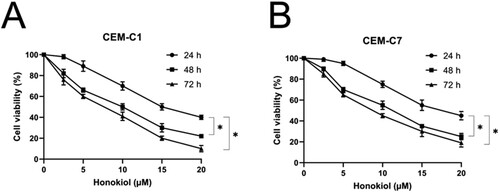
Table 2. IC50 values in CEM-C1 and CEM-C7 cells after 24, 48, and 72 h-treatment by honokiol.
3.2. HNK triggers G0/G1 phase arrest by regulating cell cycle-regulated proteins
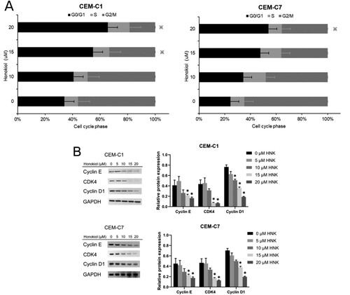
To determine whether honokiol inhibited the proliferation of T-ALL cells by inducing cell cycle arrest, we incubated DEX-resistant CEM-C1 and DEX-sensitive CEM-C7 cells with different concentrations of honokiol for 48 h, and the distribution of cell cycle was detected using PI staining and flow cytometry. As shown in (A), honokiol induced a significant increased proportion of G0/G1 phase, and a simultaneously decreased proportion of S and G2/M phase in both cell lines, indicating that honokiol arrested both DEX-resistant CEM-C1 cells and DEX-sensitive CEM-C7 cells at G0/G1 phase in a dose-dependent manner. To further understand the molecular mechanism of G0/G1 phase arrest, we determined the level of protein markers using western blot analysis. (B) shows that honokiol resulted in a dose-dependent downregulated levels of Cyclin E, CDK4, and Cyclin D1 in DEX-resistant CEM-C1 and DEX-sensitive CEM-C7 cells. Overall, these results suggested that honokiol triggers G0/G1 phase arrest by regulating cell cycle-regulated proteins in T-ALL cell lines.
Figure 2. Effect of honokiol at different concentrations on cell cycle of T-ALL cell lines. DEX-resistant CEM-C1 cells and DEX – sensitive CEM-C7 cells were treated with honokiol at different concentrations (0–20 µM) for 48 h. Cell cycle distribution (A) was determined using flow cytometry and the expressions of cell cycle-regulated proteins (B) were measured by western blot. Data represent mean ± SD (n = 3), *p < 0.05 as compared to the control group.
3.3. HNK induces apoptosis by regulating pro – and anti-apoptotic proteins
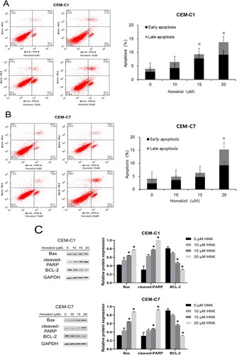
To determine the role of apoptosis in cell cycle arrest induced by honokiol, both DEX-resistant CEM-C1 cells and DEX-sensitive CEM-C7 cells treated with different concentrations of honokiol were stained using Annexin V-PE/PI and the apoptosis was subsequently quantified using flow cytometry. (A,B) depicts that in comparison with the control group, honokiol treatment for 48 h resulted in increased early, late, and total apoptosis rate in a dose-dependent manner. We then investigated the expression of downstream apoptotic proteins by western blotting. As shown in (C), honokiol activated pro-apoptotic protein Bax and significantly led to PARP cleavage (p < 0.05), while the expression of anti-apoptotic proteins BCL-2 was observed to be significantly decreased (p < 0.05). These results indicate that honokiol results in apoptosis of DEX-resistant CEM-C1 and DEX-sensitive CEM-C7 cells. Our results are in line with previous reports that cell cycle regulation is an important process in apoptosis [Citation32,Citation33].
Figure 3. Effect of honokiol at different concentrations on cell apoptosis of T-ALL cell lines. DEX-resistant CEM-C1 cells and DEX – sensitive CEM-C7 cells were treated with honokiol at different concentrations (0–20 µM) for 48 h. Cell apoptosis (A) and the rate of apoptosis (B) was measured using flow cytometry and the expressions of pro – and anti-apoptotic proteins (C) were measured by western blot. Data represent mean ± SD (n = 3), *p < 0.05 as compared to the control group.
3.4. HNK induces autophagy which contributes to cell death
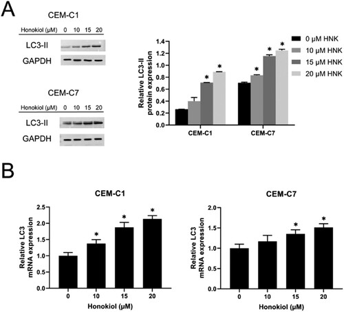
To understand the role of apoptosis in HNK-induced cell death, we investigated the formation of acidic autophagy vesicles in the cells using acridine orange staining. shows the accumulation of orange fluorescence staining in CEM-C1 and CEM-C7 cells with the increasing concentration of honokiol as compared to the control group without HNK treatment, indicating that honokiol induces the formation of intracellular autophagosomes in human T-ALL cell lines in a dose-dependent manner. We also measured the level of protein marker LC3-II to determine whether autophagic cell death was induced. As shown by the western blot analysis in (A), honokiol significantly increased the level of LC3-II protein in CEM-C1 and CEM-C7 cells (p < 0.05). Furthermore, RT-qPCR determined that the expression level of LC3 mRNA was significantly upregulated in CEM-C1 and CEM-C7 cells ((B)), providing evidence that honokiol treatment induces autophagy in human T-ALL cells. These results are clearly in good agreement with previous publications where breast cancer cells undergo cytoprotective autophagy to circumvent HNK [Citation34].
Figure 4. Acridine orange staining for the detection of autophagy in DEX-resistant CEM-C1 cells and DEX – sensitive CEM-C7 cells. Maenification.x400.Viable cells’ nuclei stain green due to permeability of only acridine orange whereas, apoptotic cells appear red/orange due to co-staining of both the fluorescent dyes.
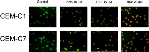
Figure 5. Effect of honokiol at different concentrations on cell autophagy of T-ALL cell lines. The expression level of the autophagy-related protein LC3-II (A) and LC3 mRNA (B) in DEX-resistant CEM-C1 cells and DEX – sensitive CEM-C7 cells upon the treatment of honokiol at different concentrations (0–20 µM) were determined using western blot and RT-qPCR, respectively. Data represent mean ± SD (n = 3), *p < 0.05 as compared to the control group.
3.5. HNK increases sensitivity of DEX-resistant CEM-C1 cells to DEX by downregulation of c-Myc expression
It has been previously reported that T-ALL cell line CEM-C1 were resistant to dexamethasone, whereas CEM-C7 cell lines were highly sensitive to the treatment of dexamethasone [Citation35,Citation36]. To further confirm whether honokiol increases the sensitivity of DEX-resistant CEM-C1 cells to dexamethasone, the DEX-resistant CEM-C1 cells and DEX-sensitive CEM-C7 cells were incubated with different concentrations of dexamethasone (0–400 µM) in combination with honokiol (1.5 µM) for 48 h. Our preliminary drug concentration screening results showed that the cell proliferation inhibition rate on the CEM-C1 and CEM-C7 cells treated with 1.5 µM honokiol was <4% (Fig. S1). Therefore, 1.5 µM honokiol was selected. As shown in , dexamethasone with or without honokiol showed cytotoxicity to both cell lines in a dose-dependent manner. The IC50 values are shown in . Upon treatment with dexamethasone only, CEM-C1 cells showed significantly higher IC50 than CEM-C7 cell lines (364.1 ± 29.5 versus 1.30 ± 0.13 µM; p < 0.05) with a resistance index of 280, confirming the high resistance of CEM-C1 cell lines to dexamethasone. Upon treatment with dexamethasone in combination with 1.5 µM of honokiol, the IC50 of CEM-C1 cells significantly decreased from 364.1 ± 29.5 to 126.2 ± 12.3 µM, and the reversal fold was 2.88, whereas the combination of dexamethasone and honokiol did not show significant impact on the IC50 of CEM-C7 cells as compared to that of CEM-C7 cells treated with dexamethasone alone (1.30 ± 0.13 versus 1.25 ± 0.14 µM; p > 0.05).
Figure 6. Effect of dexamethasone at different concentrations on cell viability of T-ALL cell lines. Cell viability of DEX-resistant CEM-C1 cells (A) and DEX – sensitive CEM-C7 cells (B) exposed to dexamethasone at different concentrations (0–400 µM) with or without 1.5 µM honokiol for 48 h. Data represent mean ± SD (n = 3), *p < 0.05.
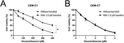
Table 3. IC50 values in CEM-C1 and CEM-C7 cells after 48 h-treatment by dexamethasone with or without 1.5 µM honokiol.
To investigate the underlying molecular mechanism that honokiol reduces the resistance of DEX-resistant CEM-C1 cells to dexamethasone, we first determined the expression level of c-Myc protein using western blot analysis. As shown in (A), DEX-resistant CEM-C1 cells exhibited significantly higher c-Myc protein expression than DEX-sensitive CEM-C7 cells, suggesting an essential role of c-Myc protein in the dexamethasone resistance. We further compared the gene expression level of c-Myc mRNA using RT-qPCR. The c-Myc mRNA was significantly upregulated in DEX-resistant CEM-C1 cells as compared to that in DEX-sensitive CEM-C7 cells, which corroborates the findings of western blot that the overexpression of c-Myc reduces the sensitivity CEM-C1 cells to dexamethasone leading to drug resistance in human acute lymphoblastic leukemia ((B)). Our results further prove the previous publications that c-Myc gene plays an essential role in apoptotic human leukemic cells [Citation37,Citation38].
Figure 7. Effect of honokiol on c-Myc protein and mRNA expression levels in T-ALL cell lines. The expression level of c-Myc protein (A) and c-Myc mRNA (B) in DEX-resistant CEM-C1 cells and DEX – sensitive CEM-C7 cells. CEM-C1 cells were treated with honokiol at different concentrations (0–20 µM) for 48 h and the expression level of c-Myc protein (C) and c-Myc mRNA (D) were determined using western blot and RT-qPCR, respectively. Data represent mean ± SD (n = 3), *p < 0.05 as compared to the control group.
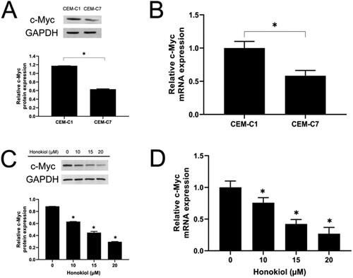
Furthermore, we studied the c-Myc protein expression level after the treatment of different concentrations of honokiol in DEX-resistant CEM-C1 cells. (C) shows that the expression of protein c-Myc was decreased with increasing amount of honokiol treated with CEM-C1 cells, indicating that HNK reduces c-Myc protein expression in a dose-dependent manner. RT-qPCR analysis in (D) further confirmed the downregulated c-Myc mRNA expression after honokiol treatment in a dose-dependent manner. These results show the evidence that mechanism of T-ALL-related dexamethasone resistance may be associated with c-Myc. However, the precise mechanisms of the dexamethasone resistance have yet to be fully understood, and there is a need to develop efficient methods of augmenting the response to glucocorticoids and overcome resistance to the dexamethasone treatment [Citation39]. The apoptosis induction and malignancy control role of honokiol has received much attention in recent times. It has shown various degree of efficacy towards pancreatic cancer, prostate cancer gastric cancer, oral cancer, glioblastoma or brain cancer, skin cancer, ovarian cancer, bone cancer/osteosarcoma [Citation40], and in different cancers, honokiol inhibits cancer cells by affecting different molecules. In the present study, honokiol may regulate T-ALL-related dexamethasone resistance by affecting c-Myc, which provides new information for the application and mechanism of honokiol.
4. Conclusion
The present study demonstrates that honokiol inhibited DEX-resistant CEM-C1 cells growth, induced cell cycle arrest at the G0/G1 phase, and triggered CEM-C1 cells to apoptosis and autophagy by suppression of c-Myc mRNA expression. These results strongly suggest the combination of honokiol and DEX were better than DEX alone in DEX-resistant CEM-C1 cell lines. Overall, the current study displays the possibilities to modulate the DEX resistance and provides new insights into dexamethasone treatment in combination with honokiol as a promising treatment strategy in pediatric T-ALL.
Supplementary material
Download TIFF Image (230.5 KB)Disclosure statement
No potential conflict of interest was reported by the author(s).
Data availability statement
The datasets used and/or analyzed during the present study are available from the corresponding author on reasonable request.
References
- Marcus KJ, Sandlund JT. Pediatric leukemias and lymphomas. In: Leonard L. Gunderson Joel E. Tepper, editor. Clinical radiation oncology. 3rd ed.. Amsterdam: Elsevier; 2012. p. 1479–1488.
- Szczepan T, Van Der Velden VHJ, Van Dongen JJM. Classification systems for acute and chronic leukaemias. Best Pract Res Clin Haematol. 2003;16:561–582.
- Brown PA, Wieduwilt M, Logan A, et al. Guidelines insights: acute lymphoblastic leukemia, version 1.2019. J Natl Compr Cancer Netw. 2019;17:414–423. doi:10.6004/jnccn.2019.0024
- Raetz EA, Teachey TD. T-cell acute lymphoblastic leukemia. Am Soc Hematol. 2016;2016:580–588. doi:10.1182/asheducation-2016.1.580
- Möricke A, Zimmermann M, Valsecchi MG, et al. Dexamethasone vs prednisone in induction treatment of pediatric ALL: results of the randomized trial AIEOP-BFM ALL 2000. Blood. 2016;127:2101–2112. doi:10.1182/blood-2015-09-670729.
- Vora A, Goulden N, Wade R, et al. Treatment reduction for children and young adults with low-risk acute lymphoblastic leukaemia defined by minimal residual disease (UKALL 2003): a randomised controlled trial. Lancet Oncol. 2013;14:199–209. doi:10.1016/S1470-2045(12)70600-9
- Tissing WJE, Meijerink JPP, Den Boer ML, et al. Molecular determinants of glucocorticoid sensitivity and resistance in acute lymphoblastic leukemia. Leukemia. 2003;17:17–25. doi:10.1038/sj.leu.2402733
- Tsai SY, Carlstedt-duke J, Weigel NL, et al. Molecular interactions of steroid hormone receptor with its enhancer element: evidence for receptor dimer formation. Cell. 1987;55:361–369. doi:10.1016/0092-8674(88)90059-1.
- Ray A, Prefontaine KE. Physical association and functional antagonism between the p65 subunit of transcription factor NF-kappa B and the glucocorticoid receptor. Proc Natl Acad Sci USA. 1994;91:752–756. doi:10.1073/pnas.91.2.752
- Harmon JM, Norman MR, Fowlkes BJ, et al. Dexamethasone induces irreversible G1 arrest and death of a human lymphoid cell line. J Cell Physiol. 1979;98:267–278. doi:10.1002/jcp.1040980203
- Wyllie AH. Glucocorticoid-induced thymocyte apoptosis is associated with endogenous endonuclease activation. Nature. 1980;284:555–556. doi:10.1038/284555a0.
- Beesley AH, Palmer M-L, Ford J, et al. Authenticity and drug resistance in a panel of acute lymphoblastic leukaemia cell lines. Br J Cancer. 2006;95:1537–1544. doi:10.1038/sj.bjc.6603447
- Wandler AM, Huang BJ, Craig JW, et al. Loss of glucocorticoid receptor expression mediates in vivo dexamethasone resistance in T-cell acute lymphoblastic leukemia. Leukemia. 2020;34:2025–2037. doi:10.1038/s41375-020-0748-6
- Gao J, Liu W. Prognostic value of the response to prednisone for children with acute lymphoblastic leukemia: a meta-analysis. Eur Rev Med Pharmacol Sci. 2018;22:7858–7866. doi:10.26355/eurrev_201811_16411.
- Inaba H, Mullighan CG. Pediatric acute lymphoblastic leukemia. Haematologica. 2020;105:2524–2539. doi:10.3324/haematol.2020.247031
- Tsai T-H, Chen C-F. Identification and determination of honokiol and magnolol from Magnolia officinalis by high-performance liquid chromatography with photodiode-array UV detection. J Chromatogr. 1992;598:143–146. doi:10.1016/0021-9673(92)85125-D
- Ho K, Tsai C, Chen C, et al. Antimicrobial activity of honokiol and magnolol isolated from Magnolia officinalis. Phyther Res. 2001;141:139–141. doi:10.1002/ptr.736
- Ou H-C, Chou F-P, Lin T-M, et al. Protective effects of honokiol against oxidized LDL-induced cytotoxicity and adhesion molecule expression in endothelial cells. Chem Biol Interact. 2006;161:1–13. doi:10.1016/j.cbi.2006.02.006.
- Liu J, Zhang T, Zhu J, et al. Honokiol attenuates lipotoxicity in hepatocytes via activating SIRT3 – AMPK mediated lipophagy. Chin Med. 2021;16:1–13. doi:10.1186/s13020-021-00528-w.
- Usach I, Alaimo A, Fern J, et al. Magnolol and honokiol: two natural compounds with similar chemical structure but different physicochemical and stability properties. Pharmaceutics. 2021;13:1–14. doi:10.3390/pharmaceutics13020224.
- Lee Y, Mo Y, Lee C, et al. Therapeutic applications of compounds in the Magnolia family. Pharmacol Ther. 2011;130:157–176. doi:10.1016/j.pharmthera.2011.01.010
- Li HY., Ye HG, Chen CQ, et al. Honokiol induces cell cycle arrest and apoptosis via inhibiting claa I histone deacetylases in acute myeloid leukemia. J Cell Biochem. 2015;116:287–298. doi:10.1002/jcb.24967.
- Pan J, Lee Y, Zhang Q, et al. Honokiol decreases lung cancer metastasis through inhibition of the STAT3 signaling pathway. Cancer Prev Res. 2018;10:133–141. doi:10.1158/1940-6207.CAPR-16-0129.
- Prasad R, Singh T, Katiyar KS. Honokiol inhibits ultraviolet immunosuppression through inhibition of ultraviolet-induced inflammation and DNA hypermethylation in mouse skin. Sci Rep. 2017;7(1):1–14. doi:10.1038/s41598-017-01774-5.
- Lahans T. Integrating conventional and Chinese medicine in cancer care. 229–251, 2007.
- Wang X, Beitler JJ, Wang H, et al. Honokiol enhances paclitaxel efficacy in multi-drug resistant human cancer model through the induction of apoptosis. PLoS One. 2014;9(2):e86369. doi:10.1371/journal.pone.0086369
- Ishitsuka K, Hideshima T, Hamasaki M, et al. Honokiol overcomes conventional drug resistance in human multiple myeloma by induction of caspase-dependent and -independent apoptosis. Blood. 2005;106:1794–1800. doi:10.1182/blood-2005-01-0346
- Eliaz I, Weil E. Intravenous honokiol in drug-resistant cancer: two case reports. Integr Cancer Ther. 2020;19:1–5. doi:10.1177/1534735420922615.
- Ong CP, Lee WL, Tang YQ, et al. Honokiol: a review of its anticancer potential and mechanisms. Cancers (Basel). 2020;12:1–44. doi:10.3390/cancers12010048.
- Kim DW, Ko SM, Jeon YJ, et al. Anti-proliferative effect of honokiol in oral squamous cancer through the regulation of specificity protein 1. Int J Oncol. 2013;43:1103–1110. doi:10.3892/ijo.2013.2028
- Dai X, Li RZ, Jiang ZB, et al. Honokiol inhibits proliferation, invasion and induces apoptosis through targeting Lyn kinase in human lung adenocarcinoma cells. Front Pharmacol. 2018;9:1–8. doi:10.3389/fphar.2018.00558.
- Vermeulen K, Berneman ZN, Van Bockstaele RD. Cell cycle and apoptosis. Cell Prolif. 2003;36:165–175. doi:10.1046/j.1365-2184.2003.00267.x
- Wiman KG, Zhivotovsky B. Understanding cell cycle and cell death regulation provides novel weapons against human diseases. J Intern Med. 2017;281:483–495. doi:10.1111/joim.12609
- Muniraj N, Siddharth S, Shriver M, et al. Induction of STK11-dependent cytoprotective autophagy in breast cancer cells upon honokiol treatment. Cell Death Discov. 2020;6:1–15. doi:10.1038/s41420-020-00315-w.
- Medh RD, Saeed MF, Johnson BH, et al. Resistance of human leukemic CEM-C1 cells is overcome by synergism between glucocorticoid and protein kinase a pathways: correlation with c-Myc suppression. Cancer Res. 1998;58:3684–3693.
- Thompson EB, Thulasi R, Saeed MF, et al. Glucocorticoid antagonist RU 486 reverses agonist-induced apoptosis and C-Myc repression in human leukemic CEM-C7 cells. Ann Ny Acad Sci. 1995;761:261–275. doi:10.1111/j.1749-6632.1995.tb31383.x
- Zhou F, Medh RD, Thompson EB. Glucocorticoid mediated transcriptional repression of c-myc in apoptotic human leukemic CEM cells. J Steroid Biochem Mol Biol. 2000;73:195–202. doi:10.1016/S0960-0760(00)00080-7
- Medh RD, Wang A, Zhou F, et al. Constitutive expression of ectopic c-Myc delays glucocorticoid-evoked apoptosis of human leukemic CEM-C7 cells. Oncogene. 2001;20:4629–4639. doi:10.1038/sj.onc.1204680
- Kośmider K, Karska K, Kozakiewicz A, et al. Overcoming steroid resistance in pediatric acute lymphoblastic leukemia—the state-of-the-art knowledge and future prospects. Int J Mol Sci. 2022;23(7):3795. doi:10.3390/ijms23073795
- Rauf A, Patel S, Imran M, et al. Honokiol: an anticancer lignan. Biomed Pharmacother. 2018;107:555–562. doi:10.1016/j.biopha.2018.08.054