
Abstract
Carbon nanotubes (CNTs) have been extensively investigated, and several studies have shown that multi-walled CNTs can trigger inflammation and fibrosis in animal models. However, while neutrophils are involved in inflammation, most in vitro studies have addressed macrophages. Here we explored the impact of three MWCNTs with varying morphology (i.e. long and rigid versus short and/or tangled) on primary human macrophages and macrophage-differentiated THP-1 cells versus primary human neutrophils and neutrophil-differentiated HL-60 cells. We found that long and rigid MWCNTs triggered caspase-dependent cell death in macrophages, accompanied by NLRP3 inflammasome activation and gasdermin D (GSDMD)-mediated release of pro-inflammatory IL-1β. The release of IL-1β was suppressed by disulfiram, an FDA-approved drug known to act as an inhibitor of membrane pore formation by GSDMD. Evidence of autophagic cell death was noted in macrophages exposed to higher concentrations of the long and rigid MWCNTs. Furthermore, lysosomal damage with cytosolic release of cathepsin B was observed in macrophages exposed to the latter MWCNTs. On the other hand, there was little evidence of uptake of MWCNTs in neutrophils and the cells failed to undergo MWCNT-triggered cell death. Our studies have demonstrated that long and rigid MWCNTs trigger pyroptosis in human macrophages.
Introduction
Carbon nanotubes (CNTs) are seamless cylinders of one or more layers of graphene, and they are denoted as single-walled or multi-walled CNTs, respectively (De Volder et al. Citation2013). These materials are extensively studied and exploited for their many remarkable properties including electrical and thermal conductivity as well as exceptional tensile strength (Rao et al. Citation2018). However, the increasing number of applications of CNTs has also raised concerns regarding potential adverse effects on human health, especially in relation to occupational exposure (Castranova, Schulte, and Zumwalde Citation2013). The International Agency for Research on Cancer (IARC) previously evaluated the potential carcinogenicity of CNTs including both SWCNTs and MWCNTs and arrived at the conclusion that only one distinct type of MWCNTs designated MWCNT-7 (also known as Mitsui-7) could be classified as possibly carcinogenic to humans (Group 2B) whereas SWCNTs and MWCNTs excluding MWCNT-7 were categorized as ‘not classifiable’ with respect to their carcinogenicity to humans (Grosse et al. Citation2014). Notwithstanding, evidence has been provided, using susceptible animal models, that long and ‘fiber-like’ CNTs are more pathogenic as compared to short or tangled CNTs (Chernova et al. Citation2017). Indeed, it is well-known that fibers such as asbestos that are sufficiently long and biopersistent can cause inflammation when deposited in the lungs, culminating in malignancies in the lungs and the pleura surrounding the lungs (Donaldson et al. Citation2013). Nagai et al. (Citation2011) showed that varying the diameter of MWCNTs imparted varying degrees of mesothelial injury by MWCNTs, and other investigators have provided evidence that rigidity is strongly correlated with the inflammogenic potential of MWCNTs (Lee et al. Citation2018). In addition to animal studies, there is ample evidence that certain CNTs could cause cytotoxicity in macrophages and other cell types, and the induction of oxidative stress has been cited as a potential mechanism (Liu et al. Citation2013). However, there is still a lack of understanding concerning the specific cell death pathways (Andón and Fadeel Citation2013). Indeed, most studies of CNTs to date have addressed apoptosis or necrosis (Yuan et al. Citation2019), but it is important to recognize that other forms of regulated cell death occur in the immune system, including several different forms of regulated necrosis (Vanden Berghe et al. Citation2014).
Several previous studies have explored the interactions of CNTs with macrophages. Boyles et al. (Citation2015) demonstrated a length-dependent cytotoxicity of MWCNTs using the murine macrophage cell line J774A.1. Similarly, a length- and shape-dependent cytotoxicity of MWCNTs was observed using the rat alveolar cell line NR8383 insofar as the most pronounced effects were noted for the long and rigid or ‘needle-like’ MWCNTs (Westphal et al. Citation2019). Evidence of lysosomal damage has been provided in THP-1 cells exposed to MWCNTs (Sun et al. Citation2015). Furthermore, MWCNTs were shown to trigger IL-1β production in THP-1 cells and BEAS-2B cells (Li et al. Citation2013) and in primary alveolar macrophages from C57BL/6 mice (Hamilton et al. Citation2013; Jessop and Holian Citation2015). Palomäki et al. (Citation2011) showed that long and rigid MWCNTs and crocidolite asbestos both triggered NLRP3-dependent IL-1β production in primary human monocyte-derived macrophages. Moreover, surface modification has been shown to mitigate the pro-inflammatory effects of MWCNTs in vitro and in vivo (Li et al. Citation2013; Yang et al. Citation2013; Sager et al. Citation2014). However, the molecular link between cell death and the production of IL-1β in macrophages exposed to MWCNTs has not yet been elucidated. Furthermore, numerous studies have shown that neutrophil influx, measured as the neutrophil count in bronchoalveolar lavage fluid serves as an important marker of pulmonary inflammation in animals exposed to CNTs (Shvedova et al. Citation2012). However, very few studies have addressed the specific interactions of CNTs with neutrophils and it remains unclear whether neutrophil responses to CNTs differ from those of macrophages, even though both cell types are capable of phagocytosis. Using a whole blood system to study the impact of various forms of asbestos versus MWCNTs on neutrophils ex vivo, Funahashi et al. (Citation2015) concluded that crocidolite and amosite induced significant ROS generation, whereas chrysotile triggered a less pronounced response (both crocidolite and amosite contained large amounts of Fe). On the other hand, MWCNTs of varying diameters failed to induce ROS production in peripheral blood, while asbestos and MWCNTs both induced ROS production in the murine RAW264.7 macrophage cell line (Funahashi et al. Citation2015). Tabei et al. (Citation2019) recently showed that MWCNTs decreased the viability of neutrophil-like differentiated HL-60 cells but not undifferentiated HL-60 cells. Because neutrophil-like differentiated HL-60 cells are capable of phagocytosis, the authors assumed that the cytotoxicity of MWCNTs is dependent on cellular uptake of MWCNTs. The authors found that the cytotoxicity of MWCNTs in the latter model correlated with the Fe content. Using fluorescently labeled SWCNTs, Kagan et al. (Citation2010) observed a significant increase in the uptake of IgG-coated SWCNTs by neutrophils compared to that of non-functionalized SWCNTs. These findings were confirmed by Lu et al. (Citation2018) who also showed that SWCNTs coated with IgG triggered MPO release in neutrophils whereas pre-coating with human serum albumin or fibrinogen did not significantly affect MPO release. Overall, the picture that emerges is one in which macrophages are capable of indiscriminately ingesting foreign particles, whereas phagocytosis appears to be regulated in a different manner in (primary) neutrophils (Keshavan et al. Citation2019).
The objective of the present study was to apply three different ‘benchmark’ MWCNTs (NM400, NM401, NM402) obtained from the Joint Research Center (JRC) of the European Commission using human macrophage and neutrophil models to determine whether these cell types differ in terms of their responses to MWCNTs, and whether or not cell lines are able to recapitulate the responses evidenced in primary cells. We also aimed to identify the molecular mechanism underlying the cytotoxicity of the MWCNTs.
Materials and methods
MWCNT characterization and dispersion
MWCNTs (NM400, NM401, and NM402) were obtained from the nanomaterial repository of the Joint Research Center (JRC) of the European Commission. For the dispersion of stock concentrations of MWCNTs, the protocol developed under the NANOGENOTOX Joint Action was used (Totaro et al. Citation2016). Briefly, MWCNTs were weighed in glass vials under inert conditions, and pre-wetted by adding ethanol drop-by-drop. The volume of the samples was adjusted by adding 0.05% endotoxin-free bovine serum albumin (BSA) (Sigma). Sonication of the MWCNT suspensions was performed in ice water using Soniprep 150 (MSE, UK) for 16 min. The characterization of the MWCNTs is reported in Supplementary Table S1 (and refer to: Rasmussen, et al. Citation2014). The samples were endotoxin-free as determined by the LAL assay (data not shown).
Primary human macrophages and neutrophils
Peripheral blood mononuclear cells (PBMCs) were isolated from buffy coats obtained from healthy human blood donors (Karolinska University Hospital, Stockholm, Sweden) by density gradient centrifugation using Lymphoprep™ (Axis Shield, Oslo, Norway), as described previously (Kunzmann et al. Citation2011). Then, the PBMCs were positively selected for CD14 expression using CD14 MicroBeads (Miltenyi Biotec, Sweden). To obtain human monocyte-derived macrophages (HMDMs), CD14+ monocytes were cultured in RPMI-1640 cell medium supplemented with 2 mM L-glutamine, 100 IU/mL penicillin, 100 μg/mL streptomycin, and 10% heat-inactivated FBS, supplemented with 50 ng/mL recombinant M-CSF (PeproTech, UK) for three days. For some experiments, HMDMs were primed with LPS (0.1 µg/mL) for 3 h (Mukherjee, Kostarelos, and Fadeel Citation2018) and then exposed to the MWCNTs at the indicated concentrations, and in some cases, the cells were pre-incubated with disulfiram (10 µM), Ac-YVAD-cmk (50 µM), CaO74Me (10 µM), and MCC-950 (10 µM) (all from Sigma-Aldrich, Sweden) for 60 min before exposing the cells to MWCNTs. Polymorphonuclear neutrophils (PMNs) were isolated from buffy coats of healthy human blood donors as described (Jitkaew et al. Citation2009). The samples are completely anonymized and cannot be traced back to the individual donors by the investigators. Briefly, neutrophils were isolated by density gradient centrifugation using Lymphoprep™ (Axis Shield) followed by gradient sedimentation in a 5% dextran solution and hypotonic lysis of residual erythrocytes. For subsequent studies, freshly isolated neutrophils (0.3 × 10⁶ cells/mL) were incubated in phenol red-free RPMI-1640 culture medium (Sigma Aldrich) supplemented with 2 mM L-glutamine, 100 U/mL penicillin and 100 mg/mL streptomycin with 10% FBS and incubated in 5% CO2 at 37 °C.
Macrophage- and neutrophil-like cell lines
The human acute monocytic leukemia cell line THP-1 was purchased from the American Type Culture Collection (ATCC). Cells were grown in RPMI-1640-Glutamax™ medium containing HEPES (Gibco, Sweden) and supplemented with 100 U/mL penicillin, 100 mg/mL streptomycin and 10% heat-inactivated FBS (Sigma). The cells were passaged at a cell density of up to max. 8.0 × 10⁵ cells/mL every 3–4 days. For subsequent experiments, the THP-1 cells were plated at the cell density of 60,000 cells/well and incubated for 24 h in the presence of 0.5 µM PMA. Then, the differentiated THP-1 cells were washed with lukewarm sterile PBS and exposed to MWCNTs. The cells were only used for up to 30 passages and cells were tested regularly using MycoAlert® mycoplasma detection kit (Lonza). The cell death mechanism in THP-1 cells was evaluated using the pan-caspase inhibitor, zVAD-fmk (20 µM), caspase-1 inhibitor, Ac-YVAD-cmk (50 µM), receptor-interacting serine/threonine-protein kinase 1 (RIPK1) inhibitor, necrostatin-1 (nec-1) (5 µM), and the PI3 kinase inhibitors, 3-methyladenine (3-MA) (10 µM) and wortmannin (1 µM). In all experiments, cells were preincubated with the inhibitor for at least 60 min and then exposed to MWCNTs for 24 h at 37 °C and 5% CO2. After the exposure, viability of the cells was determined as described below. Additionally, cells were preincubated with disulfiram (30 µM; Sigma-Aldrich, Sweden) to evaluate the role of gasdermin D pore formation. The human acute promyelocytic leukemia cell line HL‐60 (ATCC) was maintained in phenol red‐free RPMI‐1640 medium supplemented with 2 mM l‐glutamine and 10% heat‐inactivated FBS (Sigma). To allow for differentiation into neutrophil‐like cells, the cells were seeded at 0.5 × 10⁶ cells/mL in the above‐mentioned cell medium supplemented with 1.25% DMSO for 5 days, changing the medium after 3 days. Cells were tested regularly using MycoAlert® mycoplasma detection kit (Lonza). To assess the role of cell adhesion, the individual wells in 96 well-plates were precoated with cell culture grade extracellular matrix (ECM) proteins, collagen type IV (25 µg/mL), laminin (5 µg/mL), and fibronectin (25 µg/mL) for 1 h, as described (Borgquist, Quinn, and Swain Citation2002). The ECM proteins were obtained from Sigma-Aldrich (Germany). After coating, the wells were washed twice with sterile endotoxin-free water (Merck, Germany). Then, primary neutrophils or differentiated HL-60 cells were seeded in ECM coated or uncoated 96-well plates at a density of 60,000 cells/well and allowed to attach for 1 h. After 1 h, the cells were exposed to the MWCNTs as indicated. Thereafter, cellular uptake was measured by performing light microscopy (ZOETM Fluorescent cell imager, BIO-RAD) or by flow cytometer. In addition, cell viability and IL-1β release was determined by using the CytoTox 96® Non-Radioactive Cytotoxicity Assay kit (Promega) and the IL1beta ELISA kit (Invitrogen, Sweden), respectively (see below).
Macrophage autophagy reporter cell line
The murine RAW-Difluo™ mLC3 reporter cell line (InvivoGen) was maintained in DMEM supplemented with 10% FBS, 100 U/mL penicillin, 100 µg/mL streptomycin, glutamine (2 mM), and 100 µg/mL of ZeocinTM (InvivoGen). For confocal analysis, cells (1.0 × 10⁴) were seeded on glass coverslips placed in 24 well-plates one day prior to the experiment. Then, cells were exposed to MWCNTs for 24 h. Bafilomycin A1 (25 nM, Sigma-Aldrich, Sweden) was used as positive control. After exposure, the cells were washed with PBS, fixed with 4% formaldehyde and counterstained and mounted using Vectashield® Antifade Mounting Medium with DAPI (Vector Laboratories, Burlingame, CA). The images were captured at 630× magnification on a Zeiss LSM880 confocal microscope.
Human gasdermin D knockdown cell line
The wild-type human monocytic THP-1 cell line (designated Null-1) and gasdermin D knockout THP-1 cell line (THP1-KO-GSDMD) were both obtained from InvivoGen (Toulouse, France). The cells were cultured in RPMI1640 supplemented with 10% FBS, 100 U/mL penicillin, 100 µg/mL streptomycin, glutamine (2 mM), NormocinTM (100 µg/mL) and ZeocinTM (100 µg/mL) selection antibiotics mixture (InvivoGen, France). THP-Null 1 and THP1-KO-GSDMD cells were differentiated into macrophage-like cells for 72 h using 50 nM phorbol 12-myristate 13-acetate (PMA; Sigma, Sweden). After differentiation, cells were exposed to MWCNTs (NM401) at 25 µg/mL for 24 h. After exposure, the cell supernatant was collected and IL-1β release was quantified using the IL1beta ELISA kit (Invitrogen, Sweden). To further validate the role of GSDMD in wild-type THP1-cells, cells were differentiated for 72 h with 50 nM PMA and then preincubated with 30 µM disulfiram (Sigma, Sweden) for 1 h and exposed to MWCNTs for 24 h. In parallel, nigericin (50 µM, Sigma-Aldrich, Sweden) was applied as a positive control to induce inflammasome mediated IL-1β release in LPS-primed (0.1 µg/mL) cells.
Flow cytometry for cell differentiation
Macrophage-differentiated THP-1 and neutrophil-differentiated HL-60 cells were incubated with FITC-conjugated anti-CD11b antibody, or its isotype-matched control antibody (both from BD Biosciences), and cells were incubated for 30 min at room temperature in the dark. Cells were then washed with cold Hank’s balanced salt solution (HBSS)/0.5% BSA. Flow cytometry was carried out on a BD LSRFortessa™ flow cytometer (BD Biosciences) and data were analyzed using FCS Express™ software.
Flow cytometry for uptake of MWCNTs
The cellular uptake of MWCNTs in macrophage-differentiated THP-1 and neutrophil-differentiated HL-60 cells was determined by measuring changes in the side scatter (SSC) intensity of MWCNT-exposed cells with respect to control using flow cytometry. To this end, cells were exposed to MWCNTs for 3 h at the indicated concentrations. The HL-60 cells were cultured on non-coated or ECM-coated substrates (see above). After exposure, cells were washed at least three times and resuspended in HBSS medium (Lonza, Sweden) and analyzed using a BD LSRFortessa™ flow cytometer operating with BD FACS DIVATM software (BD Biosciences). Data were analyzed using FCS Express™ software. The median SSC-intensity values of three independent measurements are shown and data are presented as percent increase with respect to control.
Cytotoxicity/cell viability assessment
LDH assay
Cells were seeded in 96-well plates in RPMI-1640 cell medium at a density of 60,000 cells/well and exposed to MWCNTs at the indicated concentrations or were maintained in cell medium alone (negative control) at 37 °C, in a humidified 5% CO2 incubator. The lactate dehydrogenase (LDH) release assay to assess the loss of cellular (plasma membrane) integrity was performed using the CytoTox96® non-radioactive cytotoxicity kit (Promega). The samples were analyzed using a spectrophotometer (Tecan Infinite® F200) (Männedorf, Switzerland). The experiments were performed with at least three biological replicates and three technical replicates for each concentration of MWCNTs. Results were expressed as percentage cell viability versus maximum LDH release. To control for interference, MWCNTs were maintained in cell-free medium with the substrate; no interference was observed (data not shown).
Alamar blue
Cells were seeded in 96-well plates in RPMI-1640 cell medium at a density of 106 cells/mL and exposed to MWCNTs at the indicated concentrations or were maintained in cell medium alone (negative control). The assay reagent (Thermo Scientific, Sweden) (10% [v/v] solution of AlamarBlue® reagent) was added to each well to monitor the cellular metabolic function. The samples were analyzed using a spectrophotometer (Tecan Infinite® F200). The experiment was performed with at least three biological and three technical replicates were applied for each concentration of MWCNTs.
ATP assay
Cells were seeded in 96-well plates in phenol red-free RPMI-1640 cell medium at a density of 60.000 cells/well and exposed to MWCNTs at the indicated concentrations or were maintained in cell medium alone (negative control) at 37 °C in a humidified 5% CO2 incubator. After the exposure, cells were lysed and total cellular ATP content was quantified with a luminescence-based cell viability assay, CellTiter-Glo® (Promega). The luminescence measurements were performed using a Tecan Infinite® F200 plate reader. The experiment was performed with at least three biological replicates and three technical replicates for each concentration of MWCNTs. Results were expressed as percentage cell viability versus control. To control for interference, MWCNTs were maintained in cell-free medium and mixed with CellTiter-Glo® reagent (data not shown).
Transmission electron microscopy
MWCNT-exposed cells were analyzed as described previously (Witasp et al. Citation2009). Briefly, HMDMs were exposed to MWCNTs for 24 h and PMNs were exposed for 3 h. After exposure, the cells were fixed in 2.5% glutaraldehyde in 0.1 M phosphate buffer, pH 7.4 at room temperature for 30 min and further fixed overnight in the refrigerator. Samples were rinsed in 0.1 M phosphate buffer and centrifuged. The pellets were then post-fixed in 2% osmium tetroxide in 0.1 M phosphate buffer, pH 7.4 at 4 °C for 2 h, dehydrated in ethanol followed by acetone and embedded in LX-112. Ultrathin sections (50–60 nm) were cut by using a Leica ultracut UCT/Leica EM UC 6. Sections were contrasted with uranyl acetate followed by lead citrate and examined using a Tecnai 12 Spirit Bio TWIN transmission electron microscope (FEI Company) at 100 kV/Hitachi HT 7700. Digital images were taken using a Veleta camera (Olympus Soft Imaging Solutions).
Confocal microscopy of cathepsin B
Differentiated THP-1 and HL-60 cells were seeded at a density of 0.3 × 10⁶cells/mL in chamber slides from ibidi GmbH and on poly-L-lysine coated coverslips, respectively. The cells were fixed in 4% paraformaldehyde (PFA) solution for 30 min at room temperature, followed by 60 min blocking with 5% normal serum. Samples were incubated overnight with rabbit anti-human cathepsin B antibody (D1C7Y XP®) (Cell Signaling Technology) followed by staining with Alexa 594-conjugated goat anti-rabbit secondary antibody for 1 h at room temperature. The slides were mounted using ProLong™ Gold antifade mountant (Thermo Fisher) with or without DAPI, while VectaShield® antifade mounting medium (Vector Laboratories) with or without DAPI was used for the chamber slides. Visualization was carried out using a ZEISS LSM900 confocal microscope (Carl Zeiss), and data were analyzed using ZEN software (Zeiss).
Cathepsin B enzyme activity assay
Lysosomal activation with release of cathepsin B was measured using the Magic Red™ cathepsin B substrate (MR-RR2) (i.e. the preferred dipeptide targeting sequence for cathepsin B) from Immuno-Chemistry Technologies (Bloomington, MN). The substrate is cleaved in the presence of cathepsin B and the cresyl violet fluorophore becomes fluorescent upon excitation. To this end, cells were exposed to MWCNTs at 25 µg/mL for the indicated time-point. Then, cell medium was removed, and cells were washed with PBS (37 °C). Then, cells were stained with the Magic Red™ substrate in PBS for 1 h at 37 °C in 5% CO2. Cell were then washed with PBS and the fluorescence was measured at excitation/emission of 540/590 nm using a Tecan Infinite® F200 plate reader.
Western blotting for gasdermin D
For western blotting, cells were collected and lysed overnight at 4 °C in RIPA buffer [50 mM Tris HCl (pH 7.4), 150 mM NaCl, 1% Triton X-100, 0.25% sodium deoxycholate, 0.1% SDS, 1 mM EDTA]. Protease and phosphatase inhibitors (Mini EDTA-free Protease Inhibitor Cocktail, Sigma Aldrich; 1 mM PMSF, Thermo Fisher; PhosSTOP, Sigma Aldrich) as well as 1 mM DTT (Sigma Aldrich) were freshly added to the RIPA buffer. Thirty µg total protein were loaded into each well of a NuPAGE 4–12% Bis–Tris gradient gel (Thermo Fisher) and subjected to electrophoretic separation of the proteins. The proteins were then transferred to a Hybond Low-fluorescent 0.2 µm PVDF membrane (Amersham), blocked for 1 h in Odyssey® Blocking Buffer (PBS) (LI-COR Biotechnology, GmbH), and stained overnight at 4 °C with primary antibodies against gasdermin D (Abcam) and GAPDH (Thermo Fisher) as a control. The goat anti-rabbit IgG (H + L) HRP-conjugated antibody (Thermo Fisher) or goat anti-mouse IRDye 680RD antibody (LI-COR Biotechnology, GmbH) were used as secondary antibodies. The proteins were detected and analyzed using the Kodak medical X-ray processor and the Clarity ECL western blotting substrates (BioRad) and Super RX-N film (Fuji), or the LI-COR Odyssey® CLx scanner operating with Image Studio software.
Profiling of cytokine production
Multi-plex assay
We employed the Meso Scale Discovery (MSD) (Rockville, MD, USA) plate-based electrochemiluminescence (ECL) assay platform to quantify cell supernatant concentrations of the specified cytokines. As a positive control, cells were exposed to 0.1 μg/mL LPS for 24 h. The collected cell supernatants were thawed on wet ice prior to use, diluted 1:2 using Diluent 2 (MSD), and each sample was run in duplicate by loading 50 μL of cell supernatant samples into the wells. The V-PLEX Human Pro-inflammatory Panel 1 Human Biomarker 10-Plex Kit was used [interferon (IFN)-γ, interleukin (IL)-1β, IL-2, IL-4, IL-6, IL-8, IL-10, IL-12 p70, IL-13, and tumor necrosis factor (TNF)-α]. Plates were processed according to the manufacturer’s instructions and analyzed using the MSD MESO Sector® S 600 instrument. The data were then analyzed using the MSD Discovery Workbench 4.0 software and exported to an Excel spreadsheet for further analysis. Cytokines with any samples below the lower limit of detection were excluded from further analysis. The cytokine expression data were analyzed by hierarchical clustering analysis as described (Bhattacharya et al. Citation2017).
ELISA:
Differentiated THP-1 cells were primed or not with LPS (0.1 μg/mL) and then exposed to NM401 (25 μg/mL). The cell supernatants were collected and stored at −80 °C for subsequent analysis. IL-1β release was determined by using a human IL-1β ELISA kit (Invitrogen, Sweden) according to the manufacturer’s instruction. Absorbance was measured at 450 nm using a Tecan Infinite® F200 plate reader. Results are expressed as pg/60,000 cells of released cytokine. To assess the underlying mechanism, cells were preincubated for 1 h with either zVAD-fmk (20 µM), CA-074 (10 µM), Ac-YVAD-cmk (50 µM), or MCC-950 (10 µM). Then, cells were exposed to NM401 for 24 h, and IL-1β quantification was performed as described above.
Statistical analysis
Experiments were performed in at least three biological replicates and duplicate or triplicate technical replicates. Data shown are average values ± S.D. Statistical analysis was performed by one-way ANOVA using Prism 8.0 (GraphPad Software, Inc.), assuming equal variances with p < 0.05. *p < 0.05, **p < 0.01, ***p < 0.005, ****p < 0.001.
Results
Characterization of ‘benchmark’ nanomaterials from JRC
The MWCNTs selected for this study (NM400, NM401, and NM402) were procured from the nanomaterial repository at the JRC. The physicochemical properties are summarized in Table S1. Hence, NM400 and NM402 were shown to have a similar average diameter of 10–17 nm and average lengths of 846 ± 446 nm and 1372 ± 836 nm, respectively. Instead, NM401 was found to have an average diameter of 60–70 nm with an average length of 4048 ± 2371 nm. The morphology of the MWCNTs also differed: NM400 and NM402 are tangled whereas NM401 are straight and rigid ().
Figure 1. TEM micrographs of (A) NM400, (B) NM401, and (C) NM402. Scale bars: 500 nm. Refer to Table S1 for detailed characterization of the three MWCNT samples.
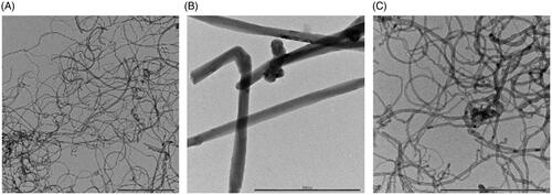
Metal impurities arising from the synthesis may potentially influence cellular responses to MWCNTs (Aldieri et al. Citation2013; Vitkina et al. Citation2016). The present test materials (NM400, NM401, and NM402) contained the following metal impurities as determined by ICP-MS: Al 9951 ± 31 ppm, 59 ± 4 ppm, 12,955 ± 1530 ppm; Fe 1988 ± 26 ppm, 379 ± 71 ppm, 16,321 ± 664 ppm. NM400 also contained Co (693 ± 26 ppm) while all other contaminants were found in negligible amounts in all samples (Rasmussen et al. Citation2014).
Cytotoxicity profiling of MWCNTs of varying morphology
For the evaluation of cytotoxicity, we used primary human immune cells versus cell lines as our model. Hence, HMDMs and macrophage-differentiated THP-1 cells were used to study macrophage effects while PMNs and neutrophil-differentiated HL-60 cells were used to study neutrophil effects. Cell differentiation was confirmed by flow cytometric analysis of the cell surface marker CD11b (data not shown). We also evaluated the performance of different cell viability assays. The results obtained using the LDH release assay are reported in and . The cells were exposed to the indicated concentrations of the three MWCNTs for 24 h. We found that NM401 triggered a dose-dependent cell death in both HMDMs and THP-1 cells, whereas NM400 and NM402 were non-cytotoxic (there was a slight trend for NM402 when tested in the THP-1 model, but this was not significant) (). On the other hand, all three MWCNTs were non-cytotoxic for PMNs as well as HL-60 cells (). For comparison, we also monitored cell viability (metabolic activity) by using the Alamar Blue assay and NM401 was found to trigger a dose-dependent loss of cell viability in macrophage-differentiated THP-1 cells, but not in neutrophil-differentiated HL-60 cells (Supplemental Figure S1), in accordance with the results obtained with the LDH release assay. However, our attempt to monitor cell viability using the ATP assay CellTiter-Glo® failed as the MWCNTs appeared to quench the luminescence signal (data not shown). In fact, a previous study showed that luminescence-based tests may yield erroneous results (Szymański et al. Citation2020); further examples are provided in Monteiro-Riviere, Inman, and Zhang (Citation2009).
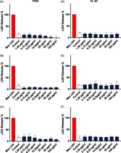
Figure 2. Cell viability assessment. Cell viability was evaluated in primary human monocyte-derived macrophages (HMDMs) (A-C) and macrophage-differentiated THP-1 cells (D–F) using the LDH assay after exposure for 24 h to NM400 (A, D), NM401 (B, E) and NM402 (C, F). Data shown are mean values ± S.D. (n = 3). **p < 0.01, ***p < 0.005. Refer to Figure S1 for corresponding results using the Alamar Blue assay.
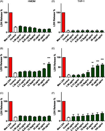
Figure 3. Cell viability assessment. Cell viability was evaluated in primary human polymorphonuclear neutrophils (PMNs) (A–C) and neutrophil-differentiated HL-60 cells (D–F) using the LDH assay after exposure for 24 h to NM400 (A, D), NM401 (B, E) and NM402 (C, F). Data shown are mean values ± S.D. (n = 3). Refer to Figure S1 for corresponding results using the Alamar Blue assay.
Cellular uptake of MWCNTs in human macrophages
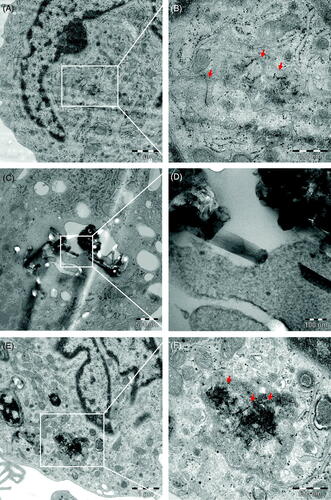
Previous studies have suggested that MWCNTs may mechanically damage HMDMs by impaling or piercing the cells (Cheng et al. Citation2009). We performed TEM of primary human macrophages following exposure to NM400, NM401, and NM402 at 25 µg/mL. Indeed, the straight and rigid NM401 fibers were found to pierce the membranes of intracellular organelles (). In contrast, NM400 () and NM402 () were present in HMDMs as individual fibers or as tangled agglomerates, but no signs of membrane piercing were observed. We also performed TEM analysis of PMNs exposed to MWCNTs but found little evidence of uptake (data not shown). TEM only affords a qualitative assessment of nanomaterial uptake, and it remains challenging to quantify the cellular uptake of MWCNTs. To evaluate cellular uptake of MWCNTs in macrophage-differentiated THP-1 cells versus neutrophil-differentiated HL-60 cells, we performed flow cytometry focusing on changes in side scatter intensity (as a proxy for cellular uptake) (Al-Jamal and Kostarelos Citation2010). We observed a dose-dependent cellular uptake of MWCNTs (NM401) in macrophage-like THP-1 cells, but not in the neutrophil-like HL-60 cells (Supplemental Figure S2). We also evaluated the uptake of MWCNTs (NM401) in HMDMs by performing light microscopy imaging at 24 h (Supplemental Figure S3). Cellular uptake was evidenced by the fact that most of the cells were black. No uptake of MWCNTs was noted in PMNs (refer to section below for results on ECM-adherent cells).
Figure 4. Cellular uptake of MWCNTs. TEM micrographs of HMDMs exposed for 24 h to MWCNTs (10 µg/mL). (A,B) NM400, (C,D) NM401, (E,F) NM402. Scale bars: 1 µm, 500 nm, and 100 nm, as indicated. Arrows in (B) point to individual MWCNT fibers; arrows in (F) point to a tangled agglomerate of fibers. Panel (D) shows NM401 penetrating intracellular membranes. Refer to Figure S3 for light microscopy of HMDMs, and to Figure S7–S9 for results on adherent versus non-adherent neutrophils.
Profiling of cytokine responses using multi-plex arrays
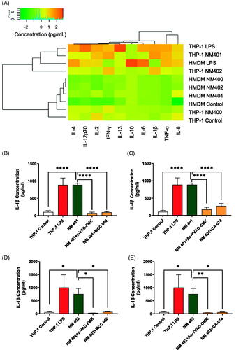
To further evaluate the impact of the different MWCNTs, we conducted a multi-plex based analysis to monitor the production of pro-inflammatory cytokines. To this end, cells were exposed to NM400, NM401, and NM402 (25 µg/mL) for 24 h. LPS (0.1 µg/mL) was used as positive control. Hierarchical clustering analysis of the data obtained in the two macrophage models showed that LPS-exposed HMDM and THP-1 samples clustered together with THP-1 samples exposed to NM401 and NM402 (). In particular, IL-1β and to a lesser degree TNF-α were induced by NM401 and NM402 in PMA-activated THP-1 cells, while the cytokine responses to MWCNTs were less pronounced or absent in HMDMs (note that the latter cells were not primed with LPS). We also examined the two neutrophil models and found low amounts of cytokines in response to all three MWCNTs. However, LPS triggered TNF-α secretion in both PMNs and neutrophil-differentiated HL-60 cells, and IL-1β release in the HL-60 model (Supplemental Figure S4). We then asked whether IL-1β release in macrophage-like THP-1 cells occurred through an active mechanism; specifically, whether IL-1β production was inflammasome-dependent. We found that NM401-triggered IL-1β release was blocked by zVAD-fmk, as well as the specific NLRP3 inhibitor, MCC-950 (Coll et al. Citation2019) (). Additionally, the caspase-1 inhibitor, YVAD-cmk, and the cathepsin B inhibitor, CA-074, also blocked IL-1β production (). Interestingly, we obtained very similar results with respect to NM402-triggered IL-1β release (). The difference, however, being that only NM401 triggered cell death () while NM402 was essentially non-cytotoxic (). For comparison, we have previously shown that inflammasome-dependent IL-1β production can occur in the absence of cell death (Andón et al. Citation2017). No IL-1β release was noted for NM400 ().
Figure 5. Inflammasome activation. (A) Cytokine production was assessed in HMDMs and THP-1 cells exposed to MWCNTs (25 µg/mL) for 24 h using the V-PLEX Human Pro-inflammatory Panel 1 Human Biomarker 10-Plex assay (MSD). As positive control, cells were exposed to 0.1 μg/mL LPS. Hierarchical cluster analysis was performed. Each branch in the dendrograms shows the similarity between samples: the shorter the branch, the more similar the samples. Association clusters for exposures and cytokines are represented by dendrograms at the left and at the top of the heat map, respectively. Refer to Figure S4 for the corresponding results in PMNs and HL-60 cells. (B) IL-1β expression was assessed by ELISA in supernatants collected from THP-1 cells exposed to NM401 in the presence or absence of the pan-caspase inhibitor, zVAD-fmk, or the NLRP3 inhibitor MCC950. As positive control, cells were exposed to 0.1 μg/mL LPS. (C) THP-1 cells exposed to NM401 in the presence or absence of the caspase-1 inhibitor, YVAD-cmk, or the cathepsin B inhibitor, CA-074. (D) IL-1β expression was assessed by ELISA in supernatants collected from THP-1 cells exposed to NM402 in the presence or absence of the pan-caspase inhibitor, zVAD-fmk, or the NLRP3 inhibitor MCC950. (E) THP-1 cells exposed to NM402 in the presence or absence of the caspase-1 inhibitor, YVAD-cmk, or the cathepsin B inhibitor, CA-074. Data in (B-E) are mean values ± S.D. (n = 3). *p < 0.05, **p < 0.01, ****p < 0.001.
Pyroptosis induction by MWCNTs: role of gasdermin D
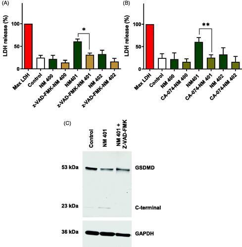
To explore the mechanism of cell death triggered by NM401, we preincubated THP-1 cells with various cell death inhibitors. To this end, cell death was monitored by using the LDH release assay. The pan-caspase inhibitor, zVAD-fmk, effectively blocked cell death (). Furthermore, the cathepsin B inhibitor, CA-074, also protected the cells from NM401-induced cell death (). Furthermore, exposure to NM401 triggered caspase-dependent cleavage of gasdermin D (). Proteolytic cleavage of gasdermin D allows for the insertion of the protein into the plasma membrane leading to the formation of pores, characteristic of pyroptosis (Broz, Pelegrín, and Shao Citation2020). Hence, long and rigid MWCNTs (NM401) triggered cathepsin- and caspase-dependent pyroptosis (a regulated necrosis) with the release of pro-inflammatory IL-1β.
Figure 6. Long and rigid MWCNTs (NM401) trigger pyroptosis. Cell viability was assessed using the LDH assay in macrophage-like THP-1 cells exposed to the indicated MWCNTs in the presence or absence of the caspase inhibitor, zVAD-fmk (A), or the cathepsin B inhibitor, CA-074 (B). Data shown are mean values ± S.D. (n = 3). *p < 0.05, **p < 0.01. (C) Western blot analysis of gasdermin D and the C-terminal cleavage product of GSDMD in cells exposed to NM401 (25 µg/mL) ± zVAD-fmk (20 µM).
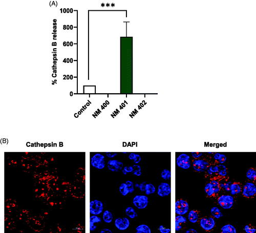
To further explore the involvement of lysosomes in MWCNT-exposed cells, we used the cathepsin B substrate, Magic Red™, to study lysosomal integrity and cytosolic release of cathepsin B. We observed significant release of cathepsin B following 24 h of exposure to NM401 whereas no cathepsin B release was noted in cells exposed to NM400 or NM402 (). Confocal microscopy confirmed that cathepsin B was abundantly present in THP-1 cells (). In contrast, HL-60 cells showed little or no expression of cathepsin B and little evidence of cathepsin B release based on the MR-RR2 substrate (Supplemental Figure S5), thus pointing to a major difference between the two cell models.
Figure 7. Long and rigid MWCNTs (NM401) trigger cathepsin B release. (A) Macrophage-differentiated THP-1 cells were exposed to MWCNTs (25 µg/mL) for 24 h and the release of cathepsin B was determined using Magic Red™, a cathepsin B substrate. (B) Representative confocal microscopy images confirmed the expression of cathepsin B in unexposed cells. Nuclei were counterstained with DAPI. Scale bars:10 µm. Refer to Figure S5 for the corresponding results in neutrophil-like HL-60 cells.
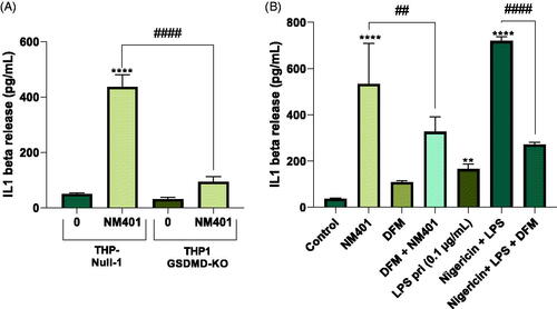
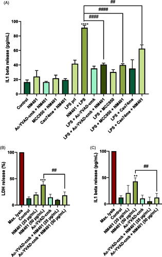
Gasdermin D is required for the secretion but not for the proteolytic maturation of IL-1β (He et al. Citation2015; Kayagaki et al. Citation2015; Shi et al. Citation2015). To further address the role of gasdermin D for NM401-triggered IL-1β release, we employed gasdermin D knockout cells. Thus, THP-Null 1 (wild-type) and THP1-KO-GSDMD cells were differentiated into macrophage-like cells with PMA and subsequently exposed to NM401 at 25 µg/mL for 24 h. The release of IL-1β was almost completely blocked in the knockdown cell line (). Additionally, we decided to test whether disulfiram, an FDA-approved drug used to treat chronic alcoholism, could block NM401-triggered IL-1β production in wild-type cells. Disulfiram was recently shown to act as an inhibitor of gasdermin D pore formation (Hu et al. Citation2020). Hence, disulfiram was shown to allow IL-1β and gasdermin D processing by caspase-1, but abrogated pore formation, thereby preventing IL-1β release. We found that disulfiram significantly reduced IL-1β release in NM401-exposed cells (). We included the bacterial toxin nigericin as a positive control for inflammasome activation. Disulfiram also prevented IL-1β release in response to nigericin.
Figure 8. Gasdermin D-dependent IL-1β release. (A) THP-Null 1 (wild-type) and THP1-KO-GSDMD cells were differentiated with PMA and exposed to NM401 at 25 µg/mL for 24 h. IL-1β release was determined by ELISA. (B) THP-1 cells were exposed to NM401 at 25 µg/mL for 24 h in the presence or absence of disulfiram (DFM) (30 µM). Nigericin was added to LPS-primed cells used as a positive control. Data shown in (A) and (B) are mean values ± S.D. (n = 3). **p < 0.01, ****p < 0.001, ##p < 0.01, ####p < 0.001.
To address whether NM401 also triggers pyroptosis in primary human macrophages, we performed additional experiments in which we compared non-primed versus LPS-primed cells. For LPS priming, HMDMs were incubated for 3 h with LPS (0.1 µg/mL) prior to the exposure to MWCNTs. We could confirm that NM401 does not trigger IL-1β production in non-primed cells (). However, in cells primed with LPS, NM401 triggered IL-1β release, as expected (Palomäki et al. Citation2011), and the cathepsin B inhibitor, CA-074, the caspase-1 inhibitor, YVAD-cmk, and the selective NLRP3 inhibitor, MCC-950, all blocked IL-1β secretion, demonstrating that IL-1β production occurred through the activation of the NLRP3 inflammasome. Moreover, YVAD-fmk, rescued HMDMs from cell death even at a higher concentration of NM401 (50 µg/mL) (), thus confirming that NM401 triggers pyroptosis in primary macrophages.
Figure 9. Pyroptosis in primary macrophages. (A) HMDM were primed or not with LPS (0.1 µg/mL) as indicated and then exposed to NM401 (25 µg/mL) in the presence or absence of the caspase-1 inhibitor, YVAD-cmk, the NLRP3 inhibitor MCC950, or the cathepsin B inhibitor, CA-074. IL-1β production was assessed by ELISA. (B,C) HMDMs primed with LPS were exposed to NM401 (25 or 50 µg/mL) and the impact of YVAD-cmk on cell death (B) and IL-1β production (C) was determined. Data shown are mean values ± S.D. (n = 3). **p < 0.01, ***p < 0.005, ****p < 0.001, ##p < 0.01, ####p < 0.001.
Dose-dependent induction of autophagic cell death
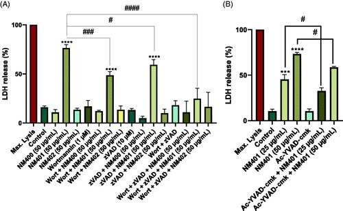
Next, we asked whether MWCNTs triggered autophagy (Wu et al. Citation2014). First, we preincubated the cells with the autophagy inhibitor, 3-MA. However, 3-MA failed to rescue the cells (data not shown). We then tested whether wortmannin blocked cell death. As shown in , NM401, but not NM400 or NM402, triggered cell death, and this was unaffected by wortmannin at the lower concentration of MWCNTs (25 µg/mL). However, when cells were exposed to a higher concentration (50 µg/mL), wortmannin suppressed cell death, arguing in favor of an autophagic cell death mechanism. Importantly, the pan-caspase inhibitor, zVAD-fmk also suppressed cell death (). Moreover, pre-incubation of THP-1 cells with a combination of zVAD-fmk and wortmannin provided a near-complete rescue when cells were exposed to 50 µg/mL (). To confirm that the higher concentration of NM401 triggered pyroptosis, cells were preincubated with the caspase-1 inhibitor, YVAD-cmk ().
Figure 10. Autophagic cell death is dose-dependent. (A) Macrophage-like THP-1 cells were exposed to NM400, NM401, or NM402 at 50 μg/mL for 24 h in the presence or absence of the autophagy inhibitor, wortmannin, or the caspase inhibitor, zVAD-fmk, or both. Cell death was determined using the LDH release assay. (B) THP-1 cells were exposed to NM401 (25 or 50 µg/mL) and the impact of YVAD-cmk on cell death was determined. Data are mean values ± S.D. (n = 3). ***p < 0.005, ****p < 0.001, #p < 0.05, ####p < 0.001. Refer to Figure S6 for results on autophagy in RAW-Difluo™ mLC3 cells.
To further address the potential involvement of autophagy, we used a murine autophagy reporter cell line (Gallud et al. Citation2019). RAW-Difluo™ mLC3 cells express the RFP::GFP::LC3B (microtubule-associated protein 1 light chain 3 beta) fusion protein to enable detection of autophagic flux; two fluorescent reporter proteins are deployed: RFP (acid-stable) and GFP (acid-sensitive). These cells were exposed to MWCNTs and autophagy was monitored by confocal microscopy. Bafilomycin A1 was used as a positive control (Supplemental Figure S6). We could not detect any signs of autophagy when cells were exposed to 25 µg/mL of NM400, NM401, or NM402. However, when RAW-Difluo™ mLC3 cells were exposed to 50 µg/mL of NM401, we observed predominantly red puncta indicating that autolysosomes are formed (because the GFP label is acid-sensitive and the RFP lable is acid-stable, autophagy can be monitored based on the progressive degradation of the GFP signal, and the concurrent increase of the RFP signal in autolysosomes). Thus, at the higher concentration, the long and rigid MWCNTs (NM401) evidently triggered pyroptosis with concomitant autophagic cell death.
ECM-adherent neutrophils remain resistant to MWCNTs
Finally, we asked whether the absence of any cell death or cytokine responses in the neutrophil models could be explained by the fact that these cells are maintained in suspension as opposed to the macrophage models that are grown as adherent cells. To address this, we coated the cell culture plates with different extracellular matrix (ECM) proteins (collagen, laminin, fibrinogen). Neutrophils encounter ECM proteins as they exit the vasculature, and the adhesion to ECM proteins is known to have a crucial impact on neutrophil function. We thus allowed freshly isolated primary human neutrophils to adhere to ECM proteins for 1 h after which time the cell cultures were exposed to MWCNTs for 3 h. These experiments clearly showed that when PMNs are plated on ECM-coated substrates, the cells became adherent (Supplemental Figure S7a). This was particularly evident in the case of laminin. However, we could not detect any uptake of MWCNTs, as determined by light microscopy (Supplemental Figure S7a). Moreover, we did not record any significant loss of cell viability up to 100 µg/mL (Supplemental Figure S8a,b,c). We performed similar experiments using neutrophil-like HL-60 cells. We did not observe any uptake of MWCNTs by flow cytometry (Supplemental Figure S7b), and there was no significant increase in cell death at 24 h and cell viability was not rescued by zVAD-fmk (Supplemental Figure S9).
Discussion
Carbon-based nanomaterials are widely studied yet there is a lack of understanding of the biological and toxicological interactions of these materials (Bhattacharya et al. Citation2016). Recent work has shown that the toxicity of MWCNTs toward human lung epithelial cells (BEAS-2B) depends on the proportion of fibers with greater nominal lengths and diameters (Fraser et al. Citation2020). Here, we showed that long and rigid MWCNTs (NM401) are cytotoxic toward macrophages, but not neutrophils, whereas short and/or tangled MWCNTs (NM400 and NM402) are non-cytotoxic. Furthermore, we found that NM401 triggered cathepsin-dependent cell death in macrophages with NLRP3-dependent production and gasdermin D-dependent release of IL-1β ().
Figure 11. Schematic representation of pyroptosis induction by long and rigid MWCNTs. Refer to main text for a discussion. The present study has also provided evidence for autophagic cell death in conjunction with pyroptosis at high doses of NM401 (not shown).
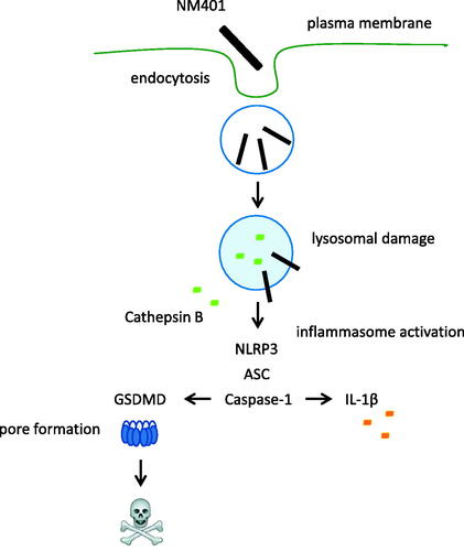
Lysosomes are ubiquitous organelles with a fundamental role in the turnover of cellular components; additionally, lysosomes (dubbed ‘suicide bags’ by Christian de Duve) act as signaling hubs in apoptosis and in different forms of regulated necrosis (Wang, Gómez-Sintes, et al. Citation2018). Lysosomes also play a critical role in cellular responses to nanoparticles (Andón and Fadeel Citation2013). Yuan et al. (Citation2020) showed that a single intratracheal instillation of carbon black nanoparticles in mice induced necrosis with lysosomal membrane permeabilization in alveolar macrophages. Cell death was partially inhibited by the cathepsin B inhibitor, CA-074. Reisetter et al. (Citation2011) demonstrated that carbon black nanoparticles triggered pyroptosis in RAW264.7 cells, characterized by caspase-1 activation and IL-1β production. In contrast, we previously reported that hollow carbon spheres triggered inflammasome-dependent IL-1β production in HMDMs and THP-1 cells in the absence of cell death (Andón et al. Citation2017). We could show, however, that IL-1β production was cathepsin B-dependent. Moreover, long and needle-like MWCNTs elicited inflammasome activation in LPS-primed HMDMs (Palomäki et al. Citation2011). Li et al. (Citation2013) studied a set of covalently functionalized MWCNTs to assess the impact of surface charge and found that polyetherimide (PEI)-modified MWCNTs triggered cathepsin B-dependent production of IL-1β in THP-1 cells. Taken together, lysosomal membrane permeabilization with release of cathepsin B appears to play a key role in macrophage responses to carbon-based nanomaterials. However, it may be relevant to distinguish between lysosome destabilizing agents and ‘classical’ pyroptosis inducing agents; in the former case, lysosomal damage precedes cell death induction whereas lysosomal rupture was shown to be a late event in macrophages exposed to prototypical pyroptosis inducing agents such as anthrax lethal toxin (LT) (Lima et al. Citation2013). In fact, it has been suggested that distinct cathepsins control necrotic cell death mediated by pyroptosis inducers and lysosome destabilizing agents (Brojatsch et al. Citation2015). Jessop et al. (Citation2017) demonstrated that MWCNTs triggered lysosomal membrane permeabilization in murine bone marrow-derived macrophages in a cathepsin B/L-independent manner; however, the subsequent lysosomal leakage of cathepsins was shown to play a role in inflammasome activation. The present study has demonstrated that long and rigid MWCNTs (NM401) trigger lysosomal release of cathepsin B, most likely through physical damage (piercing) of lysosomes. This, in turn, initiated a cascade of events involving NLRP3-dependent caspase-1 activation, with processing of pro-IL-1β and gasdermin D, leading to cell death via pyroptosis (Broz, Pelegrín, and Shao Citation2020). This result is in good agreement with the so-called nanomechanical buckling model which serves to distinguish hazardous carbon-based nanomaterials based on their geometry and inherent ‘stiffness’ (or rigidity) (Zhu et al. Citation2016). Hence, Gao and co-workers argued that nanomaterials that exceed a critical threshold in terms of length/width are capable of inducing lysosomal permeability through mechanical stress. The model also provides a plausible explanation for tangled CNTs that are internalized by macrophages yet show little or no pathogenicity (as shown here for NM400 and NM402) insofar as these may be understood as nanomaterials with intrinsic biological ‘softness’ which are more readily compressed in lysosomes (Zhu et al. Citation2016). Rigidity, which represents the ability of a material to resist deformation, can vary considerably among MWCNTs, and previous studies have provided evidence that rigidity is a key factor responsible for the pathological outcomes of exposure to these materials. Hence, Rydman et al. (Citation2014) found that only rod-like, i.e. long and straight MWCNTs, but not the flexible, tangled MWCNTs caused allergic airway inflammation in mice. Similarly, it was shown that while tangled and rod-like MWCNTs both triggered acute inflammation one day post-exposure, only the rod-like MWCNTs caused chronic inflammation in mice (Duke et al. Citation2017). Furthermore, Lee et al. (Citation2018) provided evidence that rigidity is an important factor in determining IL-1β production in THP-1 cells. In line with the latter findings, we have found that only the long and rigid MWCNTs (NM401) triggered pyroptosis in macrophages with gasdermin D-dependent IL-1β secretion. This material (NM401) obviously differed from the other test materials (NM400, NM402) insofar as the latter two materials were tangled (i.e. flexible). These results agree with those of Westphal et al. (Citation2019) who investigated the effects of a set of ‘benchmark’ materials (NM400, NM401, NM402, NM403) using the rat alveolar cell line NR8383 and found that NM401 caused the strongest effects (stronger than chrysotile asbestos) while NM400, NM402, and NM403 were non-cytotoxic. The authors argued that the strong toxicity of NM401 might be due to ‘piercing’ of the cells. As for metal impurities, NM401 was shown to have the lowest amount of Fe among the tested materials (Rasmussen, et al. Citation2014). It is important to note that while the presence of transition metals may drive oxidative stress responses, this does not necessarily translate into cell death. Indeed, we were unable to block NM401-triggered cell death in macrophage-like THP-1 cells using N-acetylcysteine (unpublished observations).
To further support the conclusion that NM401 triggers pyroptosis, we applied PMA-differentiated THP-1 cells with stable knockdown of gasdermin D. Using these knockdown cells versus wild-type cells, we provided conclusive evidence that NM401-triggered IL-1β production is gasdermin D-dependent. In fact, while previous studies have demonstrated NLRP3-dependent IL-1β production in primary human monocyte-derived macrophages (Palomäki et al. Citation2011) and in primary human bronchial epithelial cells (Hussain et al. Citation2014), no other studies have shown gasdermin D-dependent IL-1β secretion in MWCNT-exposed cells. Importantly, we also demonstrated that disulfiram, an FDA-approved drug used to treat chronic alcoholism, could block NM401-triggered IL-1β production. Disulfiram was shown in a very recent study to act as an inhibitor of gasdermin D pore formation (Hu et al. Citation2020), and the present results demonstrate for the first time that disulfiram can suppress MWCNT-induced IL-1β production. Further studies of disulfiram using relevant animal models of CNT exposure may be of interest.
Autophagy serves as a cytoprotective mechanism; however, persistent activation of autophagy can result in cell death (Orrenius, Kaminskyy, and Zhivotovsky Citation2013). Previous studies have found evidence of autophagy induction in cells exposed to MWCNTs (Ghosh et al. Citation2020), and some authors have shown that autophagy is dependent on the diameter of the MWCNTs (Zhao et al. Citation2019), but the autophagy response may also depend on the surface chemistry (Wu et al. Citation2014). Liu et al. (Citation2011) showed that SWCNTs triggered autophagic cell death in the human lung carcinoma cell line A549, a very resilient cancer cell line. In fact, exceptionally high concentrations of SWCNTs were used (1 mg/mL). In contrast, other investigators have found that exposure to MWCNTs leads to a blockade of cellular autophagic flux (Cohignac et al. Citation2018). In the present study, the phosphoinositide 3-kinase (PI3K) inhibitor, wortmannin (Wu et al. Citation2010) failed to rescue macrophages exposed to 25 µg/mL of the long and rigid MWCNTs (NM401). However, when cells were exposed to a higher concentration of MWCNTs (50 µg/mL), wortmannin significantly reduced cell death. The cell death remained zVAD-fmk-inhibitable also at the higher concentration of MWCNTs and was also reduced by the caspase-1 inhibitor, and when zVAD-fmk and wortmannin were combined, the cells were completely rescued from cell death. This is the first demonstration of a mixed cell death (i.e. autophagic cell death and pyroptosis) in macrophages exposed to MWCNTs. However, our data clearly showed that pyroptotic cell death dominates in cells exposed at the lower concentration of MWCNTs. Lysosomes are implicated in autophagy as well as in pyroptosis, and it is certainly possible that these cell death or cell survival modalities are linked (Miller, Cramer, and Thorburn Citation2020). Perhaps autophagic responses are harnessed as a protective mechanism in situations of insurmountable lysosomal damage, while this may ultimately lead to (necrotic) cell death if the cellular stress persists.
Nanomaterials from the JRC nanomaterial repository have been widely used not least in different EU-funded nanosafety projects (Farcal et al. Citation2015). The use of ‘benchmark’ nanomaterials enables the comparison of data across different laboratories (Jackson et al. Citation2015; Louro et al. Citation2016; Allegri et al. Citation2016; Rahman et al. Citation2017; Di Cristo et al. Citation2019). The picture is thus emerging that the diameter (and rigidity) of MWCNTs is a crucial determinant of their cytotoxicity (Fujita et al. Citation2020). The present study confirmed this. However, we also found that neutrophils were not killed by long and rigid MWCNTs. Thus, in addition to the physicochemical properties of the test material, the choice of the test model (in particular, whether macrophages or neutrophils) also matters. In a very recent study, Verdon et al. (Citation2021) compared the responses of the neutrophil-like HL-60 cell line and primary human neutrophils following exposure to metal (Ag) and metal oxide nanoparticles (ZnO, CuO, TiO2). The authors observed a similar pattern of responses between HL-60 cells and primary cells, although the two cell models differed with respect to certain endpoints (for instance, ZnO nanoparticles significantly affected the metabolic activity of HL-60 cells but did not impact on primary neutrophils). Overall, however, the HL-60 model was recommended for screening purposes. Based on the present studies of MWCNTs, we concur that neutrophil-differentiated HL-60 cells (and macrophage-differentiated THP-1 cells) may be useful for initial screening due to the fact that cell lines are easier to handle than the primary cells.
Neutrophils are the most abundant white blood cell and part of the first line of cellular defence against intruding pathogens (and particles) (Jhunjhunwala Citation2018), yet they have not been studied to the same extent as macrophages with respect to nanomaterial interactions (Keshavan et al. Citation2019). Neutrophils are programmed to die from the moment they are released from the bone marrow (Fadeel et al. Citation1998). Notwithstanding, the present findings showed that none of the MWCNTs (NM400, NM401, NM402) were cytotoxic toward neutrophils, i.e. not in primary human neutrophils tested at 3 h (to avoid excessive background of cell death) nor in neutrophil-differentiated HL-60 cells tested at 24 h. Furthermore, we found that cathepsin B expression was low or absent in neutrophils. Earlier work has shown that so-called ‘dense bodies’ or mature lysosomes described in other cells are not present in neutrophils (Cieutat et al. Citation1998). Instead, neutrophils express azurophilic granules (along with other granules) that may be viewed as lysosome-like organelles despite the fact that lysosomal membrane glycoproteins LAMP-1 and LAMP-2 are absent from these granules (Cieutat et al. Citation1998). It is a matter of conjecture at this point, but perhaps this may explain why MWCNTs fail to trigger lysosome-dependent cell death in these cells. However, before lysosomes are engaged, the offending (nano)material has to gain access to the cell, and our experiments suggest that the lack of toxicity toward neutrophils may instead be due to a lack of cellular uptake of the MWCNTs. To assess whether this was simply due to the fact that neutrophils are grown in suspension whereas macrophages are adherent, we decided to test whether neutrophils (neutrophil-differentiated HL-60 cells and primary neutrophils) that have been allowed to adhere to cell culture plates pre-coated with extracellular matrix (ECM) proteins are more or less susceptible to MWCNTs. The neutrophil inflammatory response can be profoundly altered by contact with ECM proteins (Nathan Citation1987; Ginis and Tauber Citation1990). Specifically, neutrophils encounter ECM proteins as they exit the vasculature, and the adhesion to ECM proteins is important for the modulation of neutrophil function. However, we found that even though the cells were attached to the substrate, there was little or no cell death following exposure to MWCNTs for 24 h (HL-60 cells) or 3 h (PMNs).
It is notable that earlier studies suggested that cellular uptake of functionalized single- and multi-walled CNTs occurs independently of cell type (Kostarelos et al. Citation2007). However, the latter study was performed using a range of non-phagocytic cancer cell lines, including A549, Jurkat, and HeLa. In contrast, the present study, using both macrophage-like and neutrophil-like cell lines as well as primary immune cells, has provided first evidence that the cellular uptake of MWCNTs differs markedly between macrophages and neutrophils. We did not investigate the mechanism of uptake in macrophages here. However, previous studies using HMDMs as a model have shown that SWCNTs were located in phagosomes and lysosomes, suggesting uptake by phagocytosis; additionally, the authors showed that SWCNTs resided in the cytoplasm and in the cell nucleus, suggesting that SWCNTs may also gain access to cells via passive diffusion across the plasma membrane, or potentially by ‘piercing’ cellular membranes (Porter et al. Citation2007; Porter et al. Citation2009). MWCNTs may also display a similar behavior, as suggested by another study by the same authors (Cheng et al. Citation2009). Notably, in the latter study, macrophages were exposed to a relatively low concentration (5 µg/mL) and cells were examined 4 days later using a range of imaging techniques. It is conceivable that the MWCNTs are first engulfed via phagocytosis, subsequently entering the cytoplasm by escaping from or piercing through the phago-lysosomal membranes, as opposed to passively diffusing across the plasma membrane. In contrast, when cells are exposed to a cytotoxic concentration of MCWCNTs, they may undergo acute (24 h), lysosome-dependent cell death, as shown here, and the release of MWCNTs into the cytoplasm (if it occurs) may not be as evident.
Gao et al. (Citation2011) could show that MWCNTs are located in phagosomes, lysosome, and in the cytoplasm in macrophage-like THP-1 cells. Furthermore, using MWCNTs with pre-adsorbed, FITC-labeled bovine serum albumin (BSA) in order to track the materials, the authors provided evidence that MWCNTs (displaying a bio-corona of BSA) deploy both scavenger receptors and mannose receptors in order to gain access to cells. Other investigators also reported, using the murine RAW264.7 macrophage cell line, that MWCNTs are internalized, at least in part, via scavenger receptors (Wang, Gómez-Sintes, et al. Citation2018). However, it is important to note that the receptor repertoire on the surface of primary macrophages varies depending on the activation state of the cells (Gallud et al. Citation2017). Furthermore, the functionalization of MWCNTs, and whether or not the MWCNTs are well-dispersed or agglomerated, may also play a role for cellular uptake (Gao et al. Citation2011; Wang, Gómez-Sintes, et al. Citation2018). In a more recent study, T cell immunoglobulin mucin 4 (Tim4) was identified as a receptor for MWCNTs (Omori et al. Citation2021). Hence, Tim4, but not Tim1, was shown to contribute to the recognition of MWCNTs by mouse peritoneal macrophages and to promote granuloma development in a mouse model of i.p. administration of MWCNTs. Furthermore, overexpression of Tim4, but not a mutant form of the receptor, significantly enhanced MWCNT recognition by THP-1 cells (Omori et al. Citation2021). However, it was previously shown that macrophage-differentiated THP-1 cells express TIM-1, but not TIM-4, on their surface suggesting that these cells differ from tissue-resident macrophages (Forrester et al. Citation2018). Thus, it seems unlikely that differences in TIM-4 could account for the differences in uptake of MWCNTs between macrophages and neutrophils observed in the present study. It is conceivable that the bio-corona, and its corresponding phagocytosis receptors on the surface of macrophages or neutrophils, might play a role. In the present study, all the cell models were maintained in the same cell culture medium supplemented with 10% FBS. Therefore, if a bio-corona is formed then it would be similar regardless of the cell model. Ge et al. (Citation2011) showed that the adsorption of serum proteins mitigated the toxicity of SWCNTs toward THP-1 cells. Moreover, the adsorption of lung surfactant proteins and lipids was found to facilitate the uptake of SWCNTs in the macrophage-like RAW264.7 cell line (Kapralov et al. Citation2012). However, while we did not formally address the role of the bio-corona in the present study, we can say with certainty that the long and rigid MWCNTs are cytotoxic for macrophages despite the presence of serum. One may speculate that the bio-corona could prevent rather than promote the uptake of MWCNTs by neutrophils, but this remains to be tested experimentally. It is worth noting in this context that while PEGylation is believed to prevent the phagocytic clearance of particles, recent work has shown that PEGylation preferentially enhances uptake of particles by neutrophils, specifically in the presence of human plasma (Kelley et al. 2018).
Conclusions
Nanotoxicological research has been largely ‘macrophage-centric’, at least with respect to in vitro studies (Bisso et al. Citation2018; Keshavan et al. Citation2019). We have shown here that: (i) macrophage- and neutrophil-differentiated human cell lines exposed to ‘benchmark’ MWCNTs displayed comparable responses to primary macrophages and neutrophils, respectively; (ii) macrophages, but not neutrophils, underwent lysosome-dependent cell death (pyroptosis) when exposed to MWCNTs; (iii) long and rigid MWCNTs, but not the short and/or tangled MWCNTS, were cytotoxic for macrophages, and the former were found to damage lysosomal membranes leading to cathepsin release. Overall, we confirmed the notion that not all CNTs are alike (Fadeel and Kostarelos Citation2020; Heller et al. Citation2020). We could also show that not all immune cells respond in the same manner to MWCNTs, possibly due to differences in uptake. This should be considered when evaluating the hazard potential of CNTs as well as other nanomaterials.
Supplemental Material
Download MS Power Point (25.2 MB)Acknowledgements
The authors are indebted to Dr. Juan Riego Sintes for providing benchmark materials from the nanomaterial repository at JRC (Ispra, Italy). We also thank Dr. Lars Haag, EM Core Facility at Karolinska Institutet, for expert assistance with TEM analysis of cells.
Disclosure statement
No potential conflict of interest was reported by the author(s).
Additional information
Funding
References
- Aldieri, E., I. Fenoglio, F. Cesano, E. Gazzano, G. Gulino, D. Scarano, A. Attanasio, G. Mazzucco, D. Ghigo, and B. Fubini. 2013. “The Role of Iron Impurities in the Toxic Effects Exerted by Short Multi-Walled Carbon Nanotubes (MWCNT) in Murine Alveolar Macrophages.” Journal of Toxicology and Environmental Health. Part A 76 (18): 1056–1071. doi:https://doi.org/10.1080/15287394.2013.834855.
- Al-Jamal, K. T., and K. Kostarelos. 2010. “Assessment of Cellular Uptake and Cytotoxicity of Carbon Nanotubes Using Flow Cytometry.” Methods in Molecular Biology 625: 123–134. doi:https://doi.org/10.1007/978-1-60761-579-8_11.
- Allegri, M., D. K. Perivoliotis, M. G. Bianchi, M. Chiu, A. Pagliaro, M. A. Koklioti, A. A. Trompeta, E. Bergamaschi, O. Bussolati, and C. A. Charitidis. 2016. “Toxicity Determinants of Multi-Walled Carbon Nanotubes: The Relationship Between Functionalization and Agglomeration.” Toxicology Reports 3: 230–243. doi:https://doi.org/10.1016/j.toxrep.2016.01.011.
- Andón, F. T., and B. Fadeel. 2013. “Programmed Cell Death: Molecular Mechanisms and Implications for Safety Assessment of Nanomaterials.” Accounts of Chemical Research 46 (3): 733–742. doi:https://doi.org/10.1021/ar300020b.
- Andón, F. T., S. P. Mukherjee, I. Gessner, L. Wortmann, L. Xiao, K. Hultenby, A. A. Shvedova, S. Mathur, and B. Fadeel. 2017. “Hollow Carbon Spheres Trigger Inflammasome-Dependent IL-1β Secretion in Macrophages.” Carbon 113: 243–251. doi:https://doi.org/10.1016/j.carbon.2016.11.049.
- Bhattacharya, K., G. Kiliç, P. M. Costa, and B. Fadeel. 2017. “Cytotoxicity Screening and Cytokine Profiling of Nineteen Nanomaterials Enables Hazard Ranking and Grouping Based on Inflammogenic Potential.” Nanotoxicology 11 (6): 809–826. doi:https://doi.org/10.1080/17435390.2017.1363309.
- Bhattacharya, K., S. P. Mukherjee, A. Gallud, S. C. Burkert, S. Bistarelli, S. Bellucci, M. Bottini, A. Star, and B. Fadeel. 2016. “Biological Interactions of Carbon-Based Nanomaterials: From Coronation to Degradation.” Nanomedicine : Nanotechnology, Biology, and Medicine 12 (2): 333–351. doi:https://doi.org/10.1016/j.nano.2015.11.011.
- Bisso, P. W., S. Gaglione, P. P. G. Guimarães, M. J. Mitchell, and R. Langer. 2018. “Nanomaterial Interactions with Human Neutrophils.” ACS Biomaterials Science & Engineering 4 (12): 4255–4265. doi:https://doi.org/10.1021/acsbiomaterials.8b01062.
- Borgquist, J. D., M. T. Quinn, and S. D. Swain. 2002. “Adhesion to Extracellular Matrix Proteins Modulates Bovine Neutrophil Responses to Inflammatory Mediators.” Journal of Leukocyte Biology 71 (5): 764–774.
- Boyles, M. S., L. Young, D. M. Brown, L. MacCalman, H. Cowie, A. Moisala, F. Smail, et al. 2015. “Multi-Walled Carbon Nanotube Induced Frustrated Phagocytosis, Cytotoxicity and Pro-Inflammatory Conditions in Macrophages Are Length Dependent and Greater than That of Asbestos.” Toxicology in Vitro 29 (7): 1513–1528. doi:https://doi.org/10.1016/j.tiv.2015.06.012.
- Brojatsch, J., H. Lima, D. Palliser, L. S. Jacobson, S. M. Muehlbauer, R. Furtado, D. L. Goldman, M. P. Lisanti, and K. Chandran. 2015. “Distinct Cathepsins Control Necrotic Cell Death Mediated by Pyroptosis Inducers and Lysosome-Destabilizing Agents.” Cell Cycle 14 (7): 964–972. doi:https://doi.org/10.4161/15384101.2014.991194.
- Broz, P., P. Pelegrín, and F. Shao. 2020. “The Gasdermins, a Protein Family Executing Cell Death and Inflammation.” Nature Reviews. Immunology 20 (3): 143–157. doi:https://doi.org/10.1038/s41577-019-0228-2.
- Castranova, V., P. A. Schulte, and R. D. Zumwalde. 2013. “Occupational Nanosafety Considerations for Carbon Nanotubes and Carbon Nanofibers.” Accounts of Chemical Research 46 (3): 642–649. doi:https://doi.org/10.1021/ar300004a.
- Cheng, C., K. H. Müller, K. K. Koziol, J. N. Skepper, P. A. Midgley, M. E. Welland, and A. E. Porter. 2009. “Toxicity and Imaging of Multi-Walled Carbon Nanotubes in Human Macrophage Cells.” Biomaterials 30 (25): 4152–4160. doi:https://doi.org/10.1016/j.biomaterials.2009.04.019.
- Chernova, T., F. A. Murphy, S. Galavotti, X.-M Sun, I. R. Powley, S. Grosso, A. Schinwald, et al. 2017. “Long-Fiber Carbon Nanotubes Replicate Asbestos-Induced Mesothelioma with Disruption of the Tumor Suppressor Gene Cdkn2a (Ink4a/Arf).” Current Biology : CB 27 (21): 3302–3314.e6. doi:https://doi.org/10.1016/j.cub.2017.09.007.
- Cieutat, A. M., P. Lobel, J. T. August, L. Kjeldsen, H. Sengeløv, N. Borregaard, and D. F. Bainton. 1998. “Azurophilic Granules of Human Neutrophilic Leukocytes Are Deficient in Lysosome-Associated Membrane Proteins but Retain the Mannose 6-Phosphate Recognition Marker.” Blood 91 (3): 1044–1058. doi:https://doi.org/10.1182/blood.V91.3.1044.
- Vanessa, C., M. J. Landry, A. Ridoux, M. Pinault, B. Annangi, A. Gerdil, N. Herlin-Boime, et al. 2018. “Carbon Nanotubes, but Not Spherical Nanoparticles, Block Autophagy by a Shape-Related Targeting of Lysosomes in Murine Macrophages.” Autophagy 14 (8): 1323–1334. doi:https://doi.org/10.1080/15548627.2018.1474993.
- Coll, R. C., J. R. Hill, C. J. Day, A. Zamoshnikova, D. Boucher, N. L. Massey, J. L. Chitty, et al. 2019. “MCC950 Directly Targets the NLRP3 ATP-Hydrolysis Motif for Inflammasome Inhibition.” Nature Chemical Biology 15 (6): 556–559. doi:https://doi.org/10.1038/s41589-019-0277-7.
- De Volder, M. F., S. H. Tawfick, R. H. Baughman, and A. J. Hart. 2013. “Carbon Nanotubes: present and Future Commercial Applications.” Science 339 (6119): 535–539. doi:https://doi.org/10.1126/science.1222453.
- Di Cristo, L., M. G. Bianchi, M. Chiu, G. Taurino, F. Donato, G. Garzaro, O. Bussolati, and E. Bergamaschi. 2019. “Comparative in Vitro Cytotoxicity of Realistic Doses of Benchmark Multi-Walled Carbon Nanotubes Towards Macrophages and Airway Epithelial Cells.” Nanomaterials 9 (7): 982. doi:https://doi.org/10.3390/nano9070982.
- Donaldson, K., C. A. Poland, F. A. Murphy, M. MacFarlane, T. Chernova, and A. Schinwald. 2013. “Pulmonary Toxicity of Carbon Nanotubes and Asbestos – Similarities and Differences.” Advanced Drug Delivery Reviews 65 (15): 2078–2086. doi:https://doi.org/10.1016/j.addr.2013.07.014.
- Duke, K. S., A. J. Taylor-Just, M. D. Ihrie, K. A. Shipkowski, E. A. Thompson, E. C. Dandley, G. N. Parsons, and J. C. Bonner. 2017. “STAT1-Dependent and -Independent Pulmonary Allergic and Fibrogenic Responses in Mice After Exposure to Tangled Versus Rod-like Multi-Walled Carbon Nanotubes.” Particle and Fibre Toxicology 14 (1): 26. doi:https://doi.org/10.1186/s12989-017-0207-3.
- Fadeel, B., A. Åhlin, J. I. Henter, S. Orrenius, and M. B. Hampton. 1998. “Involvement of Caspases in Neutrophil Apoptosis: Regulation by Reactive Oxygen Species.” Blood 92 (12): 4808–4818. doi:https://doi.org/10.1182/blood.V92.12.4808.
- Fadeel, B., and K. Kostarelos. 2020. “Grouping All Carbon Nanotubes into a Single Substance Category is Scientifically Unjustified.” Nature Nanotechnology 15 (3): 164. doi:https://doi.org/10.1038/s41565-020-0654-0.
- Farcal, L., F. T. Andón, L. D. Cristo, B. M. Rotoli, O. Bussolati, E. Bergamaschi, A. Mech, et al. 2015. “Comprehensive in Vitro Toxicity Testing of a Panel of Representative Oxide Nanomaterials: First Steps Towards an Intelligent Testing Strategy.” PLoS One 10 (5): e0127174. doi:https://doi.org/10.1371/journal.pone.0127174.
- Forrester, M. A., H. J. Wassall, L. S. Hall, H. Cao, H. M. Wilson, R. N. Barker, and M. A. Vickers. 2018. “Similarities and Differences in Surface Receptor Expression by THP-1 Monocytes and Differentiated Macrophages Polarized Using Seven Different Conditioning Regimens.” Cellular Immunology 332: 58–76. doi:https://doi.org/10.1016/j.cellimm.2018.07.008.
- Fraser, K., V. Kodali, N. Yanamala, M. Eileen Birch, L. Cena, G. Casuccio, K. Bunker, et al. 2020. “Physicochemical Characterization and Genotoxicity of the Broad Class of Carbon Nanotubes and Nanofibers Used or Produced in U.S. Facilities.” Particle and Fibre Toxicology 17 (1): 62. doi:https://doi.org/10.1186/s12989-020-00392-w.
- Fujita, K., S. Obara, J. Maru, and S. Endoh. 2020. “Cytotoxicity Profiles of Multi-Walled Carbon Nanotubes with Different Physico-Chemical Properties.” Toxicology Mechanisms and Methods 30 (7): 477–489. doi:https://doi.org/10.1080/15376516.2020.1761920.
- Funahashi, S., Y. Okazaki, D. Ito, A. Asakawa, H. Nagai, M. Tajima, and S. Toyokuni. 2015. “Asbestos and Multi-Walled Carbon Nanotubes Generate Distinct Oxidative Responses in Inflammatory Cells.” Journal of Clinical Biochemistry and Nutrition 56 (2): 111–117. doi:https://doi.org/10.3164/jcbn.14-92.
- Gallud, A., O. Bondarenko, N. Feliu, N. Kupferschmidt, R. Atluri, A. Garcia-Bennett, and B. Fadeel. 2017. “Macrophage Activation Status Determines the Internalization of Mesoporous Silica Particles: Exploring the Role of Different Pattern Recognition Receptors.” Biomaterials 121: 28–40. doi:https://doi.org/10.1016/j.biomaterials.2016.12.029.
- Gallud, A., K. Klöditz, J. Ytterberg, N. Östberg, S. Katayama, T. Skoog, V. Gogvadze, et al. 2019. “Cationic Gold Nanoparticles Elicit Mitochondrial Dysfunction: A Multi-Omics Study.” Scientific Reports 9 (1): 4366. doi:https://doi.org/10.1038/s41598-019-40579-6.
- Gao, N., Q. Zhang, Q. Mu, Y. Bai, L. Li, H. Zhou, E. R. Butch, et al. 2011. “Steering Carbon Nanotubes to Scavenger Receptor Recognition by Nanotube Surface Chemistry Modification Partially Alleviates NFκB Activation and Reduces Its Immunotoxicity.” ACS Nano 5 (6): 4581–4591. doi:https://doi.org/10.1021/nn200283g.
- Ge, C., J. Du, L. Zhao, L. Wang, Y. Liu, D. Li, Y. Yang, et al. 2011. “Binding of Blood Proteins to Carbon Nanotubes Reduces Cytotoxicity.” Proceedings of the National Academy of Sciences of the United States of America 108 (41): 16968–16973. doi:https://doi.org/10.1073/pnas.1105270108.
- Ghosh, M., S. Murugadoss, L. Janssen, S. Cokic, C. Mathyssen, K. Van Landuyt, W. Janssens, S. Carpentier, L. Godderis, and P. Hoet. 2020. “Distinct Autophagy-Apoptosis Related Pathways Activated by Multi-Walled (NM 400) and Single-Walled Carbon Nanotubes (NIST-SRM2483) in Human Bronchial Epithelial (16HBE14o-) Cells.” Journal of Hazardous Materials 387: 121691. doi:https://doi.org/10.1016/j.jhazmat.2019.121691.
- Ginis, I., and A. I. Tauber. 1990. “Activation Mechanisms of Adherent Human Neutrophils.” Blood 76 (6): 1233–1239.
- Grosse, Y., D. Loomis, K. Z. Guyton, B. Lauby-Secretan, F. E. Ghissassi, V. Bouvard, L. Benbrahim-Tallaa, et al. 2014. “Carcinogenicity of Fluoro-Edenite, Silicon Carbide Fibres and Whiskers, and Carbon Nanotubes.” The Lancet Oncology 15 (13): 1427–1428. doi:https://doi.org/10.1016/S1470-2045(14)71109-X.
- Hamilton, R. F., Z. Wu, S. Mitra, P. K. Shaw, and A. Holian. 2013. “Effect of MWCNT Size, Carboxylation, and Purification on in Vitro and in Vivo Toxicity, Inflammation and Lung Pathology.” Particle and Fibre Toxicology 10 (1): 57. doi:https://doi.org/10.1186/1743-8977-10-57.
- He, W. T., H. Wan, L. Hu, P. Chen, X. Wang, Z. Huang, Z. H. Yang, C. Q. Zhong, and J. Han. 2015. “Gasdermin D is an Executor of Pyroptosis and Required for Interleukin-1β Secretion.” Cell Research 25 (12): 1285–1298. doi:https://doi.org/10.1038/cr.2015.139.
- Heller, D. A., P. V. Jena, M. Pasquali, K. Kostarelos, L. G. Delogu, R. E. Meidl, S. V. Rotkin, et al. 2020. “Banning Carbon Nanotubes Would Be Scientifically Unjustified and Damaging to Innovation.” Nature Nanotechnology 15 (3): 164–166. doi:https://doi.org/10.1038/s41565-020-0656-y.
- Hu, J. J., X. Liu, S. Xia, Z. Zhang, Y. Zhang, J. Zhao, J. Ruan, et al. 2020. “FDA-Approved Disulfiram Inhibits Pyroptosis by Blocking Gasdermin D Pore Formation.” Nature Immunology 21 (7): 736–745. doi:https://doi.org/10.1038/s41590-020-0669-6.
- Hussain, S., S. Sangtian, S. M. Anderson, R. J. Snyder, J. D. Marshburn, A. B. Rice, J. C. Bonner, and S. Garantziotis. 2014. “Inflammasome Activation in Airway Epithelial Cells After Multi-Walled Carbon Nanotube Exposure Mediates a Profibrotic Response in Lung Fibroblasts.” Particle and Fibre Toxicology 11: 28. doi:https://doi.org/10.1186/1743-8977-11-28.
- Jackson, P., K. Kling, K. A. Jensen, P. A. Clausen, A. M. Madsen, H. Wallin, and U. Vogel. 2015. “Characterization of Genotoxic Response to 15 Multiwalled Carbon Nanotubes with Variable Physicochemical Properties Including Surface Functionalizations in the FE1-Muta(TM) Mouse Lung Epithelial Cell Line.” Environmental and Molecular Mutagenesis 56 (2): 183–203. doi:https://doi.org/10.1002/em.21922.
- Jessop, F., R. F. Hamilton, J. F. Rhoderick, P. Fletcher, and A. Holian. 2017. “Phagolysosome Acidification is Required for Silica and Engineered Nanoparticle-Induced Lysosome Membrane Permeabilization and Resultant NLRP3 Inflammasome Activity.” Toxicology and Applied Pharmacology 318: 58–68. doi:https://doi.org/10.1016/j.taap.2017.01.012.
- Jessop, F., and A. Holian. 2015. “Extracellular HMGB1 Regulates Multi-Walled Carbon Nanotube-Induced Inflammation in Vivo.” Nanotoxicology 9 (3): 365–372. doi:https://doi.org/10.3109/17435390.2014.933904.
- Jhunjhunwala, S. 2018. “Neutrophils at the Biological-Material Interface.” ACS Biomaterials Science & Engineering 4 (4): 1128–1136. doi:https://doi.org/10.1021/acsbiomaterials.6b00743.
- Jitkaew, S., E. Witasp, S. Zhang, V. E. Kagan, and B. Fadeel. 2009. “Induction of Caspase- and Reactive Oxygen Species-Independent Phosphatidylserine Externalization in Primary Human Neutrophils: Role in Macrophage Recognition and Engulfment.” Journal of Leukocyte Biology 85 (3): 427–437. doi:https://doi.org/10.1189/jlb.0408232.
- Kagan, V. E., N. V. Konduru, W. Feng, B. L. Allen, J. Conroy, Y. Volkov, I. I. Vlasova, et al. 2010. “Carbon Nanotubes Degraded by Neutrophil Myeloperoxidase Induce Less Pulmonary Inflammation.” Nature Nanotechnology 5 (5): 354–359. doi:https://doi.org/10.1038/nnano.2010.44.
- Kapralov, A. A., W. H. Feng, A. A. Amoscato, N. Yanamala, K. Balasubramanian, D. E. Winnica, E. R. Kisin, et al. 2012. “Adsorption of Surfactant Lipids by Single-Walled Carbon Nanotubes in Mouse Lung Upon Pharyngeal Aspiration.” ACS Nano 6 (5): 4147–4156. doi:https://doi.org/10.1021/nn300626q.
- Kayagaki, N., I. B. Stowe, B. L. Lee, K. O'Rourke, K. Anderson, S. Warming, T. Cuellar, et al. 2015. “Caspase-11 Cleaves Gasdermin D for Non-canonical Inflammasome Signalling.” Nature 526 (7575): 666–671. doi:https://doi.org/10.1038/nature15541.
- Kelley, W. J., C. A. Fromen, G. Lopez-Cazares, and O. Eniola-Adefeso. 2018. “PEGylation of Model Drug Carriers Enhances Phagocytosis by Primary Human Neutrophils.” Acta Biomaterialia 79: 283–293. doi:https://doi.org/10.1016/j.actbio.2018.09.001.
- Keshavan, S., P. Calligari, L. Stella, L. Fusco, L. G. Delogu, and B. Fadeel. 2019. “Nano-Bio Interactions: A Neutrophil-Centric View.” Cell Death & Disease 10 (8): 569. doi:https://doi.org/10.1038/s41419-019-1806-8.
- Kostarelos, K.,. L. Lacerda, G. Pastorin, W. Wu, S. Wieckowski, J. Luangsivilay, S. Godefroy, et al. 2007. “Cellular Uptake of Functionalized Carbon Nanotubes is Independent of Functional Group and Cell Type.” Nature Nanotechnology 2 (2): 108–113. doi:https://doi.org/10.1038/nnano.2006.209.
- Kunzmann, A., B. Andersson, C. Vogt, N. Feliu, F. Ye, S. Gabrielsson, M. S. Toprak, et al. 2011. “Efficient Internalization of Silica-Coated Iron Oxide Nanoparticles of Different Sizes by Primary Human Macrophages and Dendritic Cells.” Toxicology and Applied Pharmacology 253 (2): 81–93. doi:https://doi.org/10.1016/j.taap.2011.03.011.
- Lee, D.-K., S. Jeon, Y. Han, S.-H. Kim, S. Lee, I. J. Yu, K. S. Song, et al. 2018. “Threshold Rigidity Values for the Asbestos-Like Pathogenicity of High-Aspect Ratio Carbon Nanotubes in a Mouse Pleural Inflammation Model.” ACS Nano 12 (11): 10867–10879. doi:https://doi.org/10.1021/acsnano.8b03604.
- Li, Ruibin, Xiang Wang, Zhaoxia Ji, Bingbing Sun, Haiyuan Zhang, Chong Hyun Chang, Sijie Lin, et al. 2013. “Surface Charge and Cellular Processing of Covalently Functionalized Multiwall Carbon Nanotubes Determine Pulmonary Toxicity.” ACS Nano 7 (3): 2352–2368. doi:https://doi.org/10.1021/nn305567s.
- Lima, H., L. S. Jacobson, M. F. Goldberg, K. Chandran, F. Diaz-Griffero, M. P. Lisanti, and J. Brojatsch. 2013. “Role of Lysosome Rupture in Controlling Nlrp3 Signaling and Necrotic Cell Death.” Cell Cycle 12 (12): 1868–1878. doi:https://doi.org/10.4161/cc.24903.
- Liu, H. L., Y. L. Zhang, N. Yang, Y. X. Zhang, X. Q. Liu, C. G. Li, Y. Zhao, et al. 2011. “A Functionalized Single-Walled Carbon Nanotube-Induced Autophagic Cell Death in Human Lung Cells Through Akt-TSC2-mTOR Signaling.” Cell Death & Disease 2 (5): e159. doi:https://doi.org/10.1038/cddis.2011.27.
- Liu, Y., Y. Zhao, B. Sun, and C. Chen. 2013. “Understanding the Toxicity of Carbon Nanotubes.” Accounts of Chemical Research 46 (3): 702–713. doi:https://doi.org/10.1021/ar300028m.
- Louro, H., M. Pinhao, J. Santos, A. Tavares, N. Vital, and M. J. Silva. 2016. “Evaluation of the Cytotoxic and Genotoxic Effects of Benchmark Multi-Walled Carbon Nanotubes in Relation to Their Physicochemical Properties.” Toxicology Letters 262: 123–134. doi:https://doi.org/10.1016/j.toxlet.2016.09.016.
- Lu, N., Y. Sui, R. Tian, and Y. Y. Peng. 2018. “Adsorption of Plasma Proteins on Single-Walled Carbon Nanotubes Reduced Cytotoxicity and Modulated Neutrophil Activation.” Chemical Research in Toxicology 31 (10): 1061–1068. doi:https://doi.org/10.1021/acs.chemrestox.8b00141.
- Miller, D. R., S. D. Cramer, and A. Thorburn. 2020. “The Interplay of Autophagy and Non-Apoptotic Cell Death Pathways.” International Review of Cell and Molecular Biology 352: 159–187. doi:https://doi.org/10.1016/bs.ircmb.2019.12.004.
- Monteiro-Riviere, N. A., A. O. Inman, and L. W. Zhang. 2009. “Limitations and Relative Utility of Screening Assays to Assess Engineered Nanoparticle Toxicity in a Human Cell Line.” Toxicology and Applied Pharmacology 234 (2): 222–235. doi:https://doi.org/10.1016/j.taap.2008.09.030.
- Mukherjee, S. P., K. Kostarelos, and B. Fadeel. 2018. “Cytokine Profiling of Primary Human Macrophages Exposed to Endotoxin-Free Graphene Oxide: Size-Independent NLRP3 Inflammasome Activation.” Advanced Healthcare Materials 7 (4). doi:https://doi.org/10.1002/adhm.201700815.
- Nagai, H., Y. Okazaki, S. H. Chew, N. Misawa, Y. Yamashita, S. Akatsuka, T. Ishihara, et al. 2011. “Diameter and Rigidity of Multiwalled Carbon Nanotubes Are Critical Factors in Mesothelial Injury and Carcinogenesis.” Proceedings of the National Academy of Sciences of the United States of America 108 (49): E1330–E1338. doi:https://doi.org/10.1073/pnas.1110013108.
- Nathan, C. F. 1987. “Neutrophil Activation on Biological Surfaces. Massive Secretion of Hydrogen Peroxide in Response to Products of Macrophages and Lymphocytes.” The Journal of Clinical Investigation 80 (6): 1550–1560. doi:https://doi.org/10.1172/JCI113241.
- Omori, S., M. Tsugita, Y. Hoshikawa, M. Morita, F. Ito, S. I. Yamaguchi, Q. Xie, et al. 2021. “Tim4 Recognizes Carbon Nanotubes and Mediates Phagocytosis Leading to Granuloma Formation.” Cell Reports 34 (6): 108734. doi:https://doi.org/10.1016/j.celrep.2021.108734.
- Orrenius, S., V. O. Kaminskyy, and B. Zhivotovsky. 2013. “Autophagy in Toxicology: cause or Consequence?” Annual Review of Pharmacology and Toxicology 53: 275–297. doi:https://doi.org/10.1146/annurev-pharmtox-011112-140210.
- Palomäki, J., E. Välimäki, J. Sund, M. Vippola, P. A. Clausen, K. A. Jensen, K. Savolainen, S. Matikainen, and H. Alenius. 2011. “Long, Needle-like Carbon Nanotubes and Asbestos Activate the NLRP3 Inflammasome Through a Similar Mechanism.” ACS Nano 5 (9): 6861–6870. doi:https://doi.org/10.1021/nn200595c.
- Porter, A. E., M. Gass, J. S. Bendall, K. Muller, A. Goode, J. N. Skepper, P. A. Midgley, and M. Welland. 2009. “Uptake of Noncytotoxic Acid-Treated Single-Walled Carbon Nanotubes into the Cytoplasm of Human Macrophage Cells.” ACS Nano 3 (6): 1485–1492. doi:https://doi.org/10.1021/nn900416z.
- Porter, A. E., M. Gass, K. Muller, J. N. Skepper, P. A. Midgley, and M. Welland. 2007. “Direct Imaging of Single-Walled Carbon Nanotubes in Cells.” Nature Nanotechnology 2 (11): 713–717. doi:https://doi.org/10.1038/nnano.2007.347.
- Rahman, L., N. R. Jacobsen, S. A. Aziz, D. Wu, A. Williams, C. L. Yauk, P. White, H. Wallin, U. Vogel, and S. Halappanavar. 2017. “Multi-Walled Carbon Nanotube-Induced Genotoxic, Inflammatory and Pro-Fibrotic Responses in Mice: Investigating the Mechanisms of Pulmonary Carcinogenesis.” Mutation Research. Genetic Toxicology and Environmental Mutagenesis 823: 28–44. doi:https://doi.org/10.1016/j.mrgentox.2017.08.005.
- Rao, Rahul, Cary L. Pint, Ahmad E. Islam, Robert S. Weatherup, Stephan Hofmann, Eric R. Meshot, Fanqi Wu, et al. 2018. “Carbon Nanotubes and Related Nanomaterials: critical Advances and Challenges for Synthesis Toward Mainstream Commercial Applications.” ACS Nano 12 (12): 11756–11784. doi:https://doi.org/10.1021/acsnano.8b06511.
- Rasmussen, K., J. Mast, P.J. De Temmerman, E. Verleysen, N. Waegeneers, F. Van Steen, J.C. Pizzolon, et al. 2014. Scientific and Technical Research Reports – Multi-Walled Carbon Nanotubes, NM-400, NM-401, NM-402, NM-403: Characterisation and Physico-Chemical Properties. EUR 26796. Luxembourg: Publications Office of the European Union. doi:https://doi.org/10.2788/10986.
- Reisetter, A. C., L. V. Stebounova, J. Baltrusaitis, L. Powers, A. Gupta, V. H. Grassian, and M. M. Monick. 2011. “Induction of Inflammasome-Dependent Pyroptosis by Carbon Black Nanoparticles.” The Journal of Biological Chemistry 286 (24): 21844–21852. doi:https://doi.org/10.1074/jbc.M111.238519.
- Rydman, E. M., M. Ilves, A. J. Koivisto, P. A. Kinaret, V. Fortino, T. S. Savinko, M. T. Lehto, et al. 2014. “Inhalation of Rod-like Carbon Nanotubes Causes Unconventional Allergic Airway Inflammation.” Particle and Fibre Toxicology 11: 48. doi:https://doi.org/10.1186/s12989-014-0048-2.
- Sager, T. M., M. W. Wolfarth, M. Andrew, A. Hubbs, S. Friend, T. H. Chen, D. W. Porter, et al. 2014. “Effect of Multi-Walled Carbon Nanotube Surface Modification on Bioactivity in the C57BL/6 Mouse Model.” Nanotoxicology 8 (3): 317–327. doi:https://doi.org/10.3109/17435390.2013.779757.
- Shi, J., Y. Zhao, K. Wang, X. Shi, Y. Wang, H. Huang, Y. Zhuang, T. Cai, F. Wang, and F. Shao. 2015. “Cleavage of GSDMD by Inflammatory Caspases Determines Pyroptotic Cell Death.” Nature 526 (7575): 660–665. doi:https://doi.org/10.1038/nature15514.
- Shvedova, A. A., A. Pietroiusti, B. Fadeel, and V. E. Kagan. 2012. “Mechanisms of Carbon Nanotube-Induced Toxicity: Focus on Oxidative Stress.” Toxicology and Applied Pharmacology 261 (2): 121–133. doi:https://doi.org/10.1016/j.taap.2012.03.023.
- Sun, B., X. Wang, Z. Ji, M. Wang, Y. P. Liao, C. H. Chang, R. Li, H. Zhang, A. E. Nel, and T. Xia. 2015. “NADPH Oxidase-Dependent NLRP3 Inflammasome Activation and Its Important Role in Lung Fibrosis by Multiwalled Carbon Nanotubes.” Small 11 (17): 2087–2097. doi:https://doi.org/10.1002/smll.201402859.
- Szymański, T., M. Kempa, M. Giersig, and J. Dalibor Rybka. 2020. “Carbon Nanotubes Interference with Luminescence-Based Assays.” Materials 13 (19): 4270. doi:https://doi.org/10.3390/ma13194270.
- Tabei, Y., H. Fukui, A. Nishioka, Y. Hagiwara, K. Sato, T. Yoneda, T. Koyama, and M. Horie. 2019. “Effect of Iron Overload from Multi Walled Carbon Nanotubes on Neutrophil-like Differentiated HL-60 Cells.” Scientific Reports 9 (1): 2224. doi:https://doi.org/10.1038/s41598-019-38598-4.
- Totaro, S., G. Cotogno, K. Rasmussen, F. Pianella, M. Roncaglia, H. Olsson, J. M. Riego Sintes, and H. P. Crutzen. 2016. “The JRC Nanomaterials Repository: A Unique Facility Providing Representative Test Materials for nanoEHS Research.” Regulatory Toxicology and Pharmacology : RTP 81: 334–340. doi:https://doi.org/10.1016/j.yrtph.2016.08.008.
- Vanden Berghe, T., A. Linkermann, S. Jouan-Lanhouet, H. Walczak, and P. Vandenabeele. 2014. “Regulated Necrosis: The Expanding Network of Non-Apoptotic Cell Death Pathways.” Nature Reviews Molecular Cell Biology 15 (2): 135–147. doi:https://doi.org/10.1038/nrm3737.
- Verdon, R., S. L. Gillies, D. M. Brown, T. Henry, L. Tran, C. R. Tyler, A. G. Rossi, V. Stone, and H. J. Johnston. 2021. “Neutrophil Activation by Nanomaterials In Vitro: Comparing Strengths and Limitations of Primary Human Cells With Those of an Immortalized (HL-60) Cell Line.” Nanotoxicology 15 (1): 1–20. doi:https://doi.org/10.1080/17435390.2020.1834635.
- Vitkina, T. I., V. I. Yankova, T. A. Gvozdenko, V. L. Kuznetsov, D. V. Krasnikov, A. V. Nazarenko, V. V. Chaika, et al. 2016. “The Impact of Multi-Walled Carbon Nanotubes with Different Amount of Metallic Impurities on Immunometabolic Parameters in Healthy Volunteers.” Food and Chemical Toxicology 87: 138–147. doi:https://doi.org/10.1016/j.fct.2015.11.023.
- Wang, F., R. Gómez-Sintes, and P. Boya. 2018. “Lysosomal Membrane Permeabilization and Cell Death.” Traffic 19 (12): 918–931. doi:https://doi.org/10.1111/tra.12613.
- Wang, R., M. Lee, K. Kinghorn, T. Hughes, I. Chuckaree, R. Lohray, E. Chow, P. Pantano, and R. Draper. 2018. “Quantitation of Cell-Associated Carbon Nanotubes: Selective Binding and Accumulation of Carboxylated Carbon Nanotubes by Macrophages.” Nanotoxicology 12 (7): 677–698. doi:https://doi.org/10.1080/17435390.2018.1472309.
- Westphal, G. A., N. Rosenkranz, A. Brik, D. Weber, I. Föhring, C. Monsé, N. Kaiser, et al. 2019. “Multi-Walled Carbon Nanotubes Induce Stronger Migration of Inflammatory Cells in Vitro than Asbestos or Granular Particles but a Similar Pattern of Inflammatory Mediators.” Toxicology in Vitro 58: 215–223. doi:https://doi.org/10.1016/j.tiv.2019.03.036.
- Witasp, E., N. Kupferschmidt, L. Bengtsson, K. Hultenby, C. Smedman, S. Paulie, A. E. Garcia-Bennett, and B. Fadeel. 2009. “Efficient Internalization of Mesoporous Silica Particles of Different Sizes by Primary Human Macrophages without Impairment of Macrophage Clearance of Apoptotic or Antibody-Opsonized Target Cells.” Toxicology and Applied Pharmacology 239 (3): 306–319. doi:https://doi.org/10.1016/j.taap.2009.06.011.
- Wu, L., Y. Zhang, C. Zhang, X. Cui, S. Zhai, Y. Liu, C. Li, et al. 2014. “Tuning Cell Autophagy by Diversifying Carbon Nanotube Surface Chemistry.” ACS Nano 8 (3): 2087–2099. doi:https://doi.org/10.1021/nn500376w.
- Wu, Y. T., H. L. Tan, G. Shui, C. Bauvy, Q. Huang, M. R. Wenk, C. N. Ong, P. Codogno, and H. M. Shen. 2010. “Dual Role of 3-Methyladenine in Modulation of Autophagy via Different Temporal Patterns of Inhibition on Class I and III Phosphoinositide 3-Kinase.” The Journal of Biological Chemistry 285 (14): 10850–10861. doi:https://doi.org/10.1074/jbc.M109.080796.
- Yang, M., K. Flavin, I. Kopf, G. Radics, C. H. Hearnden, G. J. McManus, B. Moran, et al. 2013. “Functionalization of Carbon Nanoparticles Modulates Inflammatory Cell Recruitment and NLRP3 Inflammasome Activation.” Small 9 (24): 4194–4206. doi:https://doi.org/10.1002/smll.201300481.
- Yuan, X., W. Nie, Z. He, J. Yang, B. Shao, X. Ma, X. Zhang, et al. 2020. “Carbon Black Nanoparticles Induce Cell Necrosis through Lysosomal Membrane Permeabilization and Cause Subsequent Inflammatory Response.” Theranostics 10 (10): 4589–4605. doi:https://doi.org/10.7150/thno.34065.
- Yuan, X., X. Zhang, L. Sun, Y. Wei, and X. Wei. 2019. “Cellular Toxicity and Immunological Effects of Carbon-Based Nanomaterials.” Particle and Fibre Toxicology 16 (1): 18. doi:https://doi.org/10.1186/s12989-019-0299-z.
- Zhao, X., S. Chang, J. Long, J. Li, X. Li, and Y. Cao. 2019. “The Toxicity of Multi-Walled Carbon Nanotubes (MWCNTs) to Human Endothelial Cells: The Influence of Diameters of MWCNTs.” Food and Chemical Toxicology 126: 169–177. doi:https://doi.org/10.1016/j.fct.2019.02.026.
- Zhu, W., A. von Dem Bussche, X. Yi, Y. Qiu, Z. Wang, P. Weston, R. H. Hurt, A. B. Kane, and H. Gao. 2016. “Nanomechanical Mechanism for Lipid Bilayer Damage Induced by Carbon Nanotubes Confined in Intracellular Vesicles.” Proceedings of the National Academy of Sciences of the United States of America 113 (44): 12374–12379. doi:https://doi.org/10.1073/pnas.1605030113.