Abstract
Background Several methods for the treatment of acquired flexible flatfoot have been described.
Patients and methods We followed the outcome of calcaneo-cuboid distraction arthrodesis with lengthening of the lateral column prospectively in 20 patients (20 feet). The mean age of the patients was 55 (30–66) years and 16 were women. The lateral column lengthening was combined with percutaneous lengthening of the Achilles tendon and augmentation of the posterior tibial tendon in all patients. Fixed forefoot supination, hallux valgus, and/or symptomatic arthrosis, were corrected with arthrodesis of the first cuneiform-metatarsal joint (n = 8) and arthrodesis of the naviculocuneiform joint (n = 2). The Foot Function Index (FFI) and American Orthopedic Foot and Ankle Society (AOFAS) Clinical Rating Index hindfoot score (CRI) were completed preoperatively and at follow-up. Follow-up time was 25 (13–39) months. All patients were physically examined at follow-up at the outpatient clinic, and the overall satisfaction rate was registered. Standardized weight-bearing radiographs were taken preoperatively and at follow-up. The lateral and dorsoplantar talometatarsal angle was measured, together with the ground-navicular distance.
Results At follow-up, 17/20 feet had complete relief of pain or only minor symptoms. The overall patient satisfaction rate was excellent or good in 15 patients and 17 patients reported an increase in daily and/or recreational activities. 3 patients complained of pain at the distraction site and/or cuboid-MT5 joint, without signs of arthrosis. All but 1 patient would have chosen to undergo the same procedure given the same circumstances. The improvement in both the FFI and CRI was statistically significant. On radiographic examination, the lateral and dorsoplantar talometatarsal angle and the ground-navicular distance improved significantly. Nonunion developed in 2 patients and united after bone grafting. 3 patients had either paresthesia or anesthesia in the distribution area of the sural nerve.
Interpretation We found good short-term results after calcaneo-cuboid distraction arthrodesis, percutaneous tendon Achilles lengthening, and medial soft tissue augmentation for the treatment of degenerative/acquired flexible flatfoot. Pain or discomfort along the lateral aspect of the foot is the most common and worrying postoperative complaint. ▪
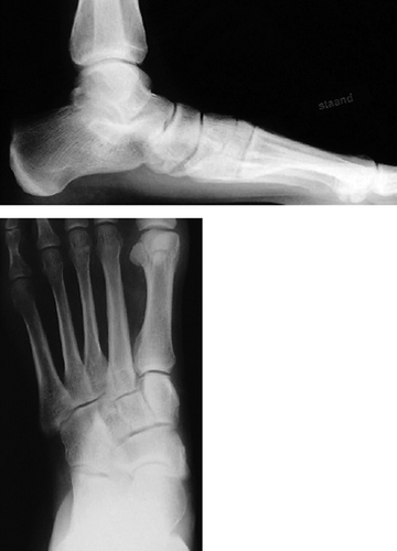
Different pathophysiological processes can cause flatfoot. In this study, we focused on those feet where the primary changes to the medial longitudinal foot arch occur at the level of the talonavicular joint. Here, dysfunction of the posterior tibial tendon seems to be important (Myerson Citation1996) andeventually leads to collapse of the medial arch, abduction and supination of the forefoot in relation to the hindfoot, and a valgus hindfoot (Myerson Citation1996, Gould Citation1997, Hintermann et al. Citation1999, Pomeroy et al. Citation1999, Toolan et al. Citation1999). Eventually, a marked subluxation in the tarsal joints can occur, leading to distinct radiographic changes with malalignment of the medial column of the foot, and dorsolateral translation or subluxation of the navicular relative to the talus with uncovering of the talar head. The matching radiographic changes have led to the introduction of the term peritalar dorsolateral subluxation of the foot (PTDLS) (Ananthakrisnan et al. Citation1999, Toolan et al. Citation1999).
Numerous procedures have been devised for the operative treatment of acquired flatfoot: tendon transfers, osteotomies, selective arthrodesis (performed independently or in combination), and finally, triple arthrodesis (Myerson Citation1996, Pomeroy et al. Citation1999, Toolan et al. Citation1999, Kitaoka et al. Citation2000, Kelly et al. Citation2001, Mosier-LaClair et al. Citation2001, Weinfeld Citation2001). Currently, triple arthrodesis is advocated for patients with a fixed deformity or painful degenerative arthrosis of the hindfoot joints (Myerson Citation1996, Flamme et al. Citation1997, Toolan et al. Citation1999, Kelly and Easley Citation2001). However, no consensus exists regarding the surgical treatment of the flexible acquired flatfoot deformity. Several techniques have been described that have the common goal of treating the osseous and soft-tissue derangements contributing to the PTDLS, without sacrificing the talonavicular joint. Thus, as much motion as possible is preserved in order to maintain mobility and postpone late onset arthrosis.
These procedures are reported to give satisfactory short-term results. The choice of procedure depends on the clinical presentation, age and weight of the patient, and the intensity of the patient's activities (Myerson Citation1996, Pomeroy et al. Citation1999, Toolan et al. Citation1999, Kitaoka et al. Citation2000). Lateral column lengthening has been advocated by several investigators, because it restores the medial arch and reduces the peritalar dorsolateral subluxation (Evans Citation1975, Mosca Citation1995, Hintermann et al. Citation1999, Toolan et al. Citation1999, Kitaoka et al. Citation2000, Mosier-LaClair et al. Citation2001). It is not yet clear whether the lengthening should be performed through the anterior calcaneus, as described by Evans (Citation1975) and Mosca (Citation1995), or by calcaneo-cuboid distraction arthrodesis as popularized by Hansen (Toolan et al. Citation1999).
We report the outcome of 20 feet treated by combined lateral column lengthening through calcaneo-cuboid distraction arthrodesis, percutaneous Achilles tendon lengthening and augmentation of the posterior tibial muscle with the flexor digitorum longus muscle.
Patients and methods
Study cohort
From February 1999 through March 2001, we treated 20 patients (22 feet) with symptomatic, acquired flatfoot and radiographic PTDLS by calcaneo-cuboid distraction arthrodesis to lengthen the lateral column, percutaneous Achilles tendon lengthening and augmentation of the posterior tibial tendon by flexor digitorum longus (FDL) transfer. 2 patients were operated on both feet, and for statistical reasons only the first foot operated on was included in the study. In 8 patients, arthrodesis of the first tarsometatarsal joint was performed in addition in order to correct fixed forefoot supination, hallux valgus, and/or symptomatic arthrosis. In 2 of these patients, the arthrodesis was extended to the naviculocuneiform joint. The mean age at surgery was 55 (30–66) years and 16 patients were women. No patient had systemic rheumatoid disease or systemic ligamentous laxity. The symptoms had existed at least 2 years before surgery. 13 patients used inlays and 4 patients wore custom-made shoes.
Pre- and postoperative evaluations
Preoperatively, all patients complained of marked pain in the medial and/or lateral hindfoot, with walking possible for only 15–30 min. On preoperative physical examination, patients exhibited a typical dorsolateral subluxation of the forefoot in relation to the hindfoot, resulting in increased valgus position of the hindfoot, and a positive ‘too many toes sign’, in comparison with the asymptomatic (or less symptomatic) contralateral foot. Typically, the patients were not able to perform the single heel rise test on the symptomatic foot due to insufficiency and/or pain of the posterior tibial tendon. In all cases, complete manual correction of the hindfoot valgus deformity and PTDLS was easily possible, thus assuring that the patient had a flexible deformity. The Clinical Rating Index hindfoot score (CRI) for the ankle-hindfoot (American Orthopedic Foot and Ankle Society; Kitaoka et al. Citation1994) was assessed. The score ranges from 0 to 100, and the higher the score the less pain and disability. The Dutch Foot Function Index with verbal rating scales (FFI-5pt) was also assessed. This is a generic self-administered instrument with good test-retest reliability for measurement of the effect of foot complaints on foot function (Kuyvenhoven et al. Citation2002). The FFI-5pt consists of 15 items divided into two subscales: pain and disability. The limitation scale, as used in the original FFI introduced by Budiman-Mak et al. (Citation1991), was excluded—as advised by Kuyvenhoven et al. (Citation2002). Similar pain and disability scales were used by Domsic and Saltzman (Citation1998) in their study assessing patients with ankle osteoarthrosis, and by Saag et al. (Citation1996) in their study on rheumatoid arthritic pain. The scores range from 0 to 100; this time, the higher the score, the more pain/disability is present. The total score is the mean of the two subscale scores.
All patients were evaluated postoperatively by one examiner (vdK). Mean follow-up time was 25 (13–39) months. The Dutch FFI and the CRI were completed. The patients were asked to comment specifically on their overall satisfaction concerning the result of the operation, and on their willingness to undergo the procedure again given similar circumstances. The position of the hindfoot and forefoot, the motion of the ankle and hindfoot joints, the single heel rise test and the status of the sural nerve were recorded. The site from which the bone graft from the iliac crest had been obtained was assessed for tenderness and wound healing.
Overall results, CRI, FFI scores and radiographic results preoperatively and at follow-up of calcaneocuboid distraction arthrodesis
Radiographic analysis
Standardized weight-bearing radiographs were taken, preoperatively and at follow-up. On the lateral view, the sagittal talometatarsal angle (angle between the talar axis and the axis of the first metatarsal) and on the AP view the dorsoplantar talometatarsal angle were measured, as described by Toolan et al. (Citation1999) The distance of the plantar aspect of the navicular bone to the weight-bearing surface was measured. Radiographs taken routinely 8 weeks and 20 weeks after surgery, were used to evaluate and confirm union of the arthrodesis.
Operative technique, postoperative care
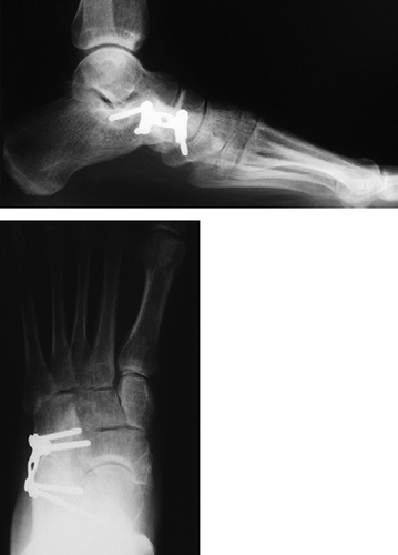
The surgical technique used in all cases has been extensively described by Toolan et al. (Citation1999). The calcaneo-cuboid joint was exposed through a longitudinal dorsolateral incision. The peroneal tendons and the sural nerve were retracted and the muscle belly of the extensor digitorum brevis was elevated dorsomedially. Accurate distraction between the calcaneus and the cuboid bone was facilitated by using a small laminaspreader or the closed-arms small Hintermann distractor (Newdeal). Distraction varied from 0.8 to 1.0 cm, which reduced the talo-navicular subluxation sufficiently, and thus, also returned the calcaneus to a neutral, slight valgus, position in relation to the lower limb. After removal of the cartilage, a tricortical iliac crest block graft was inserted. The distraction arthrodesis was internally fixated with a cervical H-plate (Synthes), secured by 2 proximal and 2 distal cortical screws ( and ). Flexor digitorum longus transfer to the posterior tibial tendon was performed routinely as described by Toolan et al. (Citation1999). In all cases, an equinus or clear loss of dorsiflexion of the ankle joint was encountered due to tightness of the m. triceps surae after lengthening between calcaneus and cuboid. A percutaneous Achilles tendon lengthening was therefore performed in all patients. In eight feet, an additional arthrodesis of the first tarsometatarsal joint (TMT1) was performed. If an elevated first ray (fixed supination of the forefoot) persisted, a plantarflecting correction arthrodesis of TMT1 was performed. TMT1 fusion was also performed in order to correct a hallux valgus deformity through correction of an increased first inter-metatarsal angle and a distal soft tissue procedure of the MP1 joint.
While lengthening the lateral column, attention was paid to maintenance or correction of the position of the forefoot in relation to the hindfoot. In order to achieve this, the foot is kept in a neutral position, counteracting the tendency to supinate during distraction by applying a careful pronating force. In one patient, a tri-calcium-phosphate block was used instead of an autologous bone graft from the anterior iliac crest.
All feet were immobilized postoperatively with a non-weight-bearing cast for 4 weeks with the foot in neutral position, followed by a weight-bearing cast for 4 weeks and a Camwalker for another 4 weeks. After 12 weeks, patients started to wean themselves off the Camwalker and to wear normal shoes as tolerated. At the 5-month follow-up, orthotics or adaptation of footwear were prescribed where necessary.
Statistics
Clinical and radiographic measurements were compared before and after the operation using the dependent paired Student’s t-test with a level of significance of < 0.05.
Results
Clinical evaluation
17 patients had complete pain relief, or only minor complaints. 2 patients had moderate complaints, and 1 patient had no pain relief. The patients with moderate complaints had discomfort over the arthrodesis site and/or cuboid-MT5 (TMT5) joint. In these patients the hardware was removed, which did not lead to reduction of the symptoms. The patient with no pain relief had diffuse complaints in the whole foot without a specific site or joint as the cause. 5 patients complained of discomfort while walking on uneven surfaces. 13 patients used inlays and/or shoe adjustments postoperatively (Table). The mean FFI score dropped from 49 (SD 14) before surgery to 22 (SD 15) at followup (p < 0.001). The mean CRI score improved from 46 (SD 13) before surgery to 79 (SD 14) at follow-up (Table).
The overall patient satisfaction was excellent in 10 cases, good in 5 cases, fair in 4 cases and poor in 1 case (Table). 17 patients reported that the procedure had resulted in an increase in their daily activities, and 10 patients described an increase regarding recreational activities as well. 18 patients had been employed before the operation and had had difficulty in coping with the workload, and 17 of them returned to work postoperatively. 12 patients reported an improvement in their ability to cope with their workload after the surgery. The person who did not return to work was the one reporting the poor result, and had been on long-term sick leave before the procedure.
The conformation of the foot, as subjectively reported by the patients themselves, improved in all cases but 1. In that case, the patient found the appearance of the foot to be unchanged. None of the patients needed to use a cane or other walking aid. All patients but 1 stated that they would choose to undergo the procedure again given the same circumstances.
Complications and subsequent procedures
2 feet had a nonunion that necessitated a reoperation with bone grafting. In one of these cases (a man weighing 110 kg), a tri-calciumphosphate block was used instead of a iliac crest bone graft. Both nonunions united within 3 months after the grafting procedure. The reoperation wound at the iliac crest site of the heavy patient mentioned above became infected, but healed after treatment with antibiotics.
3 patients had either paresthesia or anesthesia in the distribution of the sural nerve. In 5 patients, the H-plate was removed after union of the distraction arthrodesis because of local discomfort of the hardware. This group included the patients with persistent pain/discomfort along the lateral column after the lengthening procedure.
Radiographic analysis
The talo-first-metatarsal angle on the AP view was corrected from 15° (SD 8.7) preoperatively to 4.1° (SD 3.8) at follow-up, and on the lateral view from 14.2° (SD 7.1) preoperatively to 3.9° (SD 3.2) at follow-up (Table), and improvements were statistically significant (p < 0.001). The distance of the navicular to the ground on the lateral full weight-bearing radiograph improved from 1.9 (SD 0.6) cm preoperatively to 3.0 (SD 0.4) cm at follow-up, confirming a restoration of the medial arch (p < 0.001) (Table).16 feet united within 3 months, 1 foot united in 4 months and 1 in 5 months. 2 patients developed a nonunion, as previously described. None of the radiographs showed degenerative changes or signs of impingement in the TMT5 joint at follow-up.
Discussion
In this study, lengthening of the lateral column by a calcaneo-cuboid distraction arthrodesis with augmentation of the posterior tibial tendon and Achilles tendon lengthening has been found to be an effective method for the correction of an acquired flexible flatfoot deformity. The rate of complications and additional operations was less than reported by Toolan et al. (Citation1999), who have propagated this method, and thus quite acceptable. This improvement may be because of use of an H-plate for the fixation of the distraction arthrodesis, as later advised by the same authors, instead of crossed screws as used in nearly all the patients in their study (Toolan et al. Citation1999).
Hintermann et al. (Citation1999) reported the results of lengthening the lateral column by an osteotomy in the anterior calcaneal tuberosity, a modification of the ‘classic’Evans (Citation1975) method, and reconstructing the medial soft tissues in 19 patients. The patient group in their study seems to have been similar to ours. They used the AOFAS hindfoot score and talometatarsal angles as parameters. Generally, their data show a similar distribution range, but a lower standard deviation. Both the preoperative and postoperative functional scores are higher in their study, while the talometatarsal angles are less favorable both preoperatively and postoperatively. In all, their results seem more satisfying, as they report a postoperative AOFAS score of 90 and higher in 13 out of 19 patients, which was found in only 5 of 20 feet in our study. Although a secondary calcaneo-cuboid fusion was necessary after 5 months, 1 patient had pain in the ankle and the subtalar joint, 1 patient had discomfort at the calcaneo-cuboid joint and 5 patients had some difficulty in walking on uneven terrain, the patients in the Hitermann study generally reported a higher functional level. We think this may be ascribed to the possibility that more movement of the tarsal joints is maintained after calcaneal osteotomy than after calcaneo-cuboid fusion.
In our study, 3 patients complained of moderate postoperative pain along the lateral aspect of the foot, primarily at the level of the TMT5 joint, and 5 patients reported discomfort while walking on an uneven surface. 13 of our patients used orthopedic shoes and/or inlays at follow-up, while in the Hintermann study only 2 patients required some corrections in fashionable shoes. The easy access to orthotic devices in the Netherlands cannot fully explain this difference. The complaints of lateral discomfort and pain seem to be an important drawback of lateral column lengthening. They are probably due to increased mechanical stress in the adjacent joints after calcaneo-cuboid arthrodesis, partly as result of increased pressure in the TMT5 joint due the lengthening of the lateral column (Cooper et al. Citation1997, Sands et al. Citation1998), and partly because of increase and/or change of movement of the adjacent joints.
A lengthening of around 1 cm appears to have given optimal correction in the present series of patients, which is similar to the findings of others (Toolan et al. Citation1999, Kitaoka et al. Citation2000). However, exact anatomical reduction of the talocalcaneal and talonavicular joints is not controlled by lateral column lengthening. Postoperative subluxation of these joints as a result of under- or overcorrection could explain postoperative discomfort and loss of motion. Sands et al. (Citation1998) showed that the post-operative motion in the subtalar joint is strongly influenced by the position of the foot when the calcaneo-cuboid joint is fused. Only if the foot is maintained in a neutral position does the hindfoot mobility remain unchanged. Unfortunately, we were not aware of this in the period the patients were operated on, and thus no information was collected to make it possible to argue that this influenced our results.
Several other methods, preserving as much movement as possible of the talonavicular joint in order to correct this type of flexible, degenerative flatfoot are reported to provide good results. These include calcaneal osteotomy with medial displacement (Sammarco and Hockenbury Citation2001, Wacker et al. Citation2002), double osteotomy (Deland et al. Citation1999, Moseir-LaClair et al. 2001) and isolated subtalar fusion with correction of the valgus (Graves et al. Citation1997, Mangone et al. Citation1997, Mann et al. Citation1998). These procedures are most often combined with reconstruction of the medial soft tissues. A 100% rate of union and low complication rates have been reported when a subtalar fusion was performed (Graves et al. Citation1997, Mangone et al. Citation1997, Mann et al. Citation1998). The subtalar fusion causes minimal alterations of gait on flat surfaces and can lead to long-term maintenance of hindfoot alignment. An improvement of the AOFAS hindfoot score from 48 before operation to 84 at follow-up was found in 32 patients treated with medial displacement osteotomy and FDL transfer (Myerson et al. Citation1996). Wacker et al. (Citation2002) reported that 43 of 44 patients had excellent or good outcome regarding pain and 36 had good alignment, using the same procedure. Guyton et al. (Citation2001) reviewed the results of 26 patients who had undergone this procedure, also for stage 2 (Johnson and Strom Citation1989) posterior tendon dysfunction, at an average of 32 months and concluded that this procedure provides good medium-term functional and symptomatic results. However, the radiographic improvement was frequently only moderate and only half of the patients felt that the conformation of their foot had changed to any noticeable extent.
We conclude that lateral column lengthening through a calcaneo-cuboid distraction arthrodesis with augmentation of the posterior tibial tendon and percutaneous Achilles tendon lengthening is an effective method for correction of an acquired flexible flatfoot deformity (PTDLS).
No competing interests declared.
- Ananthakrisnan D, Ching R, Tencer A, Hansen S T, Sangeorzan B J. Subluxation of the talonavicular joint in adults who have symptomatic flatfoot. J Bone Joint Surg (Am) 1999; 81: 1147–54
- Budiman-Mak E, Conrad K J, Roach K E. The Foot Function Index: a measure of foot pain and disability. J Clin Epidemiol 1991; 44: 561–70
- Cooper P S, Nowak M D, Shaer J. Calcaneo-cuboid joint pressures with lateral column lengthening (Evans) procedure. Foot Ankle Int 1997; 18: 199–205
- Deland J T, Page A E, Kenneally S M. Posterior calcaneal osteotomy with wedge: cadaver testing of a new procedure for insuffiency of the posterior tibial tendon. Foot Ankle Int 1999; 20: 290–5
- Domsic R T, Saltzman C L. Ankle osteoarthritis scale. Foot Ankle Int 1998; 19: 466–71
- Evans D. Calcaneo-valgus deformity. J Bone Joint Surg (Br) 1975; 57: 270–8
- Flamme C H, Wulker N, Muller A, Wirth C J. Long-term follow-up after arthrodesis of the ankle and the hindfoot. Foot Ankle Surg 1997; 3: 21–8
- Gould J S. Direct repair of the posterior tibial tendon. Foot Ankle Clin 1997; 2: 275–9
- Graves S C, Stephenson K A. The use of subtalar and triple arthrodesis in the treatment of posterior tibial tendon dys-function. Foot Ankle Clin 1997; 2: 19–328
- Guyton G P, Jeng C, Krieger L E, Mann R A. Flexor digitorum longus transfer and medial displacement calcaneal osteotomy for posterior tibial tendon dysfunction: a middle-term clinical follow-up. Foot Ankle Int 2001; 22: 627–32
- Hintermann B, Valderrabano V, Kundert H-P. Lengthening of the lateral collumn and reconstruction of the medial soft tissue for treatment of acquired flatfoot deformity associated with insufficiency of the posterior tibial tendon. Foot Ankle Int 1999; 20: 622–9
- Johnson K A, Strom D E. Tibialis posterior tendon dysfunction. Clin Orthop 1989, 239: 196–206
- Kelly I P, Easley M E. Treatment of stage 3 adult acquired flatfoot. Foot Ankle Clin 2001; 6: 153–66
- Kitaoka H B, Alexander I J, Adelaar R S, Nunley J A, Myerson M S, Sanders M. Clinical rating systems for the anklehindfoot, midfoot, hallux and lesser toes. Foot Ankle Int 1994; 15: 349–53
- Kitaoka H B, Kura H, Luo Z-P, An K-A. Calceocuboid distraction arthrodesis for posterior tibial tendon dysfunction and flatfoot. Clin Orthop 2000, 381: 241–7
- Kuyvenhoven M M, Gorter K J, Zuithoff P, Budiman-Mak E, Conrad K J, Post M W. M. The Foot Function Index with verbal rating scales (FFI-5pt): A clinimetric evaluation and comparison with the original FFI. J Rheumatol 2002; 29: 1023–8
- Mangone H B, Fleming L L, Fleming S S. Treatment of acquired adult planovalgus deformities with subtalar fusion. Clin Orthop 1997, 345 106–12
- Mann R A, Beaman D N, Horton G A. Isolated subtalar arthrodesis. Foot Ankle Int 1998; 19: 511–9
- Mosca V S. Calcaneal lengthening for valgus deformity of the hindfoot. J Bone Joint Surg (Am) 1995; 77: 500–12
- Mosier-LaClair S, Pomeroy G, Manoli A. Operative treatment of the difficult stage 2 adult acquired flatfoot deformity. Foot Ankle Clin 2001; 6: 95–119
- Myerson M S. Adult acquired flatfoot deformity. J Bone Joint Surg (A) 1996; 78: 780–92
- Pomeroy G C, Pike H, Beals T C, Manoli A. Acquired flatfoot in adults due to dysfunction of the posterior tibial tendon. J Bone Joint Surg (Am) 1999; 81: 1173–82
- Saag K G, Saltzman C L, Brown K, Budiman-Mak E. The Foot Function Index for measuring rheumatoid arthritis pain: evaluating side-to-side reliability. Foot Ankle Int 1996; 17: 506–10
- Sammarco G J, Hockenbury R T. Treatment of stage II posterior tibial tendon dysfunction with flexor hallucis longus transfer and medial displacement calcaneal osteotomy. Foot Ankle Int 2001; 22: 305–12
- Sands A, Early J, Harrington R M, Tencer A F, Ching R P, Sangeorzan B J. Effect of variations in calcaneo-cuboid fusion technique on kinematics of the normal hindfoot. Foot Ankle Int 1998; 19: 19–25
- Toolan B C, Sangeorzan B J, Hansen S T. Complex reconstruction for the treatment of dorsolateral peritalar subluxation of the foot. J Bone Joint Surg (Am) 1999; 81: 1545–60
- Wacker J T, Hennessy M S, Saxby T S. Calcaneal osteotomy and transfer of the tendon of flexor digitorum longus for stage-II dysfunction of tibialis posterior. Three- to five-year results. J Bone Joint Surg (Am) 2002; 84: 54–8
- Weinfeld S B. Medial slide calcaneal osteotomy. Technique, patient selection, and results. Foot Ankle Clin 2001; 6: 89–94


