A 12-year-old boy was referred from the pediatric outpatient department because of a mass in his right hand, at the dorsal aspect of the first web space. The boy could not remember when he first observed the mass but his mother reported that swelling of the area had been present for at least 3 years. The mass had gradually increased in size. The boy had no pain or any other symptoms, and used his hand normally. He denied any trauma to the hand. Apart from the swelling in the hand, the boy was completely healthy.
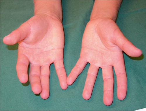
There was an obvious swelling at the area between the first and second metacarpal bones dorsally (). Palpation of the area showed a generalized swelling across the first web space dorsally. The swelling was soft to firm on palpation, but was not tender. The overlying skin was normal. A slight swelling of the distal aspect of the thenar region was also observed (). Range of motion, muscle strength and sensation were normal. Blood tests including erythrocyte sedimentation rate, Creactive protein, hemoglobin, and white blood cell count were normal.
Figure 1. Swelling of the first dorsal web space of the right hand.
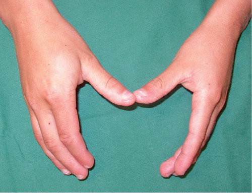
Figure 2. Slight swelling of the thenar region of the right hand.
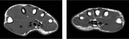
MRI with T1- and T2-weighted images in the axial and sagittal planes revealed enlarged thenar and first dorsal interosseous muscles with normal signal intensity, and no mass (). The hypertrophied muscles had normal appearance. After an interval of 5 weeks, a repeat MRI including intravenous contrast administration gave similar findings. A third MRI of both hands 6 months later showed no substantial changes in the muscle hypertrophy.
Figure 3. MRI of the right and left hands (T1-weighted images) showing hypertrophied (but otherwise normal-looking) first dorsal interosseous and thenar muscles in the right hand.
At follow-up 9 months after the boy was first seen, the swelling was unchanged and the boy was still asymptomatic.
Discussion
To our knowledge, this is the first case report of first dorsal interosseous muscle hypertrophy presenting as a tumor in a child. We could find only 2 previous reports of idiopathic enlargement of the first dorsal interosseous muscle. In one report, 2 adults who had presented with swelling in the thenar region were found on CT scan to have idiopathic thenar muscle hypertrophy (Clay and Austin Citation1988). In one of the patients, a 25-year-old man, enlargement of mainly the first dorsal interosseous muscle was shown on CT and surgical exploration revealed a grossly enlarged muscle, which was judged—based on biopsy—to be simple physiological hypertrophy. At a 7-year follow-up examination, the patient had slightly increased swelling but unchanged symptoms. No surgery was done on the second patient. In another report, a 49-year-old woman and a 38-year-old man presented with a mass in the first dorsal web space, which appears to have been similar to our case (Peh et al. Citation1999). In both patients, MRI showed a hypertrophic first dorsal interosseous muscle with normal T1 and T2 signal characteristics. Surgery was not performed in any of these patients. In another case report of idiopathic thenar hypertrophy, a 16-year-old girl presented with enlarged thenar eminence and surgery revealed a markedly hypertrophied and an accessory abductor pollicis brevis muscle (Legan et al. Citation1992).
Anomalous muscles presenting as tumors in the hand have been reported frequently, and MRI has been used successfully for the diagnosis (Coenen and Biltjes Citation1991, Sanger et al. Citation1991, Anderson et al. Citation1995, Honing et al. Citation1995, Kostakoglu et al. Citation1997).
A rare case of idiopathic muscle hypertrophy in the foot has also been reported. A child presented with a mass that was shown by MRI to be congenital hypertrophy of the abductor hallicus brevis muscle (Ringelman and Goldberg Citation1993).
- Anderson M W, Benedetti P, Walter J, Steinberg D R. MR appearance of the extensor digitorum manus brevis muscle: a pseudotumor of the hand. AJR Am J Roentgenol 1995; 164: 1477–9
- Clay N R, Austin S. Idiopathic thenar muscle hypertrophy. J Hand Surg (Br) 1988; 13: 100–1
- Coenen L, Biltjes I. Pseudotumor of the palm due to an anomalous flexor digitorum superficialis muscle belly. J Hand Surg (Am) 1991; 16: 1046–51
- Honing M L, Ritt M J, Bos K E. An anomalous flexor digitorum superficialis to the index finger. Surg Radiol Anat 1995; 17: 339–41
- Kostakoglu N, Borman H, Kecik A. Anomalous flexor digitorum superficialis muscle belly: an unusual case of mass in the palm. Br J Plast Surg 1997; 50: 654–6
- Legan J, Shepler T R, Wind G. Congenital hypertrophy of the thenar eminence with accessory head of the abductor pollicis brevis in the forearm. J Hand Surg (Am) 1992; 17: 884–6
- Peh W C, Ip W Y, Wong L L. Diagnosis of dorsal interosseous pseudotumours by magnetic resonance imaging. Australas Radiol 1999; 43: 394–6
- Ringelman P R, Goldberg N H. Hypertrophy of the abductor hallicus muscle: an unusual congenital foot mass. Foot Ankle 1993; 14: 366–9
- Sanger J R, Krasniak C L, Matloub H S, Yousif N J, Kneeland J B. Diagnosis of an anomalous superficialis muscle in the palm by magnetic resonance imaging. J Hand Surg (Am) 1991; 16: 98–101