Abstract
Background Blind injection of the subacromial-sub-deltoid bursa (SSB) for diagnostic purposes (Neer test) or therapeutic purposes (corticosteroid therapy) is frequently used. Poor response to previous blind injection or side effects may be due to a misplaced injection. It is assumed that ultrasound (US)-guided injections are more accurate than blind injections. In a randomized study, we compared the accuracy of blind injection to that of US-guided injection into the SSB.
Patients and methods 20 consecutive patients with impingement syndrome of the shoulder were randomized for blind or US-guided injection in the SSB. Injection was performed either by an experienced orthopedic surgeon or by an experienced musculoskeletal radiologist. A mixture of 1 m’L methylprednisolone acetate, 4 mL prilocaine hydrochloride and 0.02 mL (0.01 mmol) Gadolinium DTPA was injected. Immediately after injection, a 3D-gradient T1-weighted magnetic resonance scan of the shoulder was performed. The location of the injected fluid was independently assessed by 2 radiologists who were blinded as to the injection technique used.
Results The accuracy of blind and US-guided injection was the same. The fluid was injected into the bursa in all cases.
Interpretation Blind injection into the SSB is as reliable as US-guided injection and could therefore be used in daily routine. US-guided injections may offer a useful alternative in difficult cases, such as with changed anatomy postoperatively or when there is no effective clinical outcome.
Impingement of the rotator cuff and the subacro-mial-subdeltoid bursa (SSB) beneath the coracoacromial arch without associated rupture of the cuff is a well-established clinical diagnosis. Subacromial injection of corticosteroids is an effective therapy for symptomatic subacromial impingement (Petri et al. Citation1987, Blair et al. Citation1996).
Poor response to blind injection or side effects may be due to a misplaced injection (Esenyel et al. Citation2003). Potential side effects are septic arthritis, necrotizing fasciitis, a deleterious effect on intraarticular cartilage, or tendon degeneration, which may lead to late rupture of the rotator cuff and subcutaneous atrophy (Kennnedy and WillisCitation1976, Rostron and Calver Citation1979, Karpman et al. Citation1980, Birkinshaw et al. Citation1997).
Some studies have suggested that blind subacromial injection may be difficult. The incidence of injections that miss the subacromial bursa has ranged from 13% to 71% (Eustace and Brophy Citation1997, Yamakado Citation2002, Esenyel et al. Citation2003, Naredo et al. Citation2004).
Image-guided interventions—especially those using ultrasound (US)—are being increasingly used, as they can be performed quickly and reliably (Cardinal et al. Citation1998, Fessell et al. Citation2000). We hypothesized that US-guided injection of the sub-acromial-subdeltoid bursa is more accurate than blind injection and thus, in the long term, probably more successful than blind injection. There have been some studies describing the accuracy of blind injection into the SSB; the location of the injected fluid was recorded by plain radiology (Eustace and Brophy Citation1997, Yamakado Citation2002) or by ultrasound (Naredo et al. Citation2004). To our knowledge, there have been no studies that have evaluated the accuracy of US-guided bursal injections with magnetic resonance imaging (MRI). MRI provides an objective 3-dimensional view of the bursa and shoulder joint, and enables differentiation between the pre-exist-ing fluid and the injected fluid, because it contains gadolinium, which provides brighter signal intensity at T1-weighted MR images.
Using a randomized study, we compared the accuracy of blind subacromial injections with that of corresponding US-guided injections.
Patients and methods
Patients
60 consecutive patients with impingement of the shoulder of at least 2 months’ duration were considered for inclusion. They were treated by deposition of corticosteroids in the subacromial-subdel-toid bursa.
All patients were studied with ultrasound prior to injection. Patients with fluid in the bursa before injection (n = 2) or previously treated with corticosteroid injections or surgery were eliminated from the study. Patients were included when they had painful restriction of their glenohumeral mobility, had a sonographically and/or MRI-proven intact rotator cuff, and were between 18 and 65 years of age. At that time, intrabursal deposition of gadolinium was not approved officially; thus, we obtained approval from the medical ethical committee of our hospital and received written informed consent from all patients.
One of the premises of this study was that sonographic injection should improve the success rate of injection by 15%. However, because the results after 20 patients indicated that the chance of a relevant improvement would be less than 1% after 40 patients and less than 3% after 60, the study was stopped after the first 20 patients.
Randomization
20 consecutive patients were allocated to either blind or US-guided corticosteroid injection therapy by being randomly assigned a number from 1 to 20 just before injection. Patients for whom an uneven number was drawn underwent US-guided injection and those for whom an even number was drawn underwent blind injection (Table).
Study groups after random allocation to either blind or US-guided corticosteroid injection
Injection technique
The injection fluid contained 1 mL methylprednisolone acetate at 40 mg/mL (Depo-Medrol), 4 mL prilocaïnehydrochloride at 10 mg/mL (Citanest 1%), and 0.02 mL (0.01 mmol) Gadolinium DTPA (Magnevist).
All blind injections were performed by an experienced orthopedic surgeon (MWM). A commonly used puncture technique was performed. Making use of external anatomical landmarks for orientation, a lateral posterior approach (Rowe Citation1988) with a 21-gauge (0.8 × 50 mm) needle was used.
US-guided injections were performed by one radiologist (MR) using a free-hand technique. Patients lay supine on an examination table or sat upright face to face with the radiologist, with the ipsilateral arm in hyperextension and endorotation longitudinal supraspinatus view). Sonographic examinations were performed with a Toshiba Power Vision using a 7.5–14-MHz linear array transducer. Transducer and skin were disinfected with 70% ethanol and sterile gel was applied. A 21gauge needle was inserted parallel to the transducer in a semi-oblique plane from the anterior side of the shoulder. The needle was advanced under realtime US control until the needle tip entered the bursa (). To optimize visualization of the tip, it was inserted with the bevelled side facing the transducer. Rotation of the bevel of the needle 180° to the side of the rotator cuff after entering the bursa facilitated intrabursal injection. The position of the tip of the needle was also verified by the fact that touching the synovial lined bursa generates pain. Needle placement and intrabursal injection took an average of 5 min for both techniques.
Figure 1A. Ultrasonographic image showing the supraspinatus tendon (SSP) in the longitudinal plane. The advancing needle (arrows) under real-time ultrasound guidance has entered the subacromial-subdeltoid bursa (between arrowheads). D: deltoid muscle; H: humeral head.
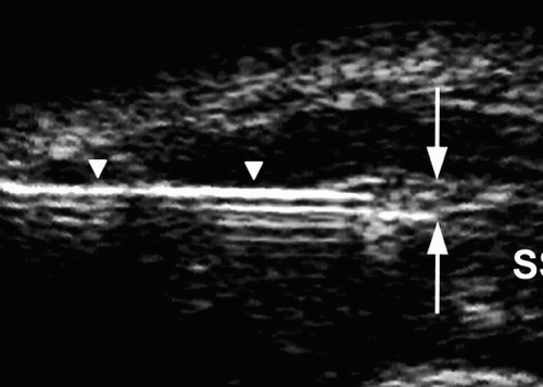
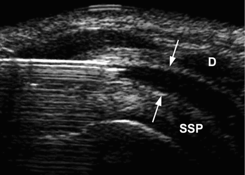
Figure 1B. Ultrasonographic image after injection of about 5 ml of fluid (arrows) into the subacromial-subdeltoid bursa. D: deltoid muscle; SSP: supraspinatus tendon.
Assessment of accuracy
MRI shows up intraarticular and periarticular fluid, soft tissues and bony structures with a high spatial resolution in 3 dimensions, and was used for evaluation () (Erickson Citation1997). All patients underwent MR imaging of the shoulder immediately after injection. Examinations were performed on a 1.5-T MRI scanner. 3D-gradient T1-weighted (FLASH 3D) oblique coronal scans were obtained and images were reconstructed in the sagittal and axial planes, and if indicated, in any other desired plane. FLASH 3D imaging (TR: 8.1 ms; TE: 4 ms; 2 acquisitions; 192 × 256 matrix; FOV: 24 cm) with 1-mm consecutive slices was used. Overall scan time was 4 min 30 sec.
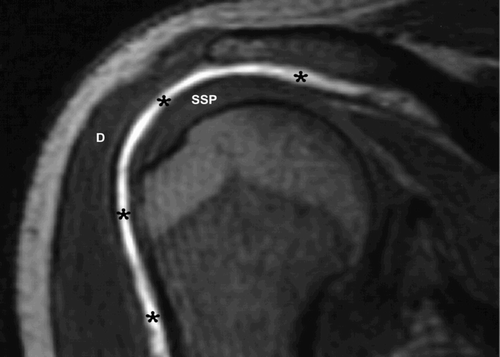
Figure 2. Semi-coronal gradient T1-weighted (FLASH 3D) MR image of the shoulder after injection of 5 mL fluid (*) into the subacromial-subdeltoid bursa. D: deltoid muscle; SSP: supraspinatus tendon.
The accuracy of the injection was assessed by 2 radiologists (BM and GJ) independently. They were blinded as to the puncture technique used. The injection fluid contains an MRI contrast agent (Gadolinium DTPA) which shortens the T1 relaxation time. This increases the signal intensity of the injected fluid on T1-weighted images, thus facilitating assessment of the precise location of the injected fluid and differentiation from other collections of fluid.
Results
With both injection techniques, the procedure was quick (average 5 min) and relatively painless. In all patients, the fluid was correctly injected into the subacromial-subdeltoid bursa. No complications or side effects occurred. In 4 patients, some leakage of fluid along the puncture canal in the deltoid muscle could be seen. 2 of them had been punctured blind and 2 had been punctured US-guided.
Discussion
Subacromial injection of corticosteroids is common. However, corticosteroid injections have some potential side effects and not all patients respond well to this therapy (Kennnedy and Willis Citation1976, Rostron and Calver Citation1979). One reason may be injection failure. In different reports, the incidence of blind subacromial injection failure has ranged from 13 to 70% (Eustace and Brophy Citation1997, Yamakado Citation2002, Esenyel et al. Citation2003, Naredo et al. Citation2004).
It is thus interesting to know how accurately USguided subacromial injections can be performed. One study involving US-guided injection has been published; however, accuracy was determined using ultrasound (Naredo et al. Citation2004). To our knowledge, there have been no studies that have objectively evaluated the targeting accuracy of USguided injections in the subacromial-subdeltoid bursa. We used MRI, because it provides an objective 3-dimensional view of the bursa and shoulder joint and enables differentiation between pre-exist-ing fluid and injected fluid, by using contrast material (gadolineum). This MRI contrast agent provides brighter signal intensity in T1-weighted MR images related to pre-existing fluid.
US-guided interventions in general, and in the shoulder joint in particular, are accurate and may reduce unnecessary and potentially traumatic or unsuccessful attempts at intervention (Cicak et al. Citation1992, Valls and Melloni Citation1997, Cardinal et al. Citation1998, Fessell et al. Citation2000). In addition, US is readily available and can be performed safely, quickly, and at low cost.
In our 20 patients, all injections using both techniques were correctly placed in the bursa. The accuracy of blind injection by one experienced orthopedic surgeon was higher than that reported in the literature (Eustace and Brophy Citation1997, Yamakado Citation2002, Esenyel et al. Citation2003, Naredo et al. Citation2004). Although this is a small group of patients, we conclude that both techniques are equally accurate and thus that blind injections should be the technique of first choice.
Contributions of authors
MR, GJ: guarantors of integrity of the entire study. All authors: conception and design of the study, data acquisition and analysis, drafting of manuscript and revision. MR, GJ, MWM: clinical studies. MR, GJ: statistical analysis.
- Birkinshaw R, O'Donnell J, Sammy I. Necrotising fasciitis as a complication of steroid injection. J Accid Emerg Med 1997; 14: 52–4
- Blair B, Rokito A S., Jarolem K, Zuckerman J D. Efficacy of injections of corticosteroids for subacromial impingement syndrome. J Bone Joint Surg (Am) 1996; 78: 1685–9
- Cardinal E, Chhem R K, Beauregard C G. Ultrasound-guided interventional procedures in the muskuloskeletal system. Radiol Clin North Am 1998; 36: 597–604
- Cicak N, Matasovic T, Bajraktarevic T. Ultrasonographic guidance of needle placement for shoulder arthrography. J Ultrasound Med 1992; 11: 135–7
- Erickson S J. High-resolution imaging of the musculoskeletal system. Radiology 1997; 205: 593–618
- Esenyel C Z, Esenyel M, Yesiltepe R, Ayanoglu S, Bulbul M, Sirvanci M, Kara A N. The correlation between the accuracy of steroid injections and subsequent shoulder pain and function in subacromial impingement syndrome. Acta Orthop Traumatol Turc 2003; 37: 41–5
- Eustace J A, Brophy D P. Comparison of the accuracy of steroid placement with clinical outcome in patients with shoulder symptoms. Ann Rheum Dis 1997; 56: 59–63
- Fessell D P, Jacobson J A, Craig J, Habra G, Prasad A, Radliff A, van Holsbeeck M T. Using sonography to reveal and aspirate joint effusions. AJR 2000; 174: 1353–62
- Karpman R R, McComb J E, Volz R G. Tendon rupture following local steroid injection: report of four cases. Postgrad Med 1980; 68: 169–74
- Kennnedy J C, Willis R B. The effects of local steroid injections on tendon: a biomechanical and microscopic correlative study. Am J Sports Med 1976; 4: 11–21
- Naredo E, Cabero F, Beneyto P, Cruz A, Mondejar B, Uson J, Palop M J, Crespo M. A randomized comparative study of short term response to blind injection versus sonographicguided injection of local corticosteroids in patients with painful shoulder. J Rheumatol 2004; 31: 308–14
- Petri M, Dobrow R, Neiman R, Whiting-O'Keefe Q, Seaman W E. Randomized, double-blind, placebo-controlled study of the treatment of the painful shoulder. Arthritis Rheum 1987; 30: 1040–5
- Rostron P K, Calver R F. Subcutaneous atrophy following methylprednisolone injection in OsGood-Schlatter epiphysitis. JBJS 1979; 61: 627–8
- Rowe C R. Injection technique for the shoulder and elbow. Orthop Clin North Am 1988; 19: 773–7
- Valls R, Melloni P. Sonographic guidance of needle position for MR arthrography of the shoulder. AJR 1997; 169: 845–7
- Yamakado K. The targeting accuracy of subacromial injection to the shoulder: an arthrographic evaluation. Arthroscopy 2002; 18: 887–91